Impact Factor
- Issue 14; 2026
- Issue 13; 2026
- Issue 12; 2026
- Issue 11; 2026
- Issue 10; 2026
- Volume 16; 2026
- Advance Articles
- Past Issues
- Cover Images
- Cover Suggestion
- Index & Coverage
- Special Issues
Introduction
Commonly used nanomaterials for...
Photo-triggered diagnosis using...
Photo-triggered therapy using...
Photo-triggered theranostic...
Medical applications of...
Conclusions and Future outlook
References
International Journal of Biological Sciences
International Journal of Medical Sciences
 Global reach, higher impact

Theranostics 2013; 3(3):152-166. doi:10.7150/thno.5327 This issue Cite
Review
Nanomaterials for Photo-Based Diagnostic and Therapeutic Applications
Jyothi U. Menon1,2, Parth Jadeja1,2, Pranjali Tambe1,2, Khanh Vu1, Baohong Yuan1,2, Kytai T. Nguyen1,2 ![]()
1. Department of Bioengineering, The University of Texas at Arlington, Arlington, TX 76019, USA;
2. Joint Biomedical Engineering Program between The University of Texas at Arlington and The University of Texas Southwestern Medical Center, Dallas, TX 75390, USA.
Received 2012-10-1; Accepted 2013-1-30; Published 2013-2-20
Abstract
Photo-based diagnosis and treatment methods are gaining prominence due to increased spatial imaging resolution, minimally invasive modalities involved as well as localized treatment. Recently, nanoparticles (NPs) have been developed and used in photo-based therapeutic applications. While some nanomaterials have inherent photo-based imaging capabilities, others including polymeric NPs act as nanocarriers to deliver various fluorescent dyes or photosensitizers for photoimaging and therapeutic applications. These applications can vary from Magnetic Resonance Imaging (MRI) and optical imaging to photothermal therapy (PTT) and chemotherapy. Materials commonly used for development of photo-based NPs ranges from metal-based (gold, silver and silica) to polymer-based (chitosan, dextran, poly ethylene glycol (PEG) and poly lactic-co-glycolic acid (PLGA)). Recent research has paved the way for multi-modal 'theranostic' (a combination of therapy and diagnosis) nano-carriers capable of active targeting using cell-specific ligands and carrying multiple therapeutic and imaging agents for accurate diagnosis and controlled drug delivery. This review summarizes the different materials used today to synthesize photo-based NPs, their diagnostic and therapeutic applications as well as the current challenges faced in bringing these novel nano-carriers into clinical practices.
Keywords: nanoparticles, theranostics, photothermal, photodynamic.
Introduction
Although treatment of various diseases using light has been in existence since ancient times, significant research on photo-based surgery and other medical applications began in the late twentieth century [1]. Specifically, pioneers such as Finsen, Raab and Von Tappeiner have played a major role in combining chemotherapy and light for drug administration, resulting in the use of photo-based chemotherapy for tumor treatment in the early 1900s [2]. Today, photodetection and photodynamic therapy are in use worldwide to diagnose and treat various diseases ranging from cancer and inflammation to skin wounds.
With the increasing incidence of fatal diseases like cancer, there is a growing need for greater efficacy of therapeutic agents and precise methods of diagnosis. As a result, researchers are increasingly turning to nanomedicine for disease imaging as well as controlled and targeted drug delivery. The term 'nanomedicine' implies the use of nanotechnology in medical applications to provide a comparatively accurate diagnosis and treatment for diseases previously considered incurable. This recent surge in nanomedical research has resulted in photo-based nanomedicine gaining prominence in the treatment of cancer and other diseases due to spatially and temporally controllable drug release, localized therapy and minimally invasive nature of treatment [3].
Several nanomaterials have been developed and investigated for photo-based nanomedicine. Of all the nanomaterials used in photo-based applications today, nanoparticles (NPs) made of noble metals such as gold and silver are prominent due to their highly desirable and tunable optical properties described in detail in later sections of this review paper. These properties make them invaluable for various biological sensing, imaging and therapeutic applications. Quantum dots (QDs) are another category of metal-based NPs that can be used for hyperthermia in the presence of light [4]. Further, biocompatible polymer-based NPs can be used for photo-based therapeutic and diagnostic applications. As most of these materials might not have imaging capabilities, they can encapsulate the aforementioned metals as well as fluorescent dyes for imaging and/or therapy [3]. The dyes that can be incorporated in these polymeric NPs include the Food Drug and Administration (FDA)-approved Indocyanine green (ICG) [5], Near Infrared (NIR) region fluorescent Cyanine7 (Cy7) [6], and dialkylcarbocyanine fluorophores (Dil, DiR) [7].
The need for efficient and biocompatible treatment methods that can simultaneously deliver imaging and therapeutic agents for combined diagnosis and therapy have resulted in the development of theranostic NPs in recent times. These multi-functional NPs can be used for disease diagnosis and imaging applications via contrast agents with modalities such as Optical Coherence Tomography (OCT), Magnetic Resonance Imaging (MRI) and fluorescence imaging. Further, therapy can be carried out using light-activated hyperthermia and by modulating the drug release rates based on the application and the polymer chosen [3]. The following sections briefly describe the different materials used in the preparation of these NPs, their various photo-based diagnostic and therapeutic applications as well as the hurdles to be overcome to translate them into clinical trials.
Commonly used nanomaterials for photo-triggered medical applications
The fabrication of different biomaterials and technologies over the last several decades has opened the door for new possibilities of therapy and diagnosis for various diseases. In the field of biomedical research, interest in 'smart' materials that can be controlled using external stimuli such as pH, temperature, and light remain high. Among these external stimuli, light-based stimulus is rapidly gaining popularity in the development of new biomaterials and drug delivery systems. The attractive qualities of light-responsive materials include the use of near-infrared range of visible light, the high spatial and focus control, minimally invasive modalities used, and the wide range of materials that can be utilized and tested. Further, light-based treatment and diagnostics are more advantageous than existing stimuli-responsive modalities due to its non-contact approach for treatment, the greater suitability of NIR light for biomedical applications, and accurate control of the release rate of encapsulated agents via external light source [8]. The light responsiveness of the materials is due to their specific light absorption properties. Due to this, most of the light-responsive materials come from plasmonic metal compounds that have high optical abilities in the near-infrared region where tissue penetration is possible [9].
Gold-based NPs. The chief material that has been used for photothermal therapy (PTT) is gold and/or gold based materials. Gold has very unique optical and physical properties, which come from the surface plasmon resonance (SPR) often observed within noble metal NPs due to their high surface area to volume ratio [10]. Other factors that make gold an ideal material are ease of synthesis, surface and structure modification as well as heat conversion using NIR light [11]. Further, gold nanocarriers are known to be cytocompatible and are also stable as they do not undergo photobleaching or blinking, unlike QDs and other fluorophores [12]. Due to these advantages, a wide range of gold materials, including gold nanorods (GNRs), nanocages, nanospheres, hollow gold nanoshells, and gold-sulfide NPs, have been developed and tested towards photo-triggered treatments [13, 14].
Of the gold-based materials, GNRs have two absorption bands from the SPR oscillations going in both directions along the rod structure, due to their specific shape. This leads to higher absorption and scattering efficiency, thus making GNRs excellent contrasting agents. By tuning the structure of the gold nano-systems, optical properties are also being tuned for better effects. Despite their many advantages, the major concern for the use of GNRs in a medical setting is the possibility of cell cytotoxicity due to the use of cetyltrimethylammonium bromide (CTAB) as a surfactant during their synthesis [15]. In the research study done by Kuo et al. [16], GNRs were coupled with other polymers and photosensitizers to create biocompatible gold- poly (styrene-alt-maleic acid) - indocyanine green (gold-(PSMA)-ICG) nanorods that served as a platform for photodynamic therapy and hyperthermia.
Besides GNRs, gold nanocages also demonstrate many attractive properties for photo-based imaging and therapeutic strategies. The hollow and porous gold nanocages are bio-inert and provide increased spatial and temporal resolution for optical imaging while overcoming the drawbacks of ultrasonic imaging [17]. Yavuz et al. [18] encapsulated these nanocages within a smart polymer poly-N-isopropylacrylamide (PNIPAAm) for temperature-dependent controlled drug release. The incident NIR light was absorbed by the nanocages and converted to heat by the photothermal effect, which resulted in collapse of polymer chains and subsequent release of encapsulated doxorubicin (DOX). In vitro studies showed that increasing the time of NIR irradiation resulted in increased cancer cell death due to greater DOX release. Also, a recent study by Chen et al. [10] demonstrated that gold nanocages can be effective light transducers for cancer treatment by showing accumulation of gold nanocages in tumors and causing irreversible damage to tumor cells. Further, thiolated PEG nanocarriers encapsulating gold and Raman reporter molecules can be synthesized for tumor targeting and surface-enhanced Raman scattering. These particles showed greater fluorescence than QDs on exposure to NIR laser and selectively accumulated within tumors in vivo when conjugated with single-chain variable fragment (ScFv) antibody specific to epidermal growth factor receptors overexpressed on tumor cells [19]. The first in vitro experimental reports on the use of gold nanoparticles for photothermal theranostics was published by Lapotko et al.[20, 21] using gold nanoparticles for both specific targeting of leukemia cells as well as nano-thermolysis of the cells by generation of microbubbles around the nanoparticle clusters on laser irradiation. This concept was later implemented by the same group for use in 'single cell theranostics', where the gold nanoparticle conjugated with anti-epidermal growth factor receptor (EGFR) antibody C225 could actively target EGFR-positive A549 lung carcinoma cells. Following cellular uptake, the gold nanoparticles could be used for providing diagnostic information as well as for generating transient plasmonic nanobubbles (PNB) during laser irradiation, to cause mechanical damage to cell membranes resulting in cell death [22]. Further, the in vivo performance of these gold nanoparticles was validated in zebra fish embryos implanted with human prostate cancer cells. These cells were previously labeled with C225-conjugated gold nanoparticles. It was observed that individual cells could be detected and ablated by generation of PNBs by gold nanoparticles when exposed to laser pulses [23]. Recently, the same group has used this concept for both localized delivery of molecular cargo as well as mechanical destruction of cells by generation of a transient PNB around the gold nanoparticles with a single incident laser pulse. Small PNBs can create a transient hole on the cell membrane to 'inject' molecular cargo without damage to the cells. Large PNBs on the other hand can cause mechanical destruction of the cells of interest [24]. Various in vitro studies on gold nanocarriers imply that the choice of materials depends on the purpose of the diagnosis and treatment. For example, large surface structures are preferred for imaging applications due to their high light scattering efficiency while smaller structures are used for photothermal effect since most of the light will be locally converted to heat for the destruction of diseased tissues [25].
Despite their multiple benefits, gold-based nanocarriers are limited by their weaker optical signals emitted compared to QDs and other fluorophores. This can be rectified by the use of NIR-absorbing gold nanocarriers, which are capable of enhancing image contrast by absorbing from a different wavelength than that of tissue chromatophores [26].
Carbon nanotubes. The choice of materials for photo-based therapy and diagnosis also extends to several novel carbon-based nanotubes incorporating inorganic materials, which have shown promise as theranostic agents [27]. The most well-known example of this class is the single-walled nanotubes (SWNTs) that have strong absorption in the NIR range. These carbon nanostructures can be further functionalized for cell selective uptake making them an ideal material for theranostic purposes [13]. In a recent study, it was shown that SWNTs can also be covalently oxygen-doped to provide clearer images due to their stronger absorption of the NIR emission wavelength [28]. Multi-walled carbon nanotubes and metal-filled carbon nanotubes have also shown some better unique physical, chemical and optical properties than SWNTs [29, 30]. A study done by Fisher et al. [29] reveals many positive aspects of multi-walled carbon nanotubes for future cancer therapy applications. In their study, multi-walled carbon nanotubes showed a decrease in viability of human prostate cancer cell line (PC3) and a decline in the heat shock protein (HSP) expression of tumors. These results indicate that these multi-walled carbon nanotubes have properties of becoming an effective drug carrier system. However, despite these positive results, more studies must be done to ensure biocompatibility of this carbon nanomaterial. In addition to SWNTs, metal-filled carbon nanotubes can also serve as a new hybrid class of carbon-based nanostructures. Due to carbon nanotubes' specific physical and chemical properties, they make ideal templates for metal attachments. The interesting aspects of these metal-filled carbon nanotubes are the unique optical properties they possess as dark field light scattering agents for imaging applications [31].
Inorganic photosensitizers. Some examples of inorganic materials used for photo-based applications are titanium oxide nanotubes, calcium phosphosilicate, nanoscintillators, copper selenide NPs and silica-cored gold NPs [32, 33]. Titanium oxide is a highly functional material, and its nanotubes are biocompatible and noted for their drug delivery applications [13]. Recently, Hong et al. [34] have shown the potential of using these titanium oxide nanotubes in PTT to treat various types of cancers such as skin, breast, and colon cancers. In the study, TiO2 nanotubes were used as a thermal coupling agent with a high photothermal effect to ensure the localized permanent destruction of tumor cells without damaging the normal cells. An important finding from this study was that the rate of in vitro cell death was highly dependent on the concentration of TiO2 nanotubes used. Furthermore, the in vivo animal study revealed that the efficiency of treatment using TiO2 nanotubes and NIR laser can be regulated by modulating the laser intensity, laser exposure, and the amount of TiO2 nanotubes in the suspension [34].
Polymeric nanocarriers for photosensitizer/dye encapsulation. Several biomaterials and their complexes are being researched for encapsulation of light-responsive agents for photo-triggered treatment and imaging [35]. These materials are advantageous as they help to overcome issues with existing photosensitizers such as toxicity, rapid clearance from the body, and low water solubility. These materials are also used to facilitate controlled delivery of encapsulated drugs/dyes for both therapy and diagnosis [36, 37]. Further, limitations of metal-based NPs such as QDs and gold NPs in terms of cellular toxicity [38] and lack of specificity can be corrected by using biocompatible polymer coatings and targeting ligands for targeted and controlled delivery. Liposomes, dendrimers, micelles, conductive materials, polymeric NPs and/or capsules are generally used under this category [13]. An example is the study by Montanari et al. [39], who synthesized ultra-deformable liposomes containing zinc phthalocyanine as a photosensitizer for treating early stages of infections by Leishmania braziliensis. In vitro studies demonstrated that the leishmanicidal effect is a result of immune reactions rather than the selective effect by photodynamic therapy. Due to these results, more studies must be performed in order to confirm the therapeutic effects of photodynamic and non-photodynamic properties of ultradeformable liposomes. Further porphysomes can be prepared from porphyrin-lipid conjugates which have intrinsic fluorescence properties for photoacoustic and fluorescence imaging [40]. Within 2 days of intravenous administration of these porphysomes into mice bearing KB tumor xenografts, strong fluorescence was observed due to their accumulation at the tumor site. Intradermal injections also resulted in strong photoacoustic signals from the porphysomes for visualization of the lymphatic system.
Dendrimers are another class of polymeric carriers being used due to their high biocompatibility, small size, ease of synthesis, and simplicity of surface functionalization [41]. A recent paper by Shen et al. [42] showed the preparation of highly photosensitive degradable polymeric nanocapsules from 4, 4′-Azobenzene dibenzoyl chloride (ADC) and B3-type monomer triethanolamine (TEA)-based dendrimer grafted nano-silica templates that produced a hollow structure. Photodynamic therapy using dendrimers is possible by adding photosensitizers to the surface of these dendrimer structures. Despite their great potential as drug delivery agents, some toxicity issues remain. Dendrimers with positively charged surface groups can cause cell death, and the toxicity of these structures is highly dependent on their generation period [41].
Besides dendrimers, Moleavin et al. [43], created amphiphilic macromolecular micelles through the bonding of micelles to amphiphilic polysiloxane or poly(chloromethyl) styrene-based azopolymers. By being amphiphilic, these micelles structures' side effects are decreased substantially. There is also the ease of screening drug and long-term stability of the structures inside the patients that allow for both targeted delivery and controlled release of the drug. Further, Zhou et al. [44] synthesized micellar nanocarriers containing pH-sensitive tetramethyl rhodamine dye which gives increased fluorescence emission within 5 minutes of pH activation, due to increased fluorophore release. Interestingly, PEG-based electron-rich micelles containing the photosensitizer 5,10,15,20-tetrakis(meso-hydroxyphenyl)porphyrin (mTHPP) synthesized by Ding et al. [45] can generate increasing amounts of O2·− by the energy transfer process, thereby competing with 1O2 production under hypoxic conditions. This results in increased photoactivation, resulting in greater phototoxicity when exposed to hypoxic cancer cells.
Many other polymer materials such as polyazobenzene, PEG, dextran, and poly(lactic-co-glycolic acid) (PLGA) are also being investigated to synthesize nanocarriers encapsulating agents that react to the external light stimulus [46, 47]. Among these polymer materials, polyazobenzene has unique physical and chemical light-induced interconversion properties that make them ideal photo-triggered switches for drug delivery systems. An example of this is shown in the study done by Umeyama et al. [48] involving polyazobenzene polymers used as dispersants for the single-walled carbon nanotubes. These nanotubes were highly tunable through intermolecular interactions within the polymer, thereby demonstrating the possibility of carbon nanotube-based polymeric drug delivery systems for therapeutic purposes.
Among commonly used polymers for light-stimulated drug delivery, PEG is frequently implemented for surface modification and formations of complex nanocarrier systems. Controlled surface modification using PEG can overcome issues in terms of stability, water solubility and cell targeting seen in gold NPs [49]. Further, studies have shown that PEGylation of QDs helped in improved biodistribution following administration, thereby preventing rapid clearance of these particles from the system [50]. Umeda et al. [51] made use of PEG in the formation of a photothermal system. By developing a PEG attached PAMAM dendrimers encapsulating gold NPs, the photothermal properties were greatly enhanced compared to the previously used gold NPs. Furthermore, the results indicate that these structures can be useful devices for specific cell targeting while maintaining effects of PTT. Another material of interest is N-(2-hydroxypropyl)methacrylamide (HPMA) which can be used by itself or as a copolymer for encapsulation or conjugation of therapeutic and/or imaging probes for imaging and drug delivery [52]. HPMA has also been used by Ren et al. [53] in the synthesis of copolymer conjugates labeled with imaging agents such as IR dye 800CW to target and treat particle-induced inflammation, suggesting aseptic implant loosening. In this study, male Swiss Webster mice models implanted with ultra-high molecular weight polyethylene particles (UHMWPE) were used to generate inflammation in vivo. The HPMA copolymers were successfully uptaken by cells at the inflammation site following administration and were observed using NIR optical imaging.
The applications of polymeric materials can vary from imaging to cell targeting. In the recent study by Bogart et al. [54], dextran polymer-coated iron oxide NPs was used for photothermal microscopy. Their results suggest increased sensitivity and resolution in imaging, indicating promising imaging abilities for live samples. PLGA polymer has also been highly utilized in different nanocarrier systems. Recently, Cheng et al. [55] prepared Fe3O4 and QD-conjugated taxol-loaded PLGA NPs onto which poly(styrenesulfonate)-coated GNRs were attached. These multi-functional NPs could be used for chemotherapy and NIR-based photothermal ablation of cancer cells. The results of their in vitro and in vivo studies indicate that the combination of chemotherapy and photothermal therapies via these NPs provide a more effective treatment against cancer compared to either of these treatments alone. These studies demonstrate the potential of use of different photo-triggered materials in combinational therapy approaches in future cancer treatment. The current research is moving towards the synthesis of multi-functional NPs that can be used for simultaneous imaging and therapy represented in Figure 1. Many new types of polymers and systems synthesized could also be used for multiple drug release and photothermal treatments as well as multiple imaging strategies [32, 35].
Multi-functional theranostic polymeric NPs for simultaneous targeting, photo-imaging and -drug delivery/therapy.
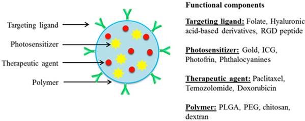
Photo-triggered diagnosis using nanocarriers
Photo-based diagnostic modalities are used extensively for several applications including in vivo imaging, targeted detection of tumors, and various diagnostic and therapeutic purposes. Commonly used photo-based techniques include molecular imaging, optical imaging, fluorescence imaging, OCT, multimodal imaging, positron emission tomography and so on. Molecular imaging is a rapidly developing field wherein optics has played a major role. The spectroscopic nature and high resolution imaging capabilities of light provides a means for characterizing biological morphology and function at molecular and cellular levels [56]. To identify and characterize various fundamental processes at the organ, tissue, cellular and molecular levels, several different NP designs using light-sensitive novel imaging agents have been developed. The modalities used to track these NPs in vivo have been summarized in Table 1. Optical imaging comprises of detection of light photons that travel through the tissues [57]. After the drug-loaded particle is administered inside the body, optical imaging is a tool that follows or tracks the delivery vehicle to determine whether the drug is effectively delivered to the desired tissue or the organ [58]. Nanomaterials play an important role in optical imaging. Metal-based NPs such as QDs, gold and silica nanoparticulates and photosensitizer-containing nanocarriers including liposomes as well as polymeric and ceramic NPs can all be used in optical imaging.
Nanomaterials for photo-based diagnostic applications.
| CORE | SHELL | MODALITY | APPLICATION | Ref. |
|---|---|---|---|---|
| Gold/Silver | Silica | Optical Sensing | Labeling cancer cells | [106] |
| Silica | Gold | Fluorescent Bioimaging | Fluorescence marking of cells/tissues for imaging | [106] |
| Magnetic NPs | Silica | Dual-imaging | Targeting, Cell sorting, Bioimaging | [107] |
| Gold/CdSe | PEG | Nanoscopic sensing | Bioimaging | [108] |
| SPIONs [17] | Amphiphilic Polymer | MRI | Cancer Imaging | [109] |
| Nanocrystals [18] | PLGA | Medical Imaging | In vivo Cancer Imaging | [110] |
| QDs/Gold NPs [18] | PLGA | CT/Optical Imaging | In vivo Cancer Imaging | [110] |
Among the different imaging techniques available, OCT is used to obtain high resolution (around 10-15 μm) cross sectional images in real time for biological tissues. OCT works on the principle of detecting reflections of a low coherence light source which is targeted into the tissue to determine at what depth the reflection has reached [59]. OCT is thus an optimal technique for examining complex structures such as tissue scaffolds to dimensionally characterize and optimize cell growth [60]. Gold and iron oxide NPs have been frequently used as OCT contrast agents [61]. Besides metal-based NPs, Au et al. [62] was the first to report a polymer-based contrast agent for OCT using polypyrrole (PPy) NPs that gave a strong contrast at around 1300 nm in an intralipid tissue phantom using OCT.
Besides OCT, quantitative spectroscopy is another light-based technique in use. For example, tracking of NPs delivered to living cancer cells can be done with the aid of fluorescence microspectroscopy. A spectral shift in the wavelength suggests the delivery of NPs in the cancer cells. The cellular uptake and aggregation of the particles can also be determined by spectroscopy. Fluorescent conjugated/encapsulated polymer NPs are often used for this technology. For example, PLGA polymeric NPs are used to encapsulate various fluorescent dyes for cellular imaging [63]. Another example includes fluorescent polydioctylfluorenebenzothiadiazole-bisthiophenyl benzothiadiazole copolymer (PFBTTB) NPs, which were synthesized and conjugated with Herceptin to make a probe suitable for targeting and imaging of HER2-overexpressing cancer cells. This type of system is also used for targeted therapy and imaging of cancer [64].
Imaging of cells can be further done by photoacoustic imaging, which is a noninvasive, nonionizing imaging modality that is a combination of high sensitivity of optical methods and outstanding resolution of the acoustic methodology. Photoacoustic imaging is done by two main techniques: photoacoustic microscopy and photoacoustic computed tomography. Photoacoustic imaging has been used to image blood vessels, tumors, hemoglobin oxygenation, and tumor angiogenesis. For photoacoustic imaging, gold nanoshells with a silica core and GNRs are frequently used. In vivo imaging of rat brain cortical blood vessels showed a significant increase in blood vessel absorption with gold nanoshells, which denotes its successful application as an NIR (Near Infrared) photoacoustic contrast agent [65]. Gold nanomaterials are commonly used for photoacoustic computed tomography as well. They have gained attention because of their plasmonic properties that are size and shape dependent and can absorb light in the visible to NIR region. This quality of bioengineered gold NPs also makes them useful for image guided therapy and photothermal ablation of the tumors. Gold surfaces can also be conjugated with biologically and chemically relevant entities for multimodal capabilities like MRI, PET, CT, and conventional microscopic optical techniques.
Multimodal Imaging is a cutting edge research which has been developed and applied because of the issues faced by biomedical technology today, including heterogeneity of the disease and patients. Personalized therapies might be more effective as the progress of disease is different for each patient, depending on genetic factors, environmental factors and the characteristic of the disease [66]. Thus multimodal imaging has several advantages over single imaging modalities, like high sensitivity, multicolor imaging, no tissue penetrating limits and so on. To overcome the limitations of the single imaging technique, researchers combined two different contrast agents into a single nanoparticle, which can be further imaged by different imaging modalities. For example, combining MRI for anatomical resolution and optical imaging for its sensitivity is a very effective procedure for finding and quantifying the size of the tumor or metastases that cannot be detected by MRI alone. This multimodal imaging is also used for monitoring enzyme activity and imaging brain tumors [67] as well as for detecting apoptosis and atherosclerosis [65]. Multimodal NPs containing up-conversion NPs as the core, layer of iron-oxide particles as the intermediate layer, and the outer-most layer of gold have been developed and used for upconversion luminescence (UCL)/magnetic resonance (MR) multimodal imaging and photothermal ablation of tumors [32]. Dual-modal fluorescent magnetic NPs by co-encapsulation of far-red/near-infrared (FR/NIR)-emissive conjugated polymer (PFVBT) and lipid coated iron-oxides into folate conjugated PLGA-PEG and PLGA NPs are other examples for nanomaterials used in the dual imaging system, in vivo fluorescence and MRI, to detect tumors [68].
Photo-triggered therapy using nanocarriers
In addition to imaging and diagnostic applications, nanomaterials have also been developed for several light-based therapeutic uses as summarized in Table 2. Photodynamic therapy (PDT) is an important approach that uses light-activated drugs for the treatment of various ailments such as cancer [69]. This method of therapy involves three essential components—light, photosensitizers and oxygen [69, 70]. The photosensitizers convert light energy into a type II chemical reaction. In clinical practice, photosensitizers are basically from three families of porphyrins, chlorophylls and dyes. Some of the photosensitizers currently in clinical trials have been summarized in Table 3. Among the different photosensitizers in use today, metallo-phthalocyanine has attracted substantial interest due to their photodynamic (PD) properties that can be easily tuned by the type of metal ion involved. For example, zinc-phthalocyanine nanowires synthesized by Moon et al. [71] showed excellent photosensitizing capabilities as well as highly efficient dual PD and photothermal (PT) effects upon irradiation of NIR laser. Studies performed by Jeong et al. [70] also demonstrated the use of photosensitizer-conjugated human serum albumin NPs as impressive photodynamic drug delivery candidates with good biocompatibility and tumor-targeting abilities.
Nanomaterials for photo-based therapeutic applications.
| Materials | Encapsulated agent | Modality | Applications | Therapy | Ref. |
|---|---|---|---|---|---|
| Gold NPs | Toluidine Blue O (TBO) | Absorption spectroscopy FTIR spectroscopy | Colon cancer treatment | PDT | [111] |
| Human serum Albumin (HAS) | Chlorin e6 (ce6) | In vivo fluorescence Imaging | Colon cancer treatment | PDT | [70] |
| CdTe &CdSe QD / silica shell | - | Fluorescence spectrophotometer | Skin cancer treatment | PTT | [4] |
| Gold-NPS | ICG | Femtosecond laser imaging system | Lung cancer treatment | PTT, PDT | [74] |
| Gold-nanorods/PEG | - | Inductively coupled plasma mass spectrophotometry (ICP-MS) | Cancer treatment | PTT | [77] |
| Pluronic NPs | Pthalocyanine dye | NIR fluorescence imaging | Liver cancer treatment | NIR therapy | [82] |
| Gold/Silica nanoshells | - | IR thermography | Cancer treatment | Laser -induced hyperthermia | [81] |
| Silica / Gold nanoshells | ICG | Absorption spectrometery, photoacoustic tomography (OAT/PAT) | Cancer surgery | Laser activate NP for tissue bonding | [112] |
| Gold NPS | PEG-Si RNA | fluorescence spectrophotometry | Cancer treatment | Photoinduced RNA interference therapy | [84] |
Photosensitizers currently in various stages of clinical trials for photo-based applications
| No. | Photosensitizer | Application | Stage completed | Ref |
|---|---|---|---|---|
| 1 | Photofrin | PDT of various carcinomas | FDA-approved | [113], [114] |
| 2 | Levulan | PDT for actinic keratosis treatment | FDA-approved | [115] |
| 3 | Metvix | PDT for actinic keratosis treatment | FDA-approved | [116] |
| 4 | Visonac(methyl aminolevulinate) | PDT of moderate acne by killing bacteria and action on sebaceous gland | Phase IIb | [117] |
| 5 | Verteporfin | PDT of neovascular age-related macular degeneration | Phase IIIb | [118] |
| 6 | δ-aminolaevulinic acid | PDT for basal cell carcinoma | Phase III | |
| 7 | Zinc phthalocyanine tetrasulfonate | PDT for naturally occurring tumors in dogs | Phase I | [119] |
| 8 | Radachlorin | PDT of skin cancer | Phase II | [120] |
| 9 | HPPH (2-[1-hexyloxyethyl]-2-devinyl pyropheophorbide-a) | PDT of obstructive esophageal tumors | Phase I/II | [121] |
| 10 | Silicon phthalocyanine | PDT of cutaneous neoplasms | Phase I | [122] |
| 11 | Hexaminolevulinate (Hexvix) | PDT of intermediate or high-risk urothelial cell | Phase I | [123] |
| 12 | BF-200 5- aminolaevulinic acid | PDT of actinic keratosis | Phase III | [124] |
| 13 | Hemoporfin | PDT for port-wine stain | Phase IIa | [125] |
PTT is another type of photo-based physical treatment which has recently attracted a lot of interest as a minimally invasive treatment modality and an alternative to currently used cancer treatments. This therapy involves irradiating the tumor with electromagnetic radiation (via NIR), which is absorbed by photoabsorbers and is converted to heat, which in turn causes thermal damage. It is very important to choose the appropriate wavelength where the absorption of the diseased tissue is greater than the surrounding tissue. Various photothermal agents like GNRs, nanoshells, nanocages as well as carbon nanotubes have been developed for PTT treatment of cancer [72]. QDs have also been recently gaining prominence chiefly due to their size-dependent fluorescence, narrow emission spectra, stability against photobleaching and stronger fluorescence than commonly used dyes [73]. Chu et al. [4] worked on CdTe and CdSe QDs which converted light energy rapidly into heat when injected into mouse melanoma tumor. Results showed a remarkable decrease in the size of the tumor. In addition to QDs, light-absorbing dyes have also been used to increase the thermal damage in the target tumors. For example, nanostructures consisting of ICG and phospholipid-polyethylene glycol (ICG-PL-PEG) were developed for PTT [74, 75]. The results suggested that ICG-PL-PEG suspension showed a more efficient NIR-temperature dependent increase compared to ICG dyes alone. Moreover, they could be retained inside the tumor when conjugated with integrin α(v)β(3) monoclonal antibody. We have previously encapsulated ICG in PLGA NPs conjugated with targeting ligands for prostate cancer-specific targeting, optical imaging and PTT [76]. Results showed that our NPs were biocompatible and uptaken significantly by PC3 prostate cancer cells. Additionally, these NPs gave significant contrast even at 3 cm depth below in the laboratory tissue phantom and showed significant temperature gains in vitro and in phantoms, demonstrating their potential for deep tissue imaging and cancer hyperthermia. Further, Lin et al. [77] developed GNRs which showed enhanced PTT when used on the soft tissue of genetically engineered mice, due to higher absorbance of these nanorods when compared to chromophores and NIR dyes. The absorption cross sections of GNRs are at least five orders larger than commonly used dyes. Further, the scattering of light by these nanorods is much larger than the light emission from highly fluorescent dyes [78].
Besides PDT and PTT, NIR also has various applications in applied biology. Due to the wavelength being longer than the red light, it can penetrate into deeper tissues easily. Moreover, it offers low scattering, energy absorption and inhibits most of the autofluorescence emitted from surrounding tissue [79]. Small diameter GNRs, gold-nanocages, gold-nanoshells have been recently gaining a lot of attention in NIR due to their stability and biocompatibility [80]. Recently, a novel near infrared photoimmunotherapy (PIT) using a new type of monoclonal antibody (mAb) has been used to monitor in vivo acute necrotic cancer cell death in real-time. This method proved to be advantageous in acutely monitoring the cytotoxicity effects of NIR mediated mAb induced PIT with the help of fluorescence time (FLT) imaging even before the morphological changes are seen in the targeted tumor [81]. Phthalocyanine-aggregated Pluronic (FPc) NPs were formulated as another novel type of NIR absorber for PTT by Lim et al. [82]. The FPc NPs were injected into SCC7 tumor-bearing mice and the NIR fluorescent images displayed a marked image contrast which was only seen at the tumor. Moreover, the tumor signal was further increased with the passage of time, while the whole-body signal decreased.
One of the present day tools using phototherapy to cure cancer is the laser, which can be used to induce hyperthermia [79]. A major drawback of this technique is the low spatial selectivity in heating of tumors and surrounding tissues. Terentyuk et al. [83] attempted to resolve this issue using plasmonic silica/gold nanoshells, which were used to produce controllable laser hyperthermia in tissues, hence increasing the photothermal effect in cancer cells. The combination uses of the laser and other minimally invasive techniques have resulted in their increasing popularity in the field of surgical medicine. This is because of their ability to reduce pain and promote faster recovery. One of the innovative approaches implemented today is the combination of NIR, laser and optical absorber for treatment. Indocyanine Green (ICG) dye has attracted attention recently for this combination approach, as it is FDA approved and has proven to be safe for surgical applications in humans [5]. Despite the vast research in this area, only a few designs have reached the clinical trials due to their failure at local heating of tissues.
In addition to drugs, genetic materials can also be incorporated in NPs for photo-based therapies. RNA interference (RNAi) has been majorly used to understand the gene functions. Photoinduced RNAi was developed in 2005, which was then modified with the help of photochemical internalization to RNAi [84]. An example is the development of a polymer using β-cyclodextrin containing polymers based on six methylene units, which entraps short interfering RNA (siRNA) and which could be controlled completely by light [84]. The human S100A4 protein in OHS cells was used as the target gene in this study. The results showed that silencing efficiency was almost 90% compared with the untreated control, and there was no effect on cell viability by the treatment. Moreover, gold NPs have been conjugated with siRNA which helps in achieving deep penetration of the tissues at NIR wavelengths [85]. Another novel design is proposed by Endoh et al. [85] for delivering of short hairpin (shRNA) by photo-stimulation of fluorescently labeled protein carriers.
Photo-triggered theranostic nanocarriers
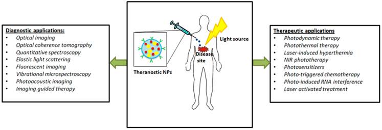
Theranostic nanomedicine is a branch of nanobiotechnology that involves state-of-the-art NPs capable of carrying out simultaneous detection/diagnosis and treatment of the disease following administration. It is derived from a combination of the two words 'therapy' and 'diagnosis'. Theranostic NPs that utilize light-based techniques for monitoring and treating diseases are of interest as they make use of minimally invasive modalities that reduce patient discomfort (Figure 2). For example, Khlebtsov et al. [86] synthesized multifunctional NPs consisting of a gold-silver nanocage core surrounded by a silica shell containing NIR photosensitizer Yb-2,4-dimethoxyhematoporphyrin (Yb-HP) for monitoring tumor as well as simultaneous therapy by PDT and plasmonic heating. Significantly higher death of HeLa cervical cancer cells occurred in vitro when they were incubated with the NPs and irradiated with light, due to plasmonic PTT by the gold-silver nanocages as well as photodynamic therapy in the presence of Yb-HP. Further, the IR luminescence of Yb-HP can be used for diagnostic purposes within the tissue transparency window. Another example of theranostic NPs are the gold layered-carbon nanotubes prepared by Galanzha et al. [87] and conjugated with folate and CD44-specific antibodies to specifically detect and target circulating breast cancer cells in vivo. Non-plasmonic silica NPs containing multiple dyes have been formulated by Singh et al. [88] for theranostic applications. These particles consisted of NIR fluorescent heptamethine cyanine dye and Si-naphthalocyanine heating dyes for NIR fluorescence imaging and increased light absorption respectively. The latter was incorporated for enhanced PTT. By direct tumor injection into mice, the authors were able to observe high fluorescence intensity and about 95% decrease in tumor viability following irradiation with NIR laser source.
Diagnostic and therapeutic applications of photo-based theranostic NPs to treat various malignancies.
With advances in nanotechnology-based research, NPs with multifunctional capabilities are gaining importance due to the combined advantages of various imaging and therapeutic modalities encapsulated within a single nanoparticle [89]. For example, Santra et al. [7] formulated novel folic acid-conjugated, poly acrylic acid-coated iron oxide NPs that are encapsulated with NIR dyes, 4-Dil and 4-DiR, and with an anticancer drug, Taxol. These NPs could aide in cancer diagnosis using both optical and MR imaging and cancer treatment via targeted drug delivery. Another novel NP formulation for dual-imaging was prepared by Huan et al. [90]. This group synthesized multi-functional folic acid-conjugated and silica-modified gold nanorods (GNR-SiO2-FA) which can be simultaneously imaged in vivo using X ray or computed tomography (CT) followed by treatment via radiation therapy (RT) or PTT. These multi-functional nanorods could specifically bind to folate receptors on the surface of MGC803 gastric cancer cells in vitro and were imaged in vivo using both X-ray and CT imaging.
The interest in dual therapy-enabled theranostic NPs has also increased due to their ability to demonstrate a greater therapeutic effect than the individual treatment alone. This is clearly demonstrated by Wu et al. [91], who had synthesized novel core-shell nanogels comprising of a silver-gold bimetallic core and a thermoresponsive poly ethylene glycol (PEG) based shell containing hyaluronic acid-based targeting ligands. These nanogels can be simultaneously used for cancer-specific targeting, fluorescence imaging, optical temperature-sensing as well as both chemo- and photothermal-therapy. The enhanced intensity of photoluminescent emissions from silver-gold core, in response to temperature-dependent shrinking of the shell, can be used for optical temperature sensing as well as cellular imaging. The hyaluronic acid strands on the surface can specifically target cluster determinant 44 (CD44) overexpressed on various cancer cells. Moreover, irradiation of B16F10 skin cancer cells in vitro using NIR light following uptake of Temozolomide (TMZ) -loaded NPs showed a photothermal effect due to the bimetallic core. The subsequent temperature increase in the region further stimulated the release of chemotherapeutic TMZ from the thermosensitive shell. Also Lee et al. [92] developed novel PEG-PLGA-gold half-shell NPs (~120-125 nm) encapsulating DOX for chemo-photothermal therapy and imaging following NIR irradiation. Intravenous and intratumoral injection of NPs in nude mice resulted in their accumulation in the tumor region. Further, the combined chemo- and photothermal therapy on NIR irradiation resulted in tumor destruction without causing weight loss in the mice and/or recurrence of the tumor. This method of cancer treatment was found to be more effective than chemotherapy or PTT alone.
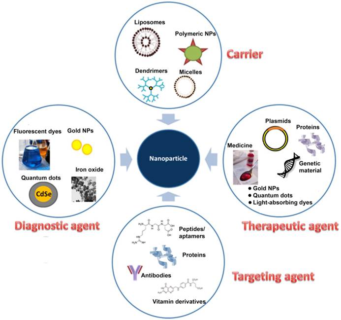
Aside from chemotherapeutic drugs, NPs can also be encapsulated with a select radionuclide for nuclear imaging and/or radiation therapy of tumors. For instance, Rossin et al. [93] synthesized 64Cu-radiolabeled shell cross-linked (SCK) NPs conjugated with folate for imaging and radiotherapy of tumors. The micelles prepared using poly(acrylic acid-b-methyl-acrylate) diblock copolymer conjugated with folic acid and radiolabeled with 64Cu were retained in the tumor in vivo and could potentially be used for imaging and treatment of early-stage small-sized tumors. Another example is a novel QD-photosensitizer (Photofrin) conjugate synthesized by Yang et al. [94] for enhancing radiation therapy. In vitro studies on H460 human lung carcinoma cell lines showed significant cell death in the presence of the QD-photofrin conjugates in combination with radiation, than in the presence of only one of the components [94]. These conjugates are safer than radiosensitizers which tend to have toxicity issues following administration. Besides QDs, gold NPs can also be used in radiation therapy as demonstrated by Hainfield et al. [95]. The gold NPs exposed to high X-ray doses of 42 Gy radiation and 157 kiloelectron volt (keV) beam energy was successful in reducing tumor volume as well as long-term survival in radio-resistant mouse head and neck squamous cell carcinoma by combined radiation therapy and hyperthermia. Thus we can see that theranostic photo-triggered NPs have opened the doors to new and advanced research that can integrate diagnostic and therapeutic components into a single particle for effective detection and treatment of various diseases (Figure 3).
The various functional components of a theranostic photo-triggered nanoparticle for effective diagnosis and treatment of the disease.
Medical applications of photo-based theranostic NPs
With the rising incidence of cancer among people in recent years, research is now extensively focused on the development of multifunctional NPs that can simultaneously be used for diagnosis as well as site-specific treatment of cancer. Photo-based theranostic NPs have been used for applications ranging from detection and treatment of circulating tumor cells to photo-based therapy of small-sized and late stage tumors [87]. For example, Bagalkot et al. [96], had developed novel aptamer-QD-DOX conjugates that can selectively target prostate-specific membrane antigen (PSMA)-overexpressing prostate cancer cells, be imaged by dual fluorescence resonance energy transfer (Bi-FRET) due to interaction between DOX-QD and DOX-aptamer, and provide cancer therapy by release of DOX within the cells. Following endocytic uptake, doxorubicin is separated from the nanoconjugates resulting in separate fluorescence from the QDs. This novel formulation thus helps in detection of NPs at a cellular level. Gao et al. [97] had also developed semiconductor QDs coated with an amphiphilic triblock polymer and conjugated with PSMA monoclonal antibody for prostate cancer targeting and spectral imaging. In vivo uptake study showed accumulation of the ligand-conjugated QDs in the PSMA-overexpressing C4-2 prostate cancer xenografts in nude mice, which could be visualized by fluorescence imaging. Recently, Cheng et al. [98] synthesized tri-functionalized mesoporous silica NPs incorporating cRGDyK peptides for αvβ3 integrin-specific targeting, an NIR fluorescent contrast agent ATTO647N for imaging, a palladium-porphyrin based photosensitizer for PDT. The NPs were found to be cytocompatible and showed a significantly higher uptake by U87-MG human glioblastoma cells overexpressing αvβ3 integrins, compared to the integrin receptor-deficient MCF-7 breast cancer cells. Similarly, Gianella et al. [6] developed multifunctional nanoemulsions encapsulating fluorescent dyes Cy7 and iron oxide nanocrystals for simultaneous NIR fluorescence and MRI for imaging-guided cancer therapy. The nanoemulsions further contained hydrophobic glucocorticoid prednisolone acetate valerate (PAV) as the therapeutic agent and RGD peptide for targeting tumor angiogenesis. The formulation was biodegradable, successfully imaged tumors in vivo and significantly reduced the tumor size.
Despite the increasing focus on a suitable nano-sized system specifically for diagnosis and treatment of several types of cancer, there has also been significant research in the development of theranostic NPs for treatment of other diseases. For example, McCarthy et al. [99] recently developed a multimodal crosslinked dextran-coated iron oxide (CLIO) nanoparticle for treatment of inflammatory atherosclerosis. The NIR fluorophore AlexaFluor 750 encapsulated within the CLIO can be used for NIR fluorescence imaging. Further, a chlorin-based photosensitizer used could be activated by light to display phototoxicity towards macrophages responsible for inflammation in the atherosclerotic lesions. Lim et al. [100] had also synthesized dextran-coated hollow-type gold NPs for imaging and treatment of macrophages. These NPs were highly uptaken by RAW274.7 macrophage cells and were capable of scattering NIR light for imaging followed by PTT. Further, the FDA has approved Visudyne as a light-activated liposome consisting of a photosensitizer (hydro-monobenzoporphyrin) and a phospholipid mixture (egg phosphatidyl glycerol (EPG) and dimyristoyl phosphatidyl choline (DMPC)) which can be used in the treatment of macular degeneration [3]. Another application of laser-activated NPs is tissue bonding as the heat generates by irradiation of the NPs via the appropriate laser resulting in reorganization of the components of the tissue, leading to wound closure and repair. For example, SPIO NPs (15 nm) have been used to produce heat and solder albumin onto rabbit aortic arteries ex vivo by applying radiowaves [101]. Also, small gold NPs are used as photothermal transducers to generate heat for non-invasive tissue bonding applications, ranging from skin lesions to explanted lens capsular tissues [102].
The applications of photo-based NPs extend to treatment of bacterial infections as well. For instance, gold NPs conjugated with anti-protein A antibodies were used to selectively target and kill Gram-positive Staphylococcus Aureus. Following uptake by the bacterium, the NPs were irradiated with focused laser pulses (420-520 nm) which were converted to heat resulting in irreparable damage of the bacterium [103]. Similarly, Huang et al. [104] developed multifunctional Fe3O4-gold nanoeggs with vancomycin immobilized on the surface, which are capable of absorbing NIR for PTT to treat bacterial infections. They further demonstrated that the use of an external magnet is useful in bacterial aggregation and more effective PTT.
Conclusions and Future outlook
This paper has attempted to summarize the existing research in the field of photo-based nanomedicine. The continuous demand for effective clinical solutions to cure diseases with minimal patient discomfort has led many researchers to turn to the field of nanotechnology in order to develop systems that can be tracked/imaged following administration and used for targeted and localized drug delivery. Although extensive research is being conducted in this area, we still have a long way to go in terms of using these nano-systems in humans. Firstly, efforts must be taken to achieve mass-production of the NPs so as to translate the system from bench to bedside. Also, a comprehensive study of the biocompatibility of the nanoparticulate system and their interaction with cells and tissues needs to be done to ensure their safety before use in humans. Each component of these NPs would have to be tested extensively for immune reactions and toxicity in vivo. Theranostic NPs are slowly transitioning into clinical trials; however, these NPs can potentially overcome limitations of existing treatment techniques and provide a safe and reliable means of diagnosing and treating diseases in a minimally invasive manner with less patient discomfort and high accuracy. Photo-based theranostic NPs have thus emerged as a possible effective system that unites different aspects of chemistry, biology, bioinformatics, medical physics and various other fields to form a joint solution that can overcome various issues in human health today [105]. However, due to the nascent stage of this field of study, there are still several issues to be addressed before these photo-based theranostic particles can be translated to clinical use. Firstly, different modalities and nanoparticle components need to be tested to optimize the depth up to which NPs containing photosensitizers and metal-based imaging agents can be imaged. This would be particularly necessary for deep tissue imaging of NPs following administration. Further, sensitive imaging techniques should be employed for accurate detection of signals from the NPs. The different photo-based therapies such as PDT and PTT using photo-based theranostic NPs should cause minimal damage to surrounding healthy tissue. Due to the toxic nature of some of the photosensitive payloads such as QDs, it is also imperative that the NPs be cleared from the system before complete degradation of the polymeric shell and subsequent release of encapsulated agents. Regardless, these nano-carriers have the potential to make significant strides towards diagnosis and treatment of different diseases due to their unique qualities such as good spatial resolution, controllable drug release and active targeting for efficient and target-specific delivery of the encapsulated agent.
Competing Interests
The authors have declared that no competing interest exists.
References
1. Ackroyd R, Kelty C, Brown N, Reed M. The history of photodetection and photodynamic therapy. Photochem Photobiol. 2001;74:656-69
2. Macdonald IJ, Dougherty TJ. Basic principles of photodynamic therapy. J Porphyrins and Phthalocyanines. 2001;5:105-29
3. Rai P, Mallidi S, Zheng X, Rahmanzadeh R, Mir Y, Elrington S. et al. Development and applications of photo-triggered theranostic agents. Adv Drug Deliv Rev. 2010;62:1094-124
4. Chu M, Pan X, Zhang D, Wu Q, Peng J, Hai W. The therapeutic efficacy of CdTe and CdSe quantum dots for photothermal cancer therapy. Biomaterials. 2012;33:7071-83
5. DeCoste SD, Farinelli W, Flotte T, Anderson RR. Dye-enhanced laser welding for skin closure. Lasers Surg Med. 1992;12:25-32
6. Gianella A, Jarzyna PA, Mani V, Ramachandran S, Calcagno C, Tang J. et al. Multifunctional nanoemulsion platform for imaging guided therapy evaluated in experimental cancer. ACS Nano. 2011;5:4422-33
7. Santra S, Kaittanis C, Grimm J, Perez JM. Drug/dye-loaded, multifunctional iron oxide nanoparticles for combined targeted cancer therapy and dual optical/magnetic resonance imaging. Small. 2009;5:1862-8
8. Liu G-Y, Chen C-J, Li D-D, Wang S-S, Ji J. Near-infrared light-sensitive micelles for enhanced intracellular drug delivery. J Mater Chem. 2012;22:16865-71
9. Katz JS, Burdick JA. Light-responsive biomaterials: development and applications. Macromol Biosci. 2010;10:339-48
10. Chen J, Glaus C, Laforest R, Zhang Q, Yang M, Giddling M. et al. Gold nanocages as photothermal transducers for cancer treatment. Small. 2010;6:811-7
11. Health Quality Ontario. Nanotechnology: an evidence-based analysis. Ont Health Technol Assess Ser. 2006;6:1-43
12. Nagesha D, Laevsky GS, Lampton P, Banyal R, Warner C, DiMarzio C. et al. In vitro imaging of embryonic stem cells using multiphoton luminescence of gold nanoparticles. Int J Nanomedicine. 2007;2:813-9
13. Young J, Figueroa E, Drezek R. Tunable nanostructures as photothermal theranostic agents. Biomedical Engineering Society. 2012;40:438-59
14. Day ES, Bickford LR, Slater JH, Riggall NS, Drezek RA, West JL. Antibody-conjugated gold-gold sulfide nanoparticles as multifunctional agents for imaging and therapy of breast cancer. Int J Nanomedicine. 2010;5:445-54
15. Choi WI, Kim JY, Kang C, Byeon CC, Kim YH, Tae G. Tumor regression in vivo by photothermal therapy based on gold-nanorod-loaded, functional nanocarriers. ACS Nano. 2011;5:1995-2003
16. Kuo W-S, Chang C-N, Yang Y-T, Yang M-H, Chien Y-H, Chen S-J. et al. Gold Nanorods in photodynamic therapy, as hyperthermia agents, and in near-infrared optical imaging. Angewandte Chemie. 2010;122:2771-5
17. Skrabalak SE, Chen J, Sun Y, Lu X, Au L, Cobley CM. et al. Gold nanocages: synthesis, properties, and applications. Acc Chem Res. 2008;41:1587-95
18. Yavuz MS, Cheng Y, Chen J, Cobley CM, Zhang Q, Rycenga M. et al. Gold nanocages covered by smart polymers for controlled release with near-infrared light. Nat Mater. 2009;8:935-9
19. Qian X, Peng XH, Ansari DO, Yin-Goen Q, Chen GZ, Shin DM. et al. In vivo tumor targeting and spectroscopic detection with surface-enhanced Raman nanoparticle tags. Nat Biotechnol. 2008;26:83-90
20. Lapotko DO, Lukianova E, Oraevsky AA. Selective laser nano-thermolysis of human leukemia cells with microbubbles generated around clusters of gold nanoparticles. Lasers Surg Med. 2006;38:631-42
21. Lapotko D, Lukianova E, Shnip A, Zheltov G, Potapnev M, Savitsky V. et al. Laser activated nanothermolysis of leukemia cells monitored by photothermal microscopy. Proc of SPIE. 2005;5697:82-9
22. Lukianova-Hleb EY, Oginsky AO, Samaniego AP, Shenefelt DL, Wagner DS, Hafner JH. et al. Tunable plasmonic nanoprobes for theranostics of prostate cancer. Theranostics. 2010;1:3-17
23. Wagner DS, Delk NA, Lukianova-Hleb EY, Hafner JH, Farach-Carson MC, Lapotko DO. The in vivo performance of plasmonic nanobubbles as cell theranostic agents in zebrafish hosting prostate cancer xenografts. Biomaterials. 2010;31:7567-74
24. Lukianova-Hleb EY, Mutonga MBG, Lapotko DO. Cell-Specific Multifunctional Processing of Heterogeneous Cell Systems in a Single Laser Pulse Treatment. ACS Nano. 2012;6:10973-81
25. Huang X, El-Sayed M. Gold Nanoparticles: Optical properties and implementations in cancer diagnosis and photothermal therapy. J Advanced Research. 2010;1:13-28
26. Cai W, Gao T, Hong H, Sun J. Applications of gold nanoparticles in cancer nanotechnology. Nanotechnology, Science and Applications. 2008;1:17-32
27. Robinson JT, Tabakman SM, Liang Y, Wang H, Casalongue HS, Vinh D. et al. Ultrasmall reduced graphene oxide with high near-infrared absorbance for photothermal therapy. J Am Chem Soc. 2011;133:6825-31
28. Ghosh S, Bachilo SM, Simonette RA, Beckingham KM, Weisman RB. Oxygen doping modifies near-infrared band gaps in fluorescent single-walled carbon nanotubes. Science. 2010;330:1656-9
29. Fisher JW, Sarkar S, Buchanan CF, Szot CS, Whitney J, Hatcher HC. et al. Photothermal response of human and murine cancer cells to multiwalled carbon nanotubes after laser irradiation. Cancer Res. 2010;70:9855-64
30. Rossella F, Soldano C, Bellani V, Tommasini M. Metal-filled carbon nanotubes as a novel class of photothermal nanomaterials. Adv Mater. 2012;24:2453-8
31. Zhang L, Zhen SJ, Sang Y, Li JY, Wang Y, Zhan L. et al. Controllable preparation of metal nanoparticle/carbon nanotube hybrids as efficient dark field light scattering agents for cell imaging. Chem Commun (Camb). 2010;46:4303-5
32. Cheng L, Yang K, Li Y, Zeng X, Shao M, Lee ST. et al. Multifunctional nanoparticles for upconversion luminescence/MR multimodal imaging and magnetically targeted photothermal therapy. Biomaterials. 2012;33:2215-22
33. Hessel CM, Pattani VP, Rasch M, Panthani MG, Koo B, Tunnell JW. et al. Copper selenide nanocrystals for photothermal therapy. Nano Lett. 2011;11:2560-6
34. Hong C, Kang J, Lee J, Zheng H, Hong S, Lee D. et al. Photothermal therapy using TiO2 nanotubes in combination with near-infrared laser. J Cancer Therapy. 2010;1:52-8
35. Stuart MA, Huck WT, Genzer J, Muller M, Ober C, Stamm M. et al. Emerging applications of stimuli-responsive polymer materials. Nat Mater. 2010;9:101-13
36. Chen Y, Gryshuk A, Achilefu S, Ohulchansky T, Potter W, Zhong T. et al. A novel approach to a bifunctional photosensitizer for tumor imaging and phototherapy. Bioconjug Chem. 2005;16:1264-74
37. Bae BC, Na K. Development of Polymeric Cargo for Delivery of Photosensitizer in Photodynamic Therapy. International Journal of Photoenergy. 2012
38. Winnik FM, Maysinger D. Quantum dot cytotoxicity and ways to reduce it. Acc Chem Res. 2012
39. Montanari J, Maidana C, Esteva MI, Salomon C, Morilla MJ, Romero EL. Sunlight triggered photodynamic ultradeformable liposomes against Leishmania braziliensis are also leishmanicidal in the dark. J Control Release. 2010;147:368-76
40. Lovell JF, Jin CS, Huynh E, Jin H, Kim C, Rubinstein JL. et al. Porphysome nanovesicles generated by porphyrin bilayers for use as multimodal biophotonic contrast agents. Nat Mater. 2011;10:324-32
41. Agrawal A, Connors M, Beylin A, Liang CP, Barton D, Chen Y. et al. Characterizing the point spread function of retinal OCT devices with a model eye-based phantom. Biomed Opt Express. 2011;3:1116-26
42. Shen R, Mu B, Du P, Liu P. Preparation of photo-sensitive degradable polymeric nanocapsules from dendrimer grafted nano-silica templates. Soft Materials. 2011;9:382-92
43. Moleavin I, Ibanescu C, Hodorog-Rusu A, Peptu E, Doroftei F, Hurduc N. Amphiphilic azopolymers capable to generate photo-sensitive micelles. Central European Journal of Chemistry. 2011;9:1117-25
44. Zhou K, Wang Y, Huang X, Luby-Phelps K, Sumer BD, Gao J. Tunable, ultrasensitive pH-responsive nanoparticles targeting specific endocytic organelles in living cells. Angewandte Chemie International Edition. 2011;50:6109-14
45. Ding H, Yu H, Dong Y, Tian R, Huang G, Boothman DA. et al. Photoactivation switch from type II to type I reactions by electron-rich micelles for improved photodynamic therapy of cancer cells under hypoxia. J Control Release. 2011;156:276-80
46. Bae BC, Na K. Self-quenching polysaccharide-based nanogels of pullulan/folate-photosensitizer conjugates for photodynamic therapy. Biomaterials. 2010;31:6325-35
47. Shim MS, Kwon YJ. Stimuli-responsive polymers and nanomaterials for gene delivery and imaging applications. Adv Drug Deliv Rev. 2012;64:1046-59
48. Umeyama T, Kawabata K, Tezuka N, Matano Y, Miyato Y, Matsushige K. et al. Dispersion of carbon nanotubes by photo- and thermal-responsive polymers containing azobenzene unit in the backbone. Chem Commun (Camb). 2010;46:5969-71
49. Qian W, Murakami M, Ichikawa Y, Che Y. Highly Efficient and Controllable PEGylation of Gold Nanoparticles Prepared by Femtosecond Laser Ablation in Water. The Journal of Physical Chemistry C. 2011;115:23293-8
50. Schipper ML, Iyer G, Koh AL, Cheng Z, Ebenstein Y, Aharoni A. et al. Particle size, surface coating, and PEGylation influence the biodistribution of quantum dots in living mice. Small. 2009;5:126-34
51. Umeda YK C, Harada A, Horinaka H, Kono K. PEG-attached PAMAM dendrimers encapsulating gold nanoparticles: growing gold nanoparticles in the dendrimers for improvement of their photothermal properties. Bioconjug Chem. 2010;21:1559-64
52. Lu Z-R. Molecular imaging of HPMA copolymers: Visualizing drug delivery in cell, mouse and man. Advanced Drug Delivery Reviews. 2010;62:246-57
53. Ren K, Purdue PE, Burton L, Quan LD, Fehringer EV, Thiele GM. et al. Early detection and treatment of wear particle-induced inflammation and bone loss in a mouse calvarial osteolysis model using HPMA copolymer conjugates. Mol Pharm. 2012;8:1043-51
54. Bogart L, Taylor A, Cesbron Y, Murray P, Levy R. Photothermal microscopy of the core of Dextran-coated iron oxide nanoparticles during cell uptake. ACS Nano. 2012;6:5961-71
55. Cheng F-Y, Su C-H, Wu P-C, Yeh C-S. Multifunctional polymeric nanoparticles for combined chemotherapeutic and near-infrared photothermal cancer therapy in vitro and in vivo. Chem Commun (Camb). 2010;46:3167-9
56. Boppart SA, Oldenburg AL, Xu C, Marks DL. Optical probes and techniques for molecular contrast enhancement in coherence imaging. J Biomed Opt. 2005;10:41208
57. Jiang S, Gnanasammandhan MK, Zhang Y. Optical imaging-guided cancer therapy with fluorescent nanoparticles. J R Soc Interface. 2010;7:3-18
58. Licha K, Olbrich C. Optical imaging in drug discovery and diagnostic applications. Adv Drug Deliv Rev. 2005;57:1087-108
59. Loo C, Lin A, Hirsch L, Lee MH, Barton J, Halas N. et al. Nanoshell-enabled photonics-based imaging and therapy of cancer. Technol Cancer Res Treat. 2004;3:33-40
60. Tomlins PH, Wang RK. Theory, developments and applications of optical coherence tomography. J Phys D: Appl Phys. 2005;38:2519
61. Aaron JS, Oh J, Larson TA, Kumar S, Milner TE, Sokolov KV. Increased optical contrast in imaging of epidermal growth factor receptor using magnetically actuated hybrid gold/iron oxide nanoparticles. Opt Express. 2006;14:12930-43
62. Au KM, Lu Z, Matcher SJ, Armes SP. Polypyrrole nanoparticles: a potential optical coherence tomography contrast agent for cancer imaging. Adv Mater. 2011;23:5792-5
63. Li K, Liu B. Polymer encapsulated conjugated polymer nanoparticles for fluorescence bioimaging. J Mater Chem. 2012;22:1257-64
64. Way T-D, Chang C-J, Lin C-W. Bioconjugated fluorescent polymeric nanoparticles for imaging and targeted therapy of HER2-overexpressing cancer cells. Journal of Fluorescence. 2011;21:1669-76
65. Hahn M, Singh A, Sharma P, Brown S, Moudgil B. Nanoparticles as contrast agents for in-vivo bioimaging: current status and future perspectives. Anal Bioanal Chem. 2011;399:3-27
66. Lee DE, Koo H, Sun IC, Ryu JH, Kim K, Kwon IC. Multifunctional nanoparticles for multimodal imaging and theragnosis. Chem Soc Rev. 2012;41:2656-72
67. Fomina N, Sankaranarayanan J, Almutairi A. Photochemical mechanisms of light-triggered release from nanocarriers. Adv Drug Deliv Rev. 2012;64:1005-20
68. Li K, Ding D, Huo D, Pu K-Y, Thao NNP, Hu Y. et al. Conjugated polymer based nanoparticles as dual-modal probes for targeted in vivo fluorescence and magnetic resonance imaging. Advanced Functional Materials. 2012;22:3107-15
69. Wilson BC, Patterson MS. The physics, biophysics and technology of photodynamic therapy. Phys Med Biol. 2008;53:R61-109
70. Jeong H, Huh M, Lee SJ, Koo H, Kwon IC, Jeong SY. et al. Photosensitizer-conjugated human serum albumin nanoparticles for effective photodynamic therapy. Theranostics. 2011;1:230-9
71. Moon HK, Son M, Park JE, Yoon SM, Lee SH, Choi HC. Significant increase in the water dispersibility of zinc phthalocyanine nanowires and applications in cancer phototherapy. NPG Asia Mater. 2012;4:e12
72. Huang X, Jain P, El-Sayed I, El-Sayed M. Plasmonic photothermal therapy (PPTT) using gold nanoparticles. Lasers in Medical Science. 2008;23:217-28
73. Smith AM, Duan H, Mohs AM, Nie S. Bioconjugated quantum dots for in vivo molecular and cellular imaging. Adv Drug Deliv Rev. 2008;60:1226-40
74. Kuo WS, Chang YT, Cho KC, Chiu KC, Lien CH, Yeh CS. et al. Gold nanomaterials conjugated with indocyanine green for dual-modality photodynamic and photothermal therapy. Biomaterials. 2012;33:3270-8
75. Zheng X, Zhou F, Wu B, Chen WR, Xing D. Enhanced tumor treatment using biofunctional indocyanine green-containing nanostructure by intratumoral or intravenous injection. Molecular Pharmaceutics. 2012;9:514-22
76. Patel RH, Wadajkar AS, Patel NL, Kavuri VC, Nguyen KT, Liu H. Multifunctionality of indocyanine green-loaded biodegradable nanoparticles for enhanced optical imaging and hyperthermia intervention of cancer. J Biomed Opt. 2012;17:046003
77. Lin KY, Bagley AF, Zhang AY, Karl DL, Yoon SS, Bhatia SN. Gold nanorod photothermal therapy in a genetically engineered mouse model of soft tissue sarcoma. Nano LIFE. 2010;01:277-87
78. Ni W, Kou X, Yang Z, Wang J. Tailoring longitudinal surface plasmon wavelengths, scattering and absorption cross sections of gold nanorods. ACS Nano. 2008;2:677-86
79. Terentyuk GS, Maslyakova GN, Suleymanova LV, Khlebtsov NG, Khlebtsov BN, Akchurin GG. et al. Laser-induced tissue hyperthermia mediated by gold nanoparticles: toward cancer phototherapy. J Biomed Opt. 2009;14:021016
80. Gobin AM, Lee MH, Halas NJ, James WD, Drezek RA, West JL. Near-infrared resonant nanoshells for combined optical imaging and photothermal cancer therapy. Nano Lett. 2007;7:1929-34
81. Conde J, Doria G, Baptista P. Noble metal nanoparticles applications in cancer. Journal of Drug Delivery. 2012
82. Lim C-K, Shin J, Lee Y-D, Kim J, Oh K, Yuk S. et al. Phthalocyanine-aggregated polymeric nanoparticles as tumor-homing near-infrared absorbers for photothermal therapy of cancer. Theranostics. 2012;2:871-9
83. Terentyuk GS, Maslyakova GN, Suleymanova LV, Khlebtsov BN, Kogan BY, Akchurin GG. et al. Circulation and distribution of gold nanoparticles and induced alterations of tissue morphology at intravenous particle delivery. J Biophotonics. 2009;2:292-302
84. Matsushita-Ishiodori Y, Ohtsuki T. Photoinduced RNA interference. Acc Chem Res. 2012;45:1039-47
85. Endoh T, Ohtsuki T. Cellular siRNA delivery using TatU1A and photo-induced RNA interference. RNA Interference: Humana Press. 2010:271-81
86. Khlebtsov B, Panfilova E, Khanadeev V, Bibikova O, Terentyuk G, Ivanov A. et al. Nanocomposites containing silica-coated gold-silver nanocages and Yb-2,4-dimethoxyhematoporphyrin: multifunctional capability of IR-luminescence detection, photosensitization, and photothermolysis. ACS Nano. 2011;5:7077-89
87. Galanzha EI, Kim J-W, Zharov VP. Nanotechnology-based molecular photoacoustic and photothermal flow cytometry platform for in-vivo detection and killing of circulating cancer stem cells. J Biophotonics. 2009;2:725-35
88. Singh AK, Hahn MA, Gutwein LG, Rule MC, Knapik JA, Moudgil BM. et al. Multi-dye theranostic nanoparticle platform for bioimaging and cancer therapy. Int J Nanomedicine. 2012;7:2739-50
89. Lee C-M, Jeong H-J, Kim E-M, Kim DW, Lim ST, Kim HT. et al. Superparamagnetic iron oxide nanoparticles as a dual imaging probe for targeting hepatocytes in vivo. Magnetic Resonance in Medicine. 2009;62:1440-6
90. Huang P, Bao L, Zhang C, Lin J, Luo T, Yang D. et al. Folic acid-conjugated Silica-modified gold nanorods for X-ray/CT imaging-guided dual-mode radiation and photo-thermal therapy. Biomaterials. 2011;32:9796-809
91. Wu W, Shen J, Banerjee P, Zhou S. Core-shell hybrid nanogels for integration of optical temperature-sensing, targeted tumor cell imaging, and combined chemo-photothermal treatment. Biomaterials. 2010;31:7555-66
92. Lee S-M, Park H, Yoo K-H. Synergistic Cancer Therapeutic Effects of Locally Delivered Drug and Heat Using Multifunctional Nanoparticles. Advanced Materials. 2010;22:4049-53
93. Rossin R, Pan D, Qi K, Turner JL, Sun X, Wooley KL. et al. 64Cu-labeled folate-conjugated shell cross-linked nanoparticles for tumor imaging and radiotherapy: Synthesis, radiolabeling, and biologic evaluation. J Nucl Med. 2005;46:1210-8
94. Yang W, Read PW, Mi J, Baisden JM, Reardon KA, Larner JM. et al. Semiconductor nanoparticles as energy mediators for photosensitizer-enhanced radiotherapy. Int J of Radiation Oncology Biol Phys. 2008;72:633-5
95. Hainfeld JF, Dilmanian FA, Zhong Z, Slatkin DN, Kalef-Ezra JA, Smilowitz HM. Gold nanoparticles enhance the radiation therapy of a murine squamous cell carcinoma. Phys Med Biol. 2010;55:3045-59
96. Bagalkot V, Zhang L, Levy-Nissenbaum E, Jon S, Kantoff PW, Langer R. et al. Quantum dot-aptamer conjugates for synchronous cancer imaging, therapy, and sensing of drug delivery based on bi-fluorescence resonance energy transfer. Nano Lett. 2007;7:3065-70
97. Gao X, Cui Y, Levenson RM, Chung LW, Nie S. In vivo cancer targeting and imaging with semiconductor quantum dots. Nat Biotechnol. 2004;22:969-76
98. Cheng S-H, Lee C-H, Chen M-C, Souris JS, Tseng F-G, Yang C-S. et al. Tri-functionalization of mesoporous silica nanoparticles for comprehensive cancer theranostics-the trio of imaging, targeting and therapy. J Mater Chem. 2010;20:6149-57
99. McCarthy JR, Korngold E, Weissleder R, Jaffer FA. A light-activated theranostic nanoagent for targeted macrophage ablation in inflammatory atherosclerosis. Small. 2010;6:2041-9
100. Lim YT, Cho MY, Choi BS, Noh Y-W, Chung BH. Diagnosis and therapy of macrophage cells using dextran-coated near-infrared responsive hollow-type gold nanoparticles. Nanotechnology. 2008;19:375105
101. Bregya A, Kohlera A, Steitzb B, Petri-Finkb A, Bognid S, Alfieria A. et al. Electromagnetic tissue fusion using superparamagnetic iron oxide nanoparticles: First experience with rabbit aorta. The Open Surgery Journal. 2008;2:3-8
102. Matteini P, Ratto F, Rossi F, de Angelis M, Cavigli L, Pini R. Hybrid nanocomposite films for laser-activated tissue bonding. J Biophotonics. 2012;5:868-77
103. Zharov VP, Mercer KE, Galitovskaya EN, Smeltzer MS. Photothermal nanotherapeutics and nanodiagnostics for selective killing of bacteria targeted with gold nanoparticles. Biophys J. 2006;90:619-27
104. Huang W-C, Tsai P-J, Chen Y-C. Multifunctional Fe3O4@Au Nanoeggs as photothermal agents for selective killing of nosocomial and antibiotic-resistant bacteria. Small. 2009;5:51-6
105. Bardhan R, Lal S, Joshi A, Halas NJ. Theranostic nanoshells: from probe design to imaging and treatment of cancer. Acc Chem Res. 2011;44:936-46
106. Sounderya N, Zhang Y. Use of core/shell structured nanoparticles for biomedical applications. Recent Patents on Biomedical Engineering. 2008;1:34-42
107. Yoon TJ, Yu KN, Kim E, Kim JS, Kim BG, Yun SH. et al. Specific targeting, cell sorting, and bioimaging with smart magnetic silica core-shell nanomaterials. Small. 2006;2:209-15
108. Haidar ZS. Bio-inspired/-functional colloidal core-shell polymeric-based nanosystems: Technology promise in tissue engineering, bioimaging and nanomedicine. Polymers. 2010;2:323-52
109. Park J, Yu MK, Jeong YY, Kim JW, Lee K, Phan VN. et al. Antibiofouling amphiphilic polymer-coated superparamagnetic iron oxide nanoparticles: synthesis, characterization, and use in cancer imaging in vivo. J Mater Chem. 2009;19:6412-7
110. Mieszawska AJ, Gianella A, Cormode DP, Zhao Y, Meijerink A, Langer R. et al. Engineering of lipid-coated PLGA nanoparticles with a tunable payload of diagnostically active nanocrystals for medical imaging. Chem Commun (Camb). 2012;48:5835-7
111. Al-Majmaie R, Alattar N, Zerulla D, Al-Rubeai M. Toluidine blue O-conjugated gold nanoparticles for photodynamic therapy of cultured colon cancer. Proc SPIE 8427, Biophotonics: Photonic Solutions for Better Health Care III. 2012:842722
112. Matteini P, Ratto F, Rossi F, Pini R. Emerging concepts of laser-activated nanoparticles for tissue bonding. J Biomed Opt. 2012;17:010701
113. He XY, Sikes RA, Thomsen S, Chung LW, Jacques SL. Photodynamic therapy with photofrin II induces programmed cell death in carcinoma cell lines. Photochem Photobiol. 1994;59:468-73
114. Overholt BF, Wang KK, Burdick JS, Lightdale CJ, Kimmey M, Nava HR. et al. Five-year efficacy and safety of photodynamic therapy with Photofrin in Barrett's high-grade dysplasia. Gastrointest Endosc. 2007;66:460-8
115. Jeffes EW. Levulan: the first approved topical photosensitizer for the treatment of actinic keratosis. J Dermatolog Treat. 2002;13(Suppl 1):S19-23
116. Wiegell SR, Fabricius S, Gniadecka M, Stender IM, Berne B, Kroon S. et al. Daylight-mediated photodynamic therapy of moderate to thick actinic keratoses of the face and scalp: a randomized multicentre study. Br J Dermatol. 2012;166:1327-32
117. PhotoCure. Efficacy and safety study with Visonac photodynamic therapy (PDT); National Institute of Health Clinical trials. Bethesda (MD): National Library of Medicine. 2012
118. Schmidt-Erfurth U, Sacu S. Randomized multicenter trial of more intense and standard early verteporfin treatment of neovascular age-related macular degeneration. Ophthalmology. 2008;115:134-40
119. Borgatti-Jeffreys A, Hooser SB, Miller MA, Lucroy MD. Phase I clinical trial of the use of zinc phthalocyanine tetrasulfonate as a photosensitizer for photodynamic therapy in dogs. American Journal of Veterinary Research. 2007;68:399-404
120. Kochneva EV, Filonenko EV, Vakulovskaya EG, Scherbakova EG, Seliverstov OV, Markichev NA. et al. Photosensitizer Radachlorin®: Skin cancer PDT phase II clinical trials. Photodiagnosis and Photodynamic Therapy. 2010;7:258-67
121. Roswell Park Cancer Institute. Photodynamic Therapy Using HPPH in Treating Patients with Obstructive Esophageal Tumor; National Institute of Health Clinical trials. Bethesda (MD): National Library of Medicine. 2012
122. Baron ED, Malbasa CL, Santo-Domingo D, Fu P, Miller JD, Hanneman KK. et al. Silicon phthalocyanine (pc 4) photodynamic therapy is a safe modality for cutaneous neoplasms: results of a phase 1 clinical trial. Lasers in Surgery and Medicine. 2010;42:728-35
123. Bader MJ, Stepp H, Beyer W, Pongratz T, Sroka R, Kriegmair M. et al. Photodynamic therapy of bladder cancer - A phase I study using Hexaminolevulinate (HAL). Urologic Oncology: Seminars and Original Investigations. 2012 doi:10.1016/j.urolonc.2012.02.007
124. Dirschka T, Radny P, Dominicus R, Mensing H, Brüning H, Jenne L. et al. Photodynamic therapy with BF-200 ALA for the treatment of actinic keratosis: results of a multicentre, randomized, observer-blind phase III study in comparison with a registered methyl-5-aminolaevulinate cream and placebo. British Journal of Dermatology. 2012;166:137-46
125. Zhao Y, Zhou Z, Zhou G, Tu P, Zheng Q, Tao J. et al. Efficacy and safety of hemoporfin in photodynamic therapy for port-wine stain: a multicenter and open-labeled phase IIa study. Photodermatology, Photoimmunology & Photomedicine. 2011;27:17-23
Author contact
![]() Corresponding author: 500 UTA Blvd, ERB-241, Arlington, TX 76010. Phone: +1-817-272-2540 Fax: +1-817-272-2251 Email: knguyenedu.
Corresponding author: 500 UTA Blvd, ERB-241, Arlington, TX 76010. Phone: +1-817-272-2540 Fax: +1-817-272-2251 Email: knguyenedu.