Impact Factor
- Issue 13; 2026
- Issue 12; 2026
- Issue 11; 2026
- Issue 10; 2026
- Issue 9; 2026
- Volume 16; 2026
- Advance Articles
- Past Issues
- Cover Images
- Cover Suggestion
- Index & Coverage
- Special Issues
Introduction
Development of First-line Pt...
Molecular Mechanism of Cisplatin
Strategies to Improve...
Pt Nanoclusters (Pt NCs) as a...
Conclusion and Perspectives
Abbreviations
Acknowledgements
References
International Journal of Biological Sciences
International Journal of Medical Sciences
 Global reach, higher impact

Theranostics 2022; 12(5):2115-2132. doi:10.7150/thno.69424 This issue Cite
Review
Platinum-based drugs for cancer therapy and anti-tumor strategies
Chunyu Zhang1,2#, Chao Xu3#, Xueyun Gao2 ![]() , Qingqiang Yao1
, Qingqiang Yao1 ![]()
1. Institute of Materia Medica, Shandong First Medical University & Shandong Academy of Medical Sciences, Jinan 250000, Shandong, China.
2. Department of Life Science and Chemistry, Faculty of Environment and Life Science, Beijing University of Technology, Beijing 100124, China.
3. College of Chemistry and Material Science, Shandong Agricultural University, Tai'an 271018, Shandong, China.
#These authors contributed equally to this work.
Received 2021-11-24; Accepted 2022-1-15; Published 2022-2-7
Abstract

Platinum-based drugs cisplatin, carboplatin, and oxaliplatin are widely used for chemotherapeutic eradication of cancer. However, the side effects of platinum drugs, such as lack of selectivity, high systemic toxicity, and drug resistance, seriously limit their clinical application. With advancements in nanotechnology and chemical synthesis, Pt-based anti-cancer drugs have made great progress in cancer therapy in recent years. Many strategies relied on the anti-cancer mechanism similar to cisplatin and achieved some success by modifying existing platinum drugs. Pt-based nanodrugs, such as platinum nanoclusters, have novel anti-cancer mechanisms and great potential in tumor-targeted therapy and have shown promising results in clinical application. In this review, we systematically explored the development of first-line platinum chemotherapy drugs in the clinic and their anti-cancer mechanisms. We also summarize the progress of Pt-based anti-cancer drug application in cancer therapy, emphasizing their modification to enhance the anti-tumor effect. Finally, we address challenges faced by platinum chemotherapy drugs, especially Pt nanocluster-based nanodrugs, in cancer treatment. The new platinum drugs and their targeted modifications undoubtedly provide a promising prospect for improving the current anti-cancer treatments.
Keywords: Platinum-based drugs, Cancer therapy, Anti-cancer mechanism, Systemic toxicity, Platinum nanoclusters
Introduction
Chemotherapy is an effective method of anti-tumor treatment [1-4]. Before the 1960s, all drugs used to treat cancer were pure organic compounds [5]. In the late 1960s, a simple coordination compound with anti-cancer properties, known as cisplatin, was accidentally discovered, and its cytostatic property to inhibit bacterial growth was detected. This discovery opened up a new possibility for cancer chemotherapy [6, 7].
The platinum-based anti-cancer drugs, including cisplatin [8], carboplatin [9], and oxaliplatin [10], with manifest therapeutic effects and well-defined mechanisms of action, are widely used in the clinic. As the first generation of the platinum anti-cancer drug, cisplatin has evident therapeutic effects on many malignant tumors, such as breast, ovarian, and colorectal [11, 12]. However, cisplatin is a non-specific chemotherapeutic drug, causing systemic toxicity besides killing tumor cells [13]. Thus, platinum anti-cancer drugs have serious undesirable effects, including dose-limiting toxicity, especially nephrotoxicity, neurotoxicity, ototoxicity, and myelosuppression [14], and long-term use of cisplatin causes serious damage to normal tissues [15]. Due to the considerable therapeutic effect of cisplatin and other first-line clinical platinum drugs on tumor tissues, various strategies have been employed to reduce the damage to normal tissues, such as liposome encapsulation [16, 17], drug delivery by nanomaterial carriers [18, 19], and bioconjunction targeting highly expressed protein moieties on tumors [20, 21].
Many reformed and new anti-cancer platinum drugs have been formulated and reported recently. Among the diverse anti-cancer platinum drugs, multifunctional high-performance platinum nanocluster-based (Pt NC-based) nanodrugs, fabricated using different biocompatible materials leading to flexible designs, have attracted much attention for cancer-specific therapy and drug delivery [22, 23]. Compared with traditional first-line clinical platinum drugs, Pt NC-based nanodrugs exhibited high stability, good water dispersibility and solubility in vitro, and low systemic toxicity and biocompatibility in vivo [24-26]. The unique advantages of Pt NC-based nanodrugs for controllable fabrication, biosafety, and anti-tumor activity provide a broad prospect for their anti-cancer applications [27].
In this review, we summarize recent scientific advances in Pt-based drugs designed for anti-cancer treatment and address other relevant issues: (1) investigation of molecular mechanisms of platinum drugs in clinical use; (2) summary of the strategies developed to avoid systemic toxicity and improve bioavailability of platinum drugs; (3) development of novel Pt NC-based nanodrugs designed for anti-cancer treatment; (4) challenges of platinum chemotherapy drugs, especially Pt NC-based nanodrugs, for the anti-cancer treatment.
Development of First-line Pt Drugs
Cisplatin, carboplatin, and oxaliplatin are clinically approved worldwide and are the first choice for malignant tumor treatment [28]. As shown in Table 1, cisplatin is the first generation of Pt-based anti-cancer drugs, discovered in the late 1960s and approved for cancer treatment in 1978 [29]. Cisplatin has a therapeutic effect on many malignant tumors, such as breast, ovarian, and colorectal cancers. However, besides killing tumor cells, it is a non-specific therapeutic drug causing systemic toxicity [30] and serious damage to normal tissues by long-term use [31]. Therefore, based on the first-generation platinum drug cisplatin, the second-generation platinum chemotherapy drug carboplatin was developed, which took more than 10 years to reach the clinic. Compared with cisplatin, carboplatin exhibits a lower hydration rate due to the bidentate cyclobutane dicarboxylic acid ligands [32, 33] and has high biosafety with greatly reduced systemic toxicity, including hepatotoxicity, nephrotoxicity, neurotoxicity, and ototoxicity [34]. Because of its lower toxicity, carboplatin can be used as high-dose chemotherapy for aggressive tumors. Nevertheless, Pt drug resistance is the main concern in platinum chemotherapy [35]. Cisplatin and carboplatin eventually produce drug resistance during treatment. Therefore, the third generation of platinum clinical drug oxaliplatin was developed. The mechanism of action of oxaliplatin is similar to cisplatin, without producing cross-resistance with cisplatin or carboplatin [36, 37]. Therefore, oxaliplatin and cisplatin can achieve a complementary effect in clinical anti-cancer treatment and have been widely used [38-40]. Although, much effort has been devoted to developing new platinum-based anti-cancer drugs, none has reached worldwide clinical application.
Timeline of major milestones in three generations of first-line Pt drug clinical application
| Generation | Pt Drug | Molecular Structure | Market Time | Listed Country |
|---|---|---|---|---|
| First | Cisplatin |  | 1978 | Japan/Italy |
| Second | Carboplatin | 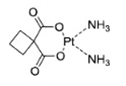 | 1986 | America |
| Third | Oxaliplatin | 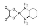 | 1996 | France |
Molecular Mechanism of Cisplatin
As Pt-based anti-cancer treatment, cisplatin, carboplatin, oxaliplatin, and other drugs are widely used in the clinic with obvious therapeutic effects and a clear mechanism of action [41-43]. Cisplatin is generally believed to be first transported into tumor cells through copper transporter 1 (CTR1). After entering the tumor cell, the platinum complex undergoes the activation step of chloro-ligand(s) replacement, generally by water molecules or other small molecules containing sulfhydryl groups. This replacement is triggered by the significantly lower intracellular chloride ion concentration (about 4 mM) compared to the extracellular matrix (about 100 mM), promoting transformation to cationic hydrate, such as cis-[Pt(NH3)2Cl(OH2)]+ and cis-[Pt(NH3)2(OH2)2]2+ [44]. Due to the chelation of the leaving ligand, carboplatin and oxaliplatin are more stable to aquation. More importantly, the rate of hydration and reaction with ammonia for transplatin is much faster than cisplatin. Following a 4-h incubation with red blood cells, transplatin reacts with 70% of the glutathione, whereas cisplatin reacts with only 35% [45]. The high reactivity of transplatin results in rapid deactivation of the complex before reaching its target, likely contributing to its lack of anti-cancer activity. After a series of chemical reactions in the cytoplasm, platinum binds to DNA by forming intra- and inter-stranded crosslinks, changing the DNA structure and causing DNA damage (Figure 1A) [46, 47]. The most nucleophilic DNA site is the N7 position of guanine, which is exposed in the major groove and is preferentially platinated. This DNA damage can prevent the cell cycle and induce apoptosis in rapidly proliferating tumor cells [48, 49].
Summary of the action mechanism of cisplatin. (A) Mechanism of action of cisplatin comprising (i) cellular uptake, (ii) aquation/activation, (iii) DNA platination, and (iv) cellular processing leading to apoptosis. Adapted with permission from ref [44], copyright 2016 American Chemical Society. (B) Alternative effects of cisplatin. Other interesting mechanisms such as acidification of the cytoplasm, ER stress, disruption of RNA transcription, inhibition of important oncogenic proteins, and decrease in metabolic plasticity of cancer cells as well as changes in their mechanobiology. Adapted with permission from ref [46], copyright 2019 Royal Society of Chemistry.
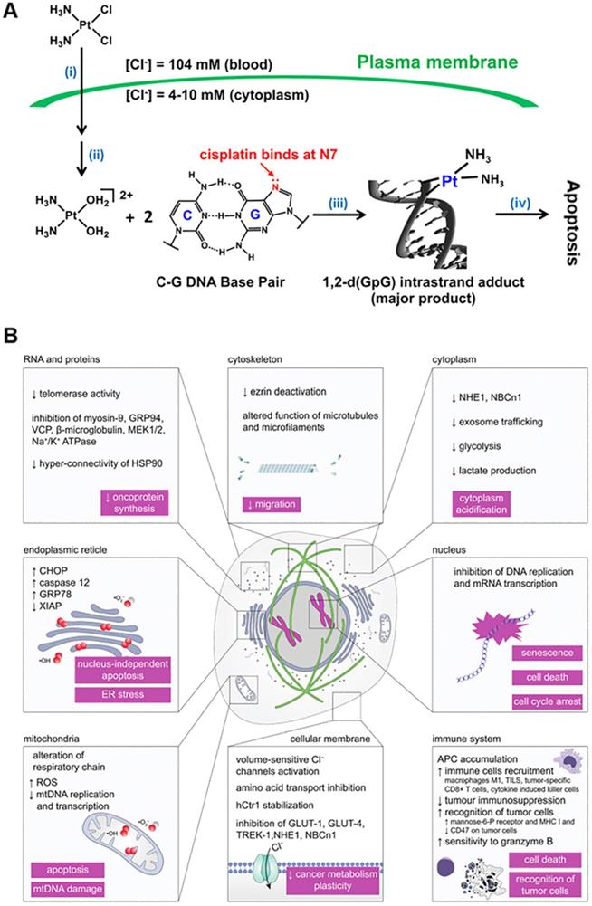
It is generally accepted that the principal mechanism of cisplatin anti-cancer action is platinum binding to DNA by forming intra-stranded and inter-stranded crosslinks [50, 51]. However, some literature reported that probably only 1-10% of intracellular cisplatin might eventually enter the nucleus and react with DNA, resulting in cell cycle arrest and apoptosis in rapidly proliferating tumor cells. In this context, other novel action mechanisms, such as acidification of the cytoplasm [52], estrogen receptor (ER) stress [53], disruption of RNA transcription [54], inhibition of key oncogenic proteins, and decrease in metabolic plasticity of cancer cells and changes in their mechanobiology [55], have also been discovered (Figure 1B). The discovery of action mechanisms that may be affected by cisplatin may provide us with an important clue to design new anti-cancer treatment strategies by finding new potential therapeutic intervention targets.
Strategies to Improve Anti-cancer Efficiency and Reduce Systemic Toxicity of Pt Drugs
Numerous studies have addressed the limitations of first-line platinum chemotherapy drugs due to potential toxicity and side effects and developed strategies to improve anti-cancer efficiency while reducing systemic toxicity [56]. Abundant evidence has demonstrated that chemical modification of first-line platinum chemotherapy drugs to achieve targeted therapy is an effective method to effectively improve drug utilization and reduce side effects [16-18]. In this section, we summarize these strategies, describing including bioconjunction targeting moiety, nanomaterials as drug carriers, and glutathione-scavenging Pt drugs.
Bioconjunction Targeting Moiety
As stated earlier, the efficacy of first-line Pt drugs is limited due to the occurrence of severe side effects (nephrotoxicity, ototoxicity, peripheral neurotoxicity, and vomiting) together with the ability of cancer cells to limit drug accumulation [56]. For Pt-based anti-cancer drugs, Pt(II) complexes are commonly used to treat malignant tumors. Photoactive Pt(IV) complexes are promising prodrug Pt(II) candidates activated by reduction in cancer cells and are being developed to lessen the side effects and improve pharmacological properties. Upon entering the cancer cells, the Pt(IV) center is reduced to Pt(II) and released [57]. Under physiological conditions, the photosensitive Pt(IV) prodrugs retain their +4 valence state in the circulation system and are selectively converted to biologically active +2 valence state via mild ultraviolet light (UVA) irradiation after they reach the tumor [58, 59].
Extensive research indicated that the killing effect of Pt-based drugs on cancer cells could be improved by integrating the cancer cell-targeting moiety into Pt(IV) prodrugs. Notably, peptide-based drug delivery systems can enhance drug targeting properties and significantly reduce side effects [60, 61] due to their bioactivity and low immune response of peptides specifically expressed on tumor cell membranes [62-65]. Peptide sequences with specific recognition characteristics of overexpressed proteins or other receptors can be introduced into Pt(IV) prodrugs to achieve the targeted function [66-68]. More importantly, polypeptide sequences could be designed to perform different targeting functions. In recent years, our research group has made significant progress in functional targeted peptide design and exploring the biological effects of these peptides [69-71]. In summary, different functional polypeptide-modified targeted platforms were developed to enhance effective utilization and reduce the side effects of Pt-based drugs.
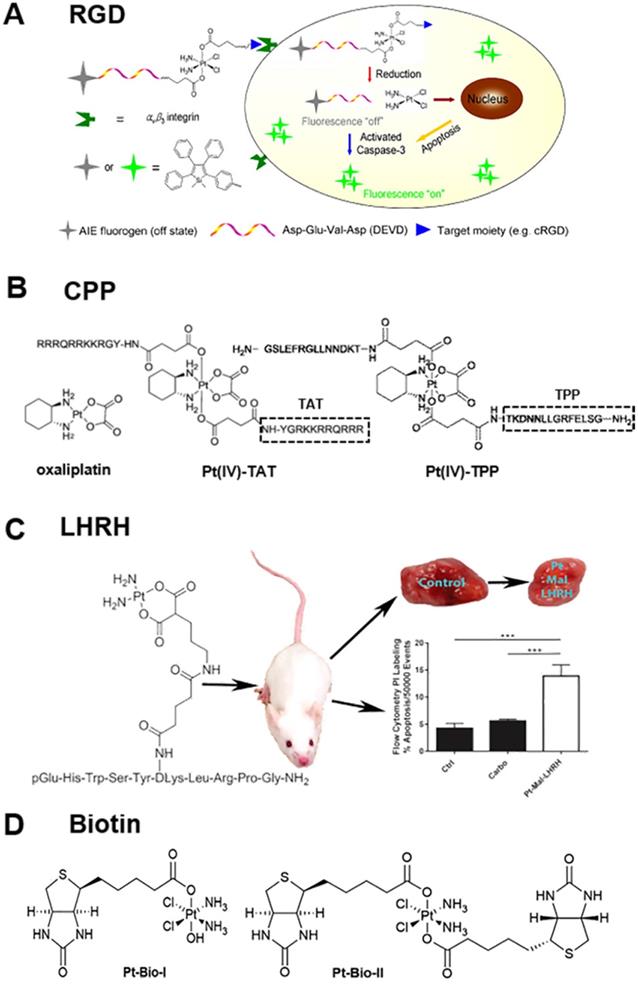
Integrins mediate cell-cell adhesion events and are overexpressed on tumor cell membranes with a key role in cancer progression [72, 73]. The tripeptide arginine-glycine-aspartic (abbreviated as RGD) recognizes integrin αvβ3 overexpressed on tumor cells [66, 74-76]. Yuan et al. reported the synthesis and biological evaluation of a chemotherapeutic Pt(IV) prodrug whose two axial positions were functionalized with the cyclic tripeptide cRGD for targeting integrin αvβ3-overexpressing cancer cells, an apoptosis sensor composed of tetraphenylsilole (TPS) fluorophore with aggregation-induced emission (AIE) characteristics, and a caspase-3 enzyme-specific Asp-Glu-Val-Asp (DEVD) peptide. The targeted Pt(IV) prodrug could selectively bind to integrin αvβ3 overexpressed on cancer cells to facilitate cellular uptake. Furthermore, the Pt(IV) prodrug was reduced to active Pt(II) drug in cells, releasing the apoptosis sensor TPS-DEVD. The reduced Pt(II) drug could induce cell apoptosis and activate the caspase-3 enzyme to cleave the DEVD peptide sequence. Due to the free rotation of the phenylene rings, TPS-DEVD was nonemissive in aqueous media. The specific cleavage of DEVD by caspase-3 generated the hydrophobic TPS residue, which aggregated, resulting in restriction of intramolecular rotations of the phenyl rings and ultimately leading to fluorescence enhancement (Figure 2A) [77].
Summary of the bio-conjunction targeting moiety to improve the anti-cancer efficiency of Pt drugs, (A) RGD peptide [77], (B) CPP peptide [81], (C) LHRH peptide [9], and (D) biotin [84]. Adapted with permission from ref [77], copyright 2014 American Chemical Society, ref [81], copyright 2017 Royal Society of Chemistry, ref [9], copyright 2017 American Chemical Society and ref [84], copyright 2017 Royal Society of Chemistry, respectively.
Similarly, Gandioso et al. reported an anti-cancer agent based on the conjugation of a photoactivatable Pt(IV) prodrug to a cyclic RGD-containing peptide [20]. Upon visible light irradiation, phototoxicity was induced preferentially in SK-MEL-28 melanoma cancer cells overexpressing αvβ3 integrin compared to control DU-145 human prostate carcinoma cells. The fact that the Pt-cyclo(RGDfK) conjugate (where f represents D-amino acids, and the others are L-amino acids) can also be internalized by αvβ5 integrin opens up the door to delivering promising anti-cancer metallodrugs to tumors overexpressing αvβ5 integrin or to tumors coexpressing αvβ3 and αvβ5 integrins. The multi-integrin targeting approach would provide new metal-based anti-cancer strategies and benefit a broader range of patients by increasing the types of tumors which can be targeted.
Furthermore, cell-penetrating peptides (CPP), such as HIV-1 TAT, which can penetrate proteins and oligoarginine, are valuable tools for transporting therapeutic macromolecules into tumor cells [78-80]. As displayed in Figure 2B, McKeon et al. reported a novel Pt(IV) tumor-penetrating peptide (TPP) conjugate, which constitutes the first example of metallodrugs to target the membrane-bound heat shock protein 70-positive (memHSP70+) phenotype in cancer cells. The conjugates exhibited superior cytotoxicity as compared to oxaliplatin alone in Pt-resistant colorectal cancer cells with relatively high memHSP70+ expression. Substitution of TPP in Pt(IV) peptide conjugates with scrambled peptide (ScP) essentially abolished the observed cytotoxicity [81].
The luteinizing hormone-releasing hormone (LHRH) peptide also acts as a targeting moiety, whose receptors are overexpressed in several types of cancer cells, such as breast, ovarian, prostate, lung, and liver [9, 82, 83]. The LHRH grafted with Pt drugs enabled selective accumulation and distribution of Pt drugs in tumor cells. Calderon et al. reported a new targeting chemotherapeutic agent, Pt-Mal-LHRH, synthesized by linking activated cisplatin to LHRH (Figure 2C) [9]. They found that Pt-Mal-LHRH significantly enhanced cellular cytotoxicity in 4T1 cells compared to the normal 3T3 cell line. Both in vitro and in vivo data suggested that Pt-Mal-LHRH elicits tumor-targeted drug delivery with increased potency, efficacy, and a possible reduction in chemotherapeutic side effects allowing the use of a high dose of chemotherapy in patients compared to other platinum drugs. Also, in vitro scratch assay data demonstrated a reduction in migration of tumor cells. Importantly, in vivo metastasis was investigated since the major cause of mortality in breast cancer patients is metastasis to distant sites, including the lungs. The in vivo data supported the in vitro data, as a significant decrease was observed in tumor volume and lung tumor colonization by Pt-Mal-LHRH treatment. Thus, the Pt-Mal-LHRH conjugate was selective for tumors overexpressing LHRH receptors while avoiding systemic distribution.
Interestingly, conjugating non-functional small molecules with platinum drugs improves the targeting ability of Pt drugs. For example, Muhammad et al. reported the design and biological properties of two Pt(IV) complexes, Pt-Bio-I and Pt-Bio-II, carrying one or two biotin moieties in the axial positions of the Pt center (Figure 2D) [84]. Tethering biotin moieties to the Pt(IV) prodrug remarkably increased cellular uptake of Pt in breast cancer cells but lowered its accumulation in breast epithelial cells. The mono-biotinylated Pt(IV) complex was more active than the di-biotinylated one in reactivity and cytotoxicity. Compared with cisplatin, Pt-Bio-I showed much stronger inhibition against cisplatin-insensitive MDA-MB-231 and MCF-7 cancer cells. Considering its low toxicity towards mammary epithelial cells, Pt-Bio-I may be superior over cisplatin in breast cancer therapy. Interestingly, Pt(IV) complexes with one hydroxyl ligand in the axial position appear to be beneficial for their interaction with DNA and cytotoxicity to cancer cells.
Nanomaterials as Drug Carriers
Gold Nanoclusters (GNCs) as Drug Carriers
Nanocarrier-based platinum drug delivery systems are promising alternatives to avoid the disadvantages of conventional platinum drugs [22, 85, 86]. In recent years, there has been much interest in GNCs as drug transport carriers due to their high photostability [87], water solubility, and biocompatibility [25, 88]. Compared with traditional nanoparticles, the particle size of GNCs is usually less than 2 nm with increased blood circulation time. Pt drugs can be loaded efficiently in GNCs with increased accumulation in tumor tissues through the enhanced permeation and retention (EPR) effect, resulting in improved therapeutic efficacy and reduced systemic toxicity. Furthermore, ultrasmall GNCs are usually filtered out of the body through effective renal clearance, indicating good metabolism and biocompatibility [89-91].
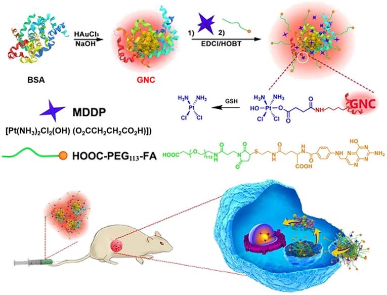
Utilizing the unique biological effects of GNCs, Zhou et al. employed BSA-protected GNCs as a dual-functional nanoplatform for drug delivery and fluorescence imaging of the tumor (Figure 3) [18]. The GNCs were first conjugated with reduction-sensitive cisplatin coupled with a prodrug (cis,cis,trans[Pt(NH3)2Cl2(OH)(O2CCH2CH2CO2H)]) (MDDP), and then functionalized with a targeting ligand folic acid (FA) [92], which can target folate receptor α (FR-α) overexpressed on the surface of cancer cells [93]. Using the highly aggressive 4T1 breast cancer cell line and its orthotopic tumor model, the investigators demonstrated selective accumulation of the prodrug and FA dual-conjugated GNCs inside the cancer cells and the tumor. The nanoparticles could efficiently inhibit the growth of the primary tumor and suppress metastasis of cancer cells to the lung. These data demonstrated the good potential of the GNC-based theranostic nanoplatform for fluorescence tumor imaging and cancer therapy and the advantages of GNCs as a drug delivery platform. First, when BSA is used for nanoparticle synthesis, GNCs are biocompatible and biodegradable. For clinical translation, BSA can be replaced with human serum protein (HSA) without changing the physicochemical properties of the nanoparticles. Second, the prodrug and FA dual-conjugated GNC nanoparticles selectively accumulated in the orthotopic 4T1 tumor model, displayed high fluorescence signals for nanoparticle tracking and tumor imaging [94] and efficiently inhibited primary tumor growth and suppressed metastasis of cancer cells to the lung. Moreover, the hydrodynamic diameter of FA-GNC-Pt was ~10 nanometers, allowing escape from the reticuloendothelial system (RES) in the liver and fast kidney clearance, avoiding liver accumulation and minimizing side effects.
Schematic illustration of GNC-based theranostic nanoplatform for tumor-targeted chemotherapy and fluorescence imaging. Adapted with permission from ref [18], copyright 2016 Ivyspring International Publisher.
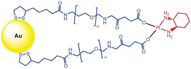
Brown et al. tethered the active component of the anti-cancer drug oxaliplatin to gold nanoparticles (AuNPs) for improved drug delivery [95]. Poly(ethylene glycol) (PEG)-modified AuNPs have been used to functionalize cisplatin or oxaliplatin. For example, the active component of oxaliplatin was tethered to AuNPs that were functionalized with a thiolated PEG monolayer capped with a carboxylate group. The platinum-tethered NPs demonstrated comparable or significantly higher cytotoxicity than oxaliplatin against the A549 epithelial lung cancer and several colon cancer cell lines. In particular, the nanoparticles showed an unusual ability to penetrate the nucleus in lung cancer cells. The platinum-tethered nanoparticles demonstrated as good as, or significantly better, cytotoxicity than oxaliplatin alone in all cell lines (HCT116, HCT15, HT29, and RKO) and an unusual ability to penetrate the nucleus in lung cancer cells (Figure 4).
Chemical synthesis of the platinum-tethered gold nanoparticles. Adapted with permission from ref [95], copyright 2010 American Chemical Society.
Magnetic Iron Oxide Nanoparticles as Drug Carriers
Superparamagnetic iron oxide nanoparticles (SPIONs) are usually employed as targeted delivery regents due to their advantages of low toxicity, biocompatibility, biodegradability, and well water dispersion. SPIONs can bind to drugs and be directed to the tissues of interest or tumors using an external magnetic field [96, 97].
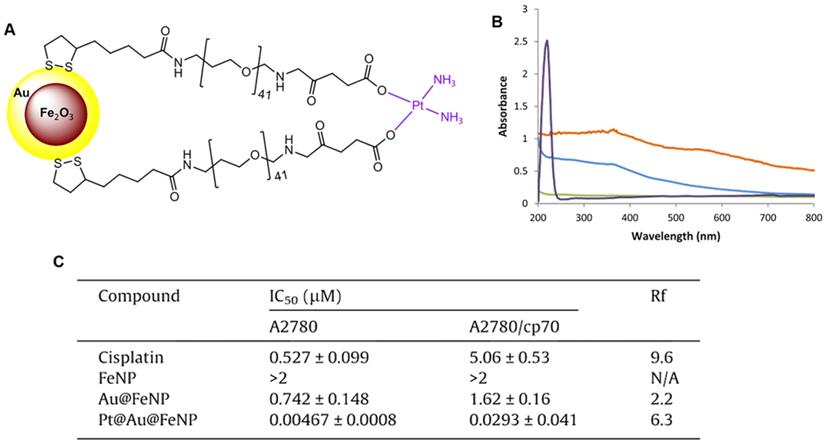
Wagstaff et al. tethered the active component of cisplatin to gold-coated iron oxide nanoparticles (Pt@Au@FeNPs) to improve their delivery to tumors and increase efficacy [98]. The nanoparticle-based drug delivery system of Pt@Au@FeNPs was functionalized with thiolated polyethylene glycol (PEG) linkers to which the active component of the anti-cancer drug cisplatin, [Pt(NH3)2]2+ was attached via the terminal carboxylate groups (Figure 5A). The successful introduction of cisplatin was confirmed by UV-visible spectra (Figure 5B). The cytotoxicity of the Pt@Au@FeNPs was examined using in vitro growth inhibition assays in the human ovarian carcinoma cell line A2780 and its cisplatin-resistant derivative A2780/cp70. Pt@Au@FeNPs demonstrated activity at nanomolar concentrations and were 110-fold more active than cisplatin in A2780 cells, while iron oxide nanoparticles (FeNP) showed no cytotoxicity at concentrations up to 2 μM. However, in this study, the cisplatin Pt@Au@FeNPs, despite having activity at nanomolar concentrations, were cross-resistant with cisplatin in A2780/cp70 cells (Figure 5C).
(A) Delivery system of gold-coated iron oxide nanoparticles functionalized with thiolated polyethylene glycol (PEG) linkers to which the active component of the anti-cancer drug cisplatin, [Pt(NH3)2]2+, is attached via the terminal carboxylate groups. (B) UV-Vis spectra of the four nanoparticles: FeNPs (blue), Au@FeNPs (orange), PEGylated Au@FeNPs (green), and Pt@Au@FeNPs (purple). (C) In vitro cytotoxicity of the nanoparticles in the human ovarian carcinoma cell line A2780 and its cisplatin-resistant sub-line A2780/cp70. Resistance factor (Rf) is defined as the IC50 of the complex in the resistant line divided by the IC50 of the complex in the sensitive line; any complex with an Rf less than 1 can overcome cisplatin resistance. Adapted with permission from ref [98], copyright 2012 Elsevier.
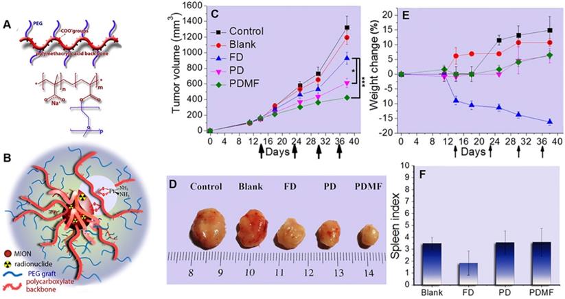
SPIONs are promising drug carriers because of the targeted delivery to tumors and increased efficacy through external magnets. Voulgari et al. synthesized a magnetic nanocarrier poly(methacrylic acid)-graft-poly(ethyleneglycol methacrylate) (p(MAA-g-EGMA)) by radical copolymerization of methoxy-PEG-methacrylate with methacrylic acid (Figure 6A). The cisplatin-loaded (PD) magnetic nanocarriers (Figure 6B) facilitated magnetically-triggered drug release and displayed in vitro anti-cancer activity comparable to free cisplatin at the same drug concentrations. In addition, they exhibited an enhanced anti-cancer effect in vivo on a cisplatin-resistant HT-29 tumor model in mice, particularly when a magnetic field was applied to the tumor area (Figure 6C-D). During the study period, a decrease in mouse weight was observed in the free cisplatin group but not in the p(MAA-g-EGMA)-treated group (Figure 6E), indicating a significant reduction of side-effects with the cisplatin-loaded magnetic nanocarriers. Moreover, spleen indices in the groups of mice injected with cisplatin-loaded magnetic nanocarriers (Figure 6F) were identical with the control group, suggesting a pronounced reduction of cisplatin systemic toxicity [99].
(A) Chemical structure of copolymers poly(methacrylic acid)-graft-poly(ethyleneglycol methacrylate) (p(MAA-g-EGMA)). (B) Schematic structure of the studied magnetic drug delivery systems. Evolution with time of (C) Tumor volume and (E)% weight change of mice (n=4) after i.v. injections with: saline (Control), nanocarriers without the drug (Blank), aqueous cisplatin solution (FD), cisplatin-loaded nanocarriers (PD), and cisplatin-loaded nanocarriers in the presence of an external magnetic field in the tumor area (PDMF). Arrows in (a) and (b) represent tail vein injection events. (D) Pictures of the tumors taken at the end of the study period are shown for comparison. (F) Spleen index of mice sacrificed at the end of the in vivo experiment. Adapted with permission from ref [99], copyright 2016 Elsevier.
Other Nanomaterials as Drug Carriers
Besides SPIONs and GNCs, other nanomaterials, such as mesoporous silica, have multiple potential applications as drug carriers [100, 101]. Mesoporous silica nanoparticles (MSNs) with large surface area and pore volume have an extraordinary ability to store drugs, and the controllable release of Pt drugs from the designed mesoporous structures is advantageous for the bioavailability of drugs [102]. Also, organic nanoparticles, such as polymeric nanoparticles, exhibit great potential in drug delivery because of their unusual properties, including simple encapsulation, high capacity, controlled release, and low toxicity. The benefits of encapsulating Pt drugs in polymeric nanoparticles to reduce side effects without affecting drug efficacy have been demonstrated in tumor-bearing mice and preclinical cancer models [103].
Glutathione-Scavenging Pt Drugs
Glutathione (GSH) is one of the most abundant non-protein thiols in tumor cells, with its intracellular content of about 0.5-10 mM [37, 104, 105] and is the most important intracellular thiol compound which participates in cellular detoxification mechanisms [106, 107]. Previous reports indicated that cancer cells could utilize endogenous GSH to chelate Pt drugs and produce inactive GSH-Pt adducts, which can be preferentially pumped out via membrane transport proteins and are non-toxic to cancer cells [12, 108, 109]. In this context, GSH-scavenging Pt drugs have been reported.
Sulforaphane (SFN) has been reported to deplete GSH by directly binding with GSH to form the GSH-SFN complex, which can be exported out of the cell. Recently, Xu et al. proposed that the therapeutic efficacy of SFN-CDDP-NPs could be significantly improved by SFN-mediated GSH depletion [110]. As displayed in Figure 7A, the investigators designed an NP-enabled codelivery system consisting of a water-soluble poly(γ, L-glutamic acid)-CDDP (γ-PGA-CDDP) conjugate and SFN for breast cancer treatment. The therapeutic efficacy of SFN-CDDP-NPs was systematically investigated and compared with free drugs both in vitro and in vivo. After efficient internalization of SFN-CDDP-NPs by tumor cells, the rapidly released SFN could notably decrease the GSH content and thus significantly increase DNA-bound Pt, resulting in severe DNA damage and cellular apoptosis. Due to the improved chemosensitivity and preferential tumor accumulation, SFN-CDDP-NPs greatly inhibited orthotopic breast cancer progression with reduced toxic side effects.
(A) Schematic diagram of the preparation of SFN-CDDP-NPs for improved anti-tumor therapy. Adapted with permission from ref [110], copyright 2020 American Chemical Society. (B) Illustration of the redox-responsive nanoplatform, composed of Pt(IV) prodrug 5, Cys-8E polymer, and lipid-PEG, for in vivo Pt delivery and treatment of cisplatin-resistant tumors. Adapted with permission from ref [111], copyright 2018 American Chemical Society. (C) Schematic illustration of phospholipid-mimic CBP-LA conjugates that self-assemble into micelle-like nanoparticles and the possible mechanism of their anti-cancer activity. Adapted with permission from ref [112], copyright 2018 Royal Society of Chemistry.
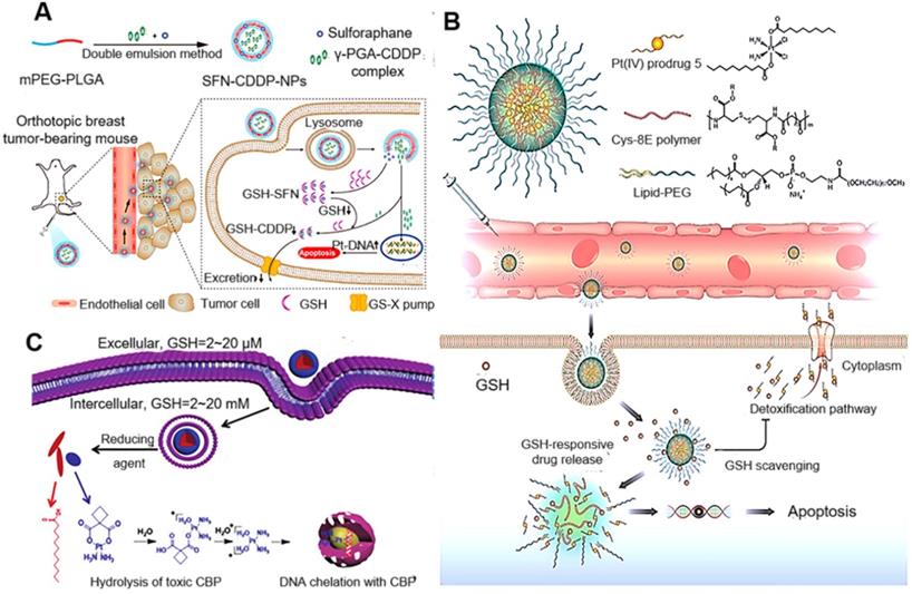
Ling et al. reported the synthesis of cysteine-based poly(disulfide amide) (Cys-PDSA) polymers (Cys-8E polymer) that readily react with GSH via disulfide-mediated reduction and their combination with a series of Pt(IV) prodrugs with tunable hydrophobicity. Optimized polymers rapidly disassembled and released Pt drugs in response to intracellular GSH while simultaneously consuming GSH to restore Pt sensitivity in cisplatin-resistant tumor cells [111]. Moreover, in vivo efficacy and safety results showed that NPs effectively inhibited the growth of cisplatin-resistant xenograft tumors with an inhibition rate of 83.32% while alleviating serious side effects associated with cisplatin. GSH-scavenging polymeric NP technology reported herein could provide a unique strategy for improving the therapeutic efficacy of current Pt drugs (Figure 7B). Liang et al. also developed novel small-molecule-based nanodrugs of carboplatin-lauric acid nanoparticles (CBP-LA NPs) to reduce GSH-mediated platinum resistance and improve the anti-tumor efficiency of Pt(II) [112]. The intracellular glutathione determination and the Pt-DNA adduct assay revealed that CBP-LA NPs could reduce intracellular GSH levels and improve the efficiency of platinum chelating with DNA to overcome GSH-mediated Pt(II) resistance (Figure 7C).
Pt Nanoclusters (Pt NCs) as a New Pt Drug for Cancer Therapy
Properties of Pt Nanoclusters
Pt NCs, similar to noble metal clusters such as GNCs, are relatively stable molecular aggregates composed of up to hundreds of metal atoms [113]. Their physical size is normally between atoms and nanoparticles, close to the Fermi wavelength of a single electron. Pt NCs have attracted much attention in bioanalysis and biomedicine due to their special physicochemical properties, such as ultra-small size, precise structure, photoluminescence, X-ray absorption, low cytotoxicity, and good biocompatibility [114, 115]. In particular, due to distinct molecular composition and a good biological safety profile, Pt NCs have great application prospects in anti-cancer treatment.
Application of Pt NC-based Drugs in Cancer Therapy
Although multiple studies of the Pt(II) complex and modified Pt(IV) prodrugs have attempted to improve the anti-cancer efficiency of Pt drugs, these strategies relied on anti-cancer mechanisms similar to cisplatin and were not very successful [116-118]. In this section, we discuss Pt NCs as anti-cancer drugs with different mechanisms for cancer therapy.
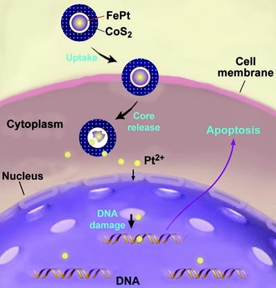
So far, the cytotoxicity mechanism of Pt NCs is still unclear because of the differences in size, shape, surface coatings, and purity of the particles [119]. Nevertheless, it is generally accepted that the cytotoxicity of Pt NCs depends primarily on abundant Pt2+ ions leaching under low pH conditions, such as in cell endosomes, to induce DNA damage [32]. As early as 2007, the inhibitory mechanism of Pt NCs on tumor cells was described. Gao et al. reported that the synthesized FePt@CoS2 yolk-shell nanoclusters exhibited an IC50 of 35.5 ng/mL (4.7 ng/mL of Pt) in HeLa cells that was much lower than cisplatin (230 ng/mL of Pt). The FePt nanoclusters were oxidized to generate Pt2+ and Fe3+ ions, especially in intracellular late lysosome with a low pH (pH < 5.5) environment, as hollow nanospheres were found in mitochondria of cancer cells, implying breakdown of the FePt core. Thus, after cellular uptake, FePt cores disintegrated to generate metal ions inside the acidic environment of secondary lysosomes. Subsequently, these metal ions could escape from endosomes and enter the cell nucleus to bind DNA, forming DNA-Pt adducts and eventually leading to tumor cell apoptosis (Figure 8). Furthermore, transmission electron microscopy (TEM) confirmed cellular uptake of FePt@CoS2 nanocrystals, and the magnetic properties analysis corroborated the release of FePt nanoparticles from yolk-shell nanostructures after cellular uptake [120].
Illustration of a possible mechanism accounting for FePt@CoS2 yolk-shell nanocrystals killing HeLa cells. After cellular uptake, FePt nanoparticles were oxidized to generate Fe3+ (omitted for clarity) and Pt2+ ions (yellow). The Pt2+ ions enter into the nucleus (and mitochondria), bind to DNA, and lead to apoptosis of the HeLa cell. Adapted with permission from ref [120], copyright 2008 American Chemical Society.
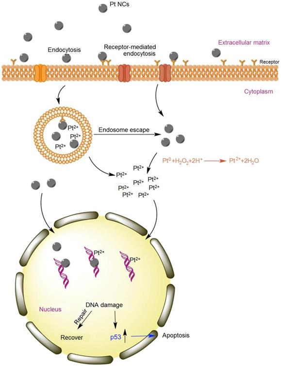
Although the anti-tumor mechanism of Pt NCs remains obscure, it is believed that most Pt atoms are exposed on the ultrasmall <2 nm size nanocluster surface [36]. These high surface-active Pt NCs are affected by intracellular acidic organelles like endosomes and lysosomes and then rapidly decompose to form oxidative states of Pt (Figure 9) that can attach to and change DNA structure, resulting in cancer cell apoptosis. In addition, ultrasmall Pt NCs can anchor onto the grooves of DNA double helix to further damage the DNA. Thus, eradicating cancer cells by Pt NC-based nanodrugs appears to be the synergistic effect of both Pt NCs and Pt ions causing DNA damage [32].
Schematic of apoptosis mechanism of Pt NCs. Abundant oxidized Pt ions and Pt NCs coordinate the DNA damage activating the p53 pathway. Adapted with permission from ref [32], copyright 2017 Elsevier.
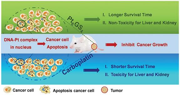
Recently, our research group explored a facile approach to develop an endogenous GSH-chelated Pt molecule containing multiple Pt atoms for efficient cancer treatment (Figure 10) [121]. These polynuclear Pt NCs were identified by electrospray ionization mass spectra (ESI-MS) and density functional theory (DFT) study as Pt6GS4. High efficacy for anti-cancer treatment was achieved by Pt6GS4 both in vitro and in vivo when compared with traditional first-line carboplatin at the same dosage. The Pt6GS4 molecule could be readily taken up by aggressive triple-negative breast cancer (TNBC) cells. Subsequently, its metabolites entered nuclei to interact with DNA, and finally, the DNA-Pt complex triggered TNBC cell apoptosis via the p53 pathway. These data revealed that Pt6GS4 was comparable to carboplatin for cancer cell uptake, nuclear localization, and cancer cell proliferation inhibition. More significantly, compared with carboplatin, Pt6GS4 was non-toxic for the liver and kidneys, and Pt6GS4-treated mice lived longer. Our study opened a new avenue to explore polynuclear Pt compounds with accurate architecture for enhancing therapeutic effects and reducing systemic toxicity.
Schematic illustration of GSH-chelated Pt molecule (Pt6GS4) as a potent anti-cancer agent. High efficacy for anti-cancer treatment and lower systemic toxicity were achieved by Pt6GS4 both in vitro and in vivo, compared to carboplatin at the same dosage. Adapted with permission from ref [121], copyright 2020 Wiley.
Targeting peptides have widely been used to synthesize Pt NCs [24, 113, 122, 123], improving the existing first-line platinum drugs in the clinic that inhibit rapid proliferation of tumor cells, and can also improve drug bioavailability. For example, Feng et al. synthesized mitochondria-targeting Pt NCs (CytcApt-Pt NCs) using cytochrome c aptamer (CytcApt) as a template. In vitro experiments showed that CytcApt-Pt NCs could kill 4T1 tumor cells in a pH-dependent manner but did not affect normal 293T cells. These results showed good therapeutic efficacy and excellent biosafety of CytcApt-Pt NCs, indicating their great potential for tumor treatment and reducing systemic toxicity [124].
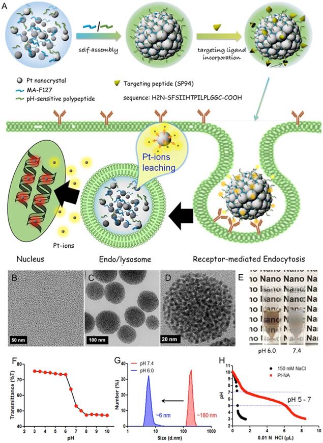
Xia et al. synthesized a Pt nanocluster assembly (Pt-NA) composed of assembled Pt NCs incorporating a pH-sensitive polymer and hepatocellular carcinoma (HCC)-targeting peptide (Figure 11) [125]. Pt-NA was latent in peripheral blood, readily targeted disseminated HCC cancer stem-like cells (CSLCs), and disassembled into small Pt NCs in acidic subcellular compartments, eventually inducing DNA damage. Moreover, the study demonstrated the underlying mechanism of these effects at the molecular level as downregulation of many genes that are highly expressed in liver cancer patients. Thus, Pt-NA has a good potential in clinical HCC treatment [125].
Design and characterization of HCC-targeted pH-sensitive Pt nanocluster assembly (Pt-NA). (A) Schematic representation of Pt-NA synthesis, targeted HCC uptake, and intracellular Pt ion release. (B) TEM image of the synthesized Pt NCs. (C) TEM image of Pt-NA. (D) High-resolution TEM image of Pt-NA. (E) Photographs of Pt-NA in pH 6.0 and 7.4. (F) The transmittance of a suspension of Pt-NA as a function of pH. (G) DLS size measurement of Pt-NA (0.1 mg mL-1) as a function of pH. (H) pH profile of Pt-NA by acid-base titration. Adapted with permission from ref [125], copyright 2016 American Chemical Society.
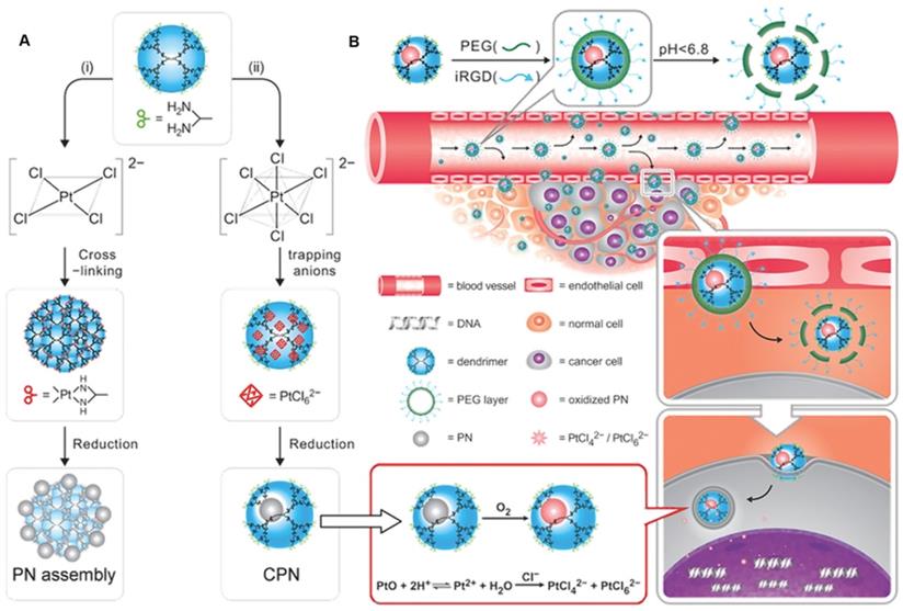
Another example is of a first-generation dendrimer-caged Pt nanocluster (CPN) with the size of an atomic level (0.93 ± 0.22 nm in diameter). CPN was endowed with targeting function by conjugating with the iRGD peptide (Figure 12A) [126]. Especially, CPN could be easily oxidized, resulting in the loss of its intrinsic chemical inertness and its surface corrodibility for further dissolution in weakly acidic organelles, such as endosomes and lysosomes, to release toxic Pt ions for DNA cross-linking (Figure 12B). Employing subcutaneous breast cancer xenografts in mice, the therapeutic effect of CPN was examined by intratumoral injection in vivo. Results indicated that this chemotherapeutic had efficacy comparable to cisplatin.
(A) Schematic representation of a novel strategy based on tuning anionic geometry for the formation of PN. (B) Schematic representation of the caged PN mixed with a tumor-penetrating peptide to target the tumor and kill malignant cells by shedding the outer PEG corona to exert tumor-inside activation. Adapted with permission from ref [126], copyright 2013 Wiley.
Conclusion and Perspectives
Traditional tumor chemotherapy employs chemo-drugs, which usually have strong side effects for normal cells and tissues [13]. Pt-based anti-cancer drugs play a vital role in clinical cancer therapy with satisfactory efficacy. The first-line clinical platinum anti-cancer drugs represented by cisplatin are relatively old drugs that have a therapeutic effect on tumors with a known molecular mechanism. However, the side effects seriously limit the application of platinum anti-cancer drugs. Therefore, modified Pt-based drugs, which could improve anti-cancer efficiency and reduce systemic toxicity have been investigated. Pt NC-based nanodrugs have attracted much attention due to the inherent higher blood circulation time, EPR effect, and facile surface functionalization. Advances in nanotechnology and nanoscience have facilitated the development of Pt NCs, representing an important research orientation for exploring platinum drugs with the precise structure to improve therapeutic effect and reduce systemic toxicity [127].
The challenges accompanying these advances provide us with future directions and efforts for designing and constructing more effective Pt-based drugs for possible clinical applications:
(1) For cisplatin and other platinum anti-cancer drugs, systemic toxicity is still the most challenging problem. Platinum drugs are modified by many methods, such as linking target molecules and adding drug delivery carriers, with the ultimate goal of reducing their toxicity.
(2) Many new platinum nanodrugs, such as Pt NCs, have been developed. Pt NCs generate platinum ions in cells and induce irreversible DNA damage [32, 37, 125, 126]. However, Pt NC-based drugs are still cytotoxic, and the possible harmful mechanisms are not entirely understood. Investigations on the role of Pt NCs size in cytotoxicity indicated that it could represent an important parameter affecting molecular mechanisms. Recent synchrotron radiation X-ray techniques may provide insights into nanomaterial biotransformation to address the anti-tumor mechanism of Pt NCs. It is crucial to study the dynamic process of Pt NCs metabolism in vivo and their interaction with biomolecules to treat malignant tumors and other diseases [128, 129], which would be critical for designing Pt NC-based nanodrugs and high efficiency.
(3) As a new type of platinum anti-cancer drug, the molecular composition of Pt NCs needs to be further improved. Mass spectrometric techniques, such as MALDI-TOF-MS and ESI-MS, would help characterize the molecular formula of clusters or the metal to ligand ratio in clusters, but the precise structural characterization methods of cluster molecules need to be further explored [130-133]. We believe that nanotechnology would be immensely helpful in addressing these issues. Besides understanding the precise molecular composition of Pt NCs, we need to study the impact of Pt NCs on their anti-tumor function in the context of the configuration of cisplatin and transplatin, which is an area worthy of in-depth exploration. The application of nanotechnology in the field of Pt NC-based nanodrugs undoubtedly provides a promising prospect for improving the current anti-cancer treatment.
(4) Combining Pt-based nanodrugs with other therapeutic methods, such as synergistic chemo-electrodynamic therapy, can maximize the bio-function of Pt NCs and strengthen their anti-tumor effect [102, 134-136]. In addition to individual Pt-based drugs, bimetallic composites, such as platinum complexes in combination with ruthenium, also showed excellent anti-cancer performance [57, 137]. This represents a new strategy to overcome the deactivation pathways during Pt drug treatment and a good option as a promising anti-cancer agent.
Abbreviations
CDDP: cisplatin; CTR1: copper transporter 1; ER: endoplasmic reticulum; RGD: arginine-glycine-aspartic; AIE: aggregation-induced emission; CPP: cell-penetrating peptides; TPP: tumor penetrating peptide; LHRH: luteinizing hormone-releasing hormone; GNC: gold nanoclusters; Pt NCs: platinum nanoclusters; mesoporous silica nanoparticles (MSNs); superparamagnetic iron oxide nanoparticles (SPIONs); FA: folic acid; HSA: human serum protein; RES: reticuloendothelial system; AuNPs: gold nanoparticle; PEG: polyethylene glycol; GSH: Glutathione; SFN: Sulforaphane; CBP-LA NPs: carboplatin-lauric acid nanoparticles; ESI-MS: electrospray ionization mass spectra; DFT: density functional theory; TNBC: triple-negative breast cancer; BSA: bovine serum albumin protein; HCC: hepatocellular carcinoma; CPN: dendrimer-caged Pt nanoclusters; PN: Pt nanoclusters; Pt-NA: Pt nanocluster assembly; MALDI-TOF-MS: matrix-assisted laser desorption/ionization-time of flight mass spectrometry.
Acknowledgements
This work was financially supported by the National Natural Science Foundation of China (21727817, U2067214, 11621505), and the Academic Promotion Program of Shandong First Medical University (2019LJ003).
Author contributions
All authors contributed to writing the manuscript and approved the final version.
Competing Interests
The authors have declared that no competing interest exists.
References
1. Florea AM, Busselberg D. Cisplatin as an anti-tumor drug: cellular mechanisms of activity, drug resistance and induced side effects. Cancers (Basel). 2011;3:1351-1371
2. Park GY, Wilson JJ, Song Y, Lippard SJ. Phenanthriplatin, a monofunctional DNA-binding platinum anticancer drug candidate with unusual potency and cellular activity profile. Proc Natl Acad Sci U S A. 2012;109:11987-11992
3. Bian M, Fan R, Zhao S, Liu W. Targeting the thioredoxin system as a strategy for cancer therapy. J Med Chem. 2019;62:7309-7321
4. Zhang J, Li X, Han X, Liu R, Fang J. Targeting the thioredoxin system for cancer therapy. Trends Pharmacol Sci. 2017;38:794-808
5. Rottenberg S, Disler C, Perego P. The rediscovery of platinum-based cancer therapy. Nat Rev Cancer. 2020;21:37-50
6. Wang D, Lippard SJ. Cellular processing of platinum anticancer drugs. Nat Rev Drug Discov. 2005;4:307-320
7. Ohmichi M, Hayakawa J, Tasaka K, Kurachi H, Murata Y. Mechanisms of platinum drug resistance. Trends Pharmacol Sci. 2005;26:113-116
8. Corte-Rodriguez M, Espina M, Sierra LM, Blanco E, Ames T, Montes-Bayon M. et al. Quantitative evaluation of cellular uptake, DNA incorporation and adduct formation in cisplatin sensitive and resistant cell lines: Comparison of different Pt-containing drugs. Biochem Pharmacol. 2015;98:69-77
9. Calderon LE, Keeling JK, Rollins J, Black CA, Collins K, Arnold N. et al. Pt-Mal-LHRH, a newly synthesized compound attenuating breast cancer tumor growth and metastasis by targeting overexpression of the LHRH receptor. Bioconjug Chem. 2017;28:461-470
10. Zayed A, Jones GD, Reid HJ, Shoeib T, Taylor SE, Thomas AL. et al. Speciation of oxaliplatin adducts with DNA nucleotides. Metallomics. 2011;3:991-1000
11. Qi L, Luo Q, Zhang Y, Jia F, Zhao Y, Wang F. Advances in toxicological research of the anticancer drug cisplatin. Chem Res Toxicol. 2019;32:1469-1486
12. Garcia Sar D, Montes-Bayon M, Blanco Gonzalez E, Sierra Zapico LM, Sanz-Medel A. Reduction of cisplatin-induced nephrotoxicity in vivo by selenomethionine: the effect on cisplatin-DNA adducts. Chem Res Toxicol. 2011;24:896-904
13. Wong. E, Giandomenico. CM. Current status of platinum-based antitumor drugs. Chem Rev. 1999;99:2451-2466
14. Wlodarczyk MT, Dragulska SA, Camacho-Vanegas O, Dottino PR, Jarzęcki AA, Martignetti JA. et al. Platinum(II) complex-nuclear localization sequence peptide hybrid for overcoming platinum resistance in cancer therapy. ACS Biomater. Sci. Eng. 2018;4:463-467
15. Mjos KD, Orvig C. Metallodrugs in medicinal inorganic chemistry. Chem Rev. 2014;114:4540-4563
16. Chen Q, Yang Y, Lin X, Ma W, Chen G, Li W. et al. Platinum(iv) prodrugs with long lipid chains for drug delivery and overcoming cisplatin resistance. Chem Commun (Camb). 2018;54:5369-5372
17. Alas M, Saghaeidehkordi A, Kaur K. Peptide-drug conjugates with different linkers for cancer therapy. J Med Chem. 2021;64:216-232
18. Zhou F, Feng B, Yu H, Wang D, Wang T, Liu J. et al. Cisplatin prodrug-conjugated gold nanocluster for fluorescence imaging and targeted therapy of the breast cancer. Theranostics. 2016;6:679-687
19. Turiel-Fernandez D, Gutierrez-Romero L, Corte-Rodriguez M, Bettmer J, Montes-Bayon M. Ultrasmall iron oxide nanoparticles cisplatin (IV) prodrug nanoconjugate: ICP-MS based strategies to evaluate the formation and drug delivery capabilities in single cells. Anal Chim Acta. 2021;1159:338356
20. Gandioso A, Shaili E, Massaguer A, Artigas G, Gonzalez-Canto A, Woods JA. et al. An integrin-targeted photoactivatable Pt(IV) complex as a selective anticancer pro-drug: synthesis and photoactivation studies. Chem Commun (Camb). 2015;51:9169-9172
21. Palchoudhury S, Xu Y, Rushdi A, Bao Y. DNA interaction of Pt-attached iron oxide nanoparticles. IEEE T Magn. 2013;49:373-376
22. Chen H, Gu Z, An H, Chen C, Chen J, Cui R. et al. Precise nanomedicine for intelligent therapy of cancer. Sci China Chem. 2018;61:1503-1552
23. Pelaz B, Alexiou C, Alvarez-Puebla RA, Alves F, Andrews AM, Ashraf S. et al. Diverse applications of nanomedicine. ACS Nano. 2017;11:2313-2381
24. Zhang P, Cui Y, Anderson CF, Zhang C, Li Y, Wang R. et al. Peptide-based nanoprobes for molecular imaging and disease diagnostics. Chem Soc Rev. 2018;47:3490-3529
25. Spicer CD, Jumeaux C, Gupta B, Stevens MM. Peptide and protein nanoparticle conjugates: versatile platforms for biomedical applications. Chem Soc Rev. 2018;47:3574-3620
26. Liu HW, Chen L, Xu C, Li Z, Zhang H, Zhang XB. et al. Recent progresses in small-molecule enzymatic fluorescent probes for cancer imaging. Chem Soc Rev. 2018;47:7140-7180
27. Fang J, Zhang B, Yao Q, Yang Y, Xie J, Yan N. Recent advances in the synthesis and catalytic applications of ligand-protected, atomically precise metal nanoclusters. Coordin Chem Rev. 2016;322:1-29
28. Wang K, Zhu C, He Y, Zhang Z, Zhou W, Muhammad N. et al. Restraining cancer cells by dual metabolic inhibition with a mitochondrion-targeted platinum(II) complex. Angew Chem Int Ed Engl. 2019;58:4638-4643
29. Rebecca A. Alderden, Matthew D. Hall, Hambley TW. The discovery and development of cisplatin. J Chem Educ. 2006;83:728-734
30. Kuwata K, Nakamura I, Ide M, Sato H, Nishikawa S, Tanaka M. Comparison of changes in urinary and blood levels of biomarkers associated with proximal tubular injury in rat models. J Toxicol Pathol. 2015;28:151-164
31. Hazlitt RA, Min J, Zuo J. Progress in the development of preventative drugs for cisplatin-induced hearing loss. J Med Chem. 2018;61:5512-5524
32. Hu X, Li F, Noor N, Ling D. Platinum drugs: from Pt(II) compounds, Pt(IV) prodrugs, to Pt nanocrystals/nanoclusters. Sci Bull. 2017;62:589-596
33. Dilruba S, Kalayda GV. Platinum-based drugs: past, present and future. Cancer Chemother Pharmacol. 2016;77:1103-1124
34. Ho GY, Woodward N, Coward JI. Cisplatin versus carboplatin: comparative review of therapeutic management in solid malignancies. Crit Rev Oncol Hematol. 2016;102:37-46
35. Kenny RG, Marmion CJ. Toward multi-targeted platinum and ruthenium drugs-a new paradigm in cancer drug treatment regimens? Chem Rev. 2019;119:1058-1137
36. Huang X, Li Z, Yu Z, Deng X, Xin Y. Recent advances in the synthesis, properties, and biological applications of platinum nanoclusters. J Nanomater. 2019;2019:1-31
37. Browning RJ, Reardon PJT, Parhizkar M, Pedley RB, Edirisinghe M, Knowles JC. et al. Drug delivery strategies for platinum-based chemotherapy. ACS Nano. 2017;11:8560-8578
38. Cheng Q, Liu Y. Multifunctional platinum-based nanoparticles for biomedical applications. Wiley Interdiscip Rev Nanomed Nanobiotechnol. 2017;9:e1410
39. Raymond E, Chaney SG, Taamma A, Cvitkovic E. Oxaliplatin: a review of preclinical and clinical studies. Ann Oncol. 1998;9:1053-1071
40. Hector S, Bolanowska-Higdon W, Zdanowicz J, Hitt S, Pendyala L. In vitro studies on the mechanisms of oxaliplatin resistance. Cancer Chemother Pharmacol. 2001;48:398-406
41. Yang Y, Adebali O, Wu G, Selby CP, Chiou YY, Rashid N. et al. Cisplatin-DNA adduct repair of transcribed genes is controlled by two circadian programs in mouse tissues. Proc Natl Acad Sci U S A. 2018;115:E4777-E4785
42. Zayed A, Shoeib T, Taylor SE, Jones GDD, Thomas AL, Wood JP. et al. Determination of Pt-DNA adducts and the sub-cellular distribution of Pt in human cancer cell lines and the leukocytes of cancer patients, following mono- or combination treatments, by inductively-coupled plasma mass spectrometry. Int J Mass Spectrom. 2011;307:70-78
43. Alessio E, Guo Z. Metal Anticancer complexes-activity, mechanism of action, future perspectives. Eur J Inorg Chem. 2017;2017:1539-1540
44. Johnstone TC, Suntharalingam K, Lippard SJ. The next generation of platinum drugs: targeted Pt(II) agents, nanoparticle delivery, and Pt(IV) prodrugs. Chem Rev. 2016;116:3436-3486
45. Johnstone TC, Suntharalingam K, Lippard SJ. Third row transition metals for the treatment of cancer. Philos Trans A Math Phys Eng Sci. 2015;373:20140185-20140185
46. Raudenska M, Balvan J, Fojtu M, Gumulec J, Masarik M. Unexpected therapeutic effects of cisplatin. Metallomics. 2019;11:1182-1199
47. Dasari S, Tchounwou PB. Cisplatin in cancer therapy: molecular mechanisms of action. Eur J Pharmacol. 2014;740:364-378
48. P. J S, Mukherjee A, Chandrasekaran N. DNA damage and mitochondria-mediated apoptosis of A549 lung carcinoma cells induced by biosynthesised silver and platinum nanoparticles. RSC Adv. 2016;6:27775-27787
49. Liu S, Wang Y. Mass spectrometry for the assessment of the occurrence and biological consequences of DNA adducts. Chem Soc Rev. 2015;44:7829-7854
50. Yimit A, Adebali O, Sancar A, Jiang Y. Differential damage and repair of DNA-adducts induced by anti-cancer drug cisplatin across mouse organs. Nat Commun. 2019;10:309
51. Hu D, Yang C, Lok CN, Xing F, Lee PY, Fung YME. et al. Anti-tumor bis(N-heterocyclic carbene)platinum(II) complex engages asparagine synthetase as an anti-cancer target. Angew Chem Int Ed Engl. 2019;58:10914-10918
52. Liang ZD, Long Y, Chen HH, Savaraj N, Kuo MT. Regulation of the high-affinity copper transporter (hCtr1) expression by cisplatin and heavy metals. J Biol Inorg Chem. 2014;19:17-27
53. Rashid HO, Yadav RK, Kim HR, Chae HJ. ER stress: Autophagy induction, inhibition and selection. Autophagy. 2015;11:1956-1977
54. Raudenska M, Kratochvilova M, Vicar T, Gumulec J, Balvan J, Polanska H. et al. Cisplatin enhances cell stiffness and decreases invasiveness rate in prostate cancer cells by actin accumulation. Sci Rep. 2019;9:1660
55. Legin AA, Schintlmeister A, Jakupec MA, Galanski M, Lichtscheidl I, Wagner M. et al. NanoSIMS combined with fluorescence microscopy as a tool for subcellular imaging of isotopically labeled platinum-based anticancer drugs. Chem Sci. 2014;5:3135-3143
56. Eskandari A, Kundu A, Ghosh S, Suntharalingam K. A triangular platinum(II) multinuclear complex with cytotoxicity towards breast cancer stem cells. Angew Chem Int Ed Engl. 2019;58:12059-12064
57. Karges J, Yempala T, Tharaud M, Gibson D, Gasser G. A multi-action and multi-target Ru(II)-Pt(IV) conjugate combining cancer-activated chemotherapy and photodynamic therapy to overcome drug resistant cancers. Angew Chem Int Ed Engl. 2020;59:7069-7075
58. Du R, Xiao H, Guo G, Jiang B, Yan X, Li W. et al. Nanoparticle delivery of photosensitive Pt(IV) drugs for circumventing cisplatin cellular pathway and on-demand drug release. Colloid Surf B Biointerfaces. 2014;123:734-741
59. Xiao H, Noble GT, Stefanick JF, Qi R, Kiziltepe T, Jing X. et al. Photosensitive Pt(IV)-azide prodrug-loaded nanoparticles exhibit controlled drug release and enhanced efficacy in vivo. J Control Release. 2014;173:11-17
60. Li C, Zhang N, Zhou J, Ding C, Jin Y, Cui X. et al. Peptide blocking of PD-1/PD-L1 interaction for cancer immunotherapy. Cancer Immunol Res. 2018;6:178-188
61. Chang HN, Liu BY, Qi YK, Zhou Y, Chen YP, Pan KM. et al. Blocking of the PD-1/PD-L1 interaction by a D-peptide antagonist for cancer immunotherapy. Angew Chem Int Ed Engl. 2015;54:11760-11764
62. Yang R, Xu J, Xu L, Sun X, Chen Q, Zhao Y. et al. Cancer cell membrane-coated adjuvant nanoparticles with mannose modification for effective anticancer vaccination. ACS Nano. 2018;12:5121-5129
63. de MRJF, de Medeiros RSS, Braghiroli MI, Galvao B, Neto JEB, Munhoz RR. et al. Expression of ERCC1, Bcl-2, Lin28a, and Ki-67 as biomarkers of response to first-line platinum-based chemotherapy in patients with high-grade extrapulmonary neuroendocrine carcinomas or small cell lung cancer. Ecancermedicalscience. 2017;11:767
64. Zhang C, Yao S, Xu C, Chang Y, Zong Y, Zhang K. et al. 3D imaging and quantification of the integrin at a single-cell base on a multisignal nanoprobe and synchrotron radiation soft X-ray tomography microscopy. Anal Chem. 2021;93:1237-1241
65. Zhai J, Wang Y, Xu C, Zheng L, Wang M, Feng W. et al. Facile approach to observe and quantify the alpha(IIb)beta3 integrin on a single-cell. Anal Chem. 2015;87:2546-2549
66. Zhang X, Liu R, Shu Q, Yuan Q, Xing G, Gao X. Quantitative analysis of multiple proteins of different invasive tumor cell lines at the same single-cell level. Small. 2018;14:e1703684
67. Li J, Zhang X, Gao F, Yuan Q, Zhang C, Yuan H. et al. Catalytic clusterbody for enhanced quantitative protein immunoblot. Anal Chem. 2021;93:10807-10815
68. Chen L, X G, L G. Advances in analytic nanotechniques for the capture and detection of circulating tumor cells. Prog Biochem Biophys. 2021;48:35-53
69. Yao Y, Lu C, Gao L, Cao K, Yuan H, Zhang X. et al. Gold cluster capped with a BCL-2 antagonistic peptide exerts synergistic antitumor activity in chronic lymphocytic leukemia cells. ACS Appl Mater Interfaces. 2021;13:21108-21118
70. Liu C, Zhang X, Han X, Fang Y, Liu X, Wang X. et al. Polypeptide-templated Au nanoclusters with red and blue fluorescence emissions for multimodal imaging of cell nuclei. ACS Appl Bio Mater. 2020;3:1934-1943
71. Zhai J, Jia Y, Zhao L, Yuan Q, Gao F, Zhang X. et al. Turning on/off the anti-Tumor effect of the Au cluster via atomically controlling its molecular size. ACS Nano. 2018;12:4378-4386
72. Zhai J, Zhao L, Zheng L, Gao F, Gao L, Liu R. et al. Peptide-Au cluster probe: precisely detecting epidermal growth factor receptor of three tumor cell lines at a single-cell level. ACS Omega. 2017;2:276-282
73. Su D, Gao L, Gao F, Zhang X, Gao X. Peptide and protein modified metal clusters for cancer diagnostics. Chem Sci. 2020;11:5614-5629
74. Gao L, Zhang Y, Zhao L, Niu W, Tang Y, Gao F et al. An artificial metalloenzyme for catalytic cancer-specific DNA cleavage and operando imaging, Sci Adv 2020; 6, eabb1421
75. Hu C, Yang X, Liu R, Ruan S, Zhou Y, Xiao W. et al. Coadministration of iRGD with multistage responsive nanoparticles enhanced tumor targeting and penetration abilities for breast cancer therapy. ACS Appl Mater Interfaces. 2018;10:22571-22579
76. Nie X, Zhang J, Xu Q, Liu X, Li Y, Wu Y. et al. Targeting peptide iRGD-conjugated amphiphilic chitosan-co-PLA/DPPE drug delivery system for enhanced tumor therapy. J Mater Chem B. 2014;2:3232
77. Yuan Y, Kwok RT, Tang BZ, Liu B. Targeted theranostic platinum(IV) prodrug with a built-in aggregation-induced emission light-up apoptosis sensor for noninvasive early evaluation of its therapeutic responses in situ. J Am Chem Soc. 2014;136:2546-2554
78. Wang Y, Cui Y, Zhao Y, Liu R, Sun Z, Li W. et al. Bifunctional peptides that precisely biomineralize Au clusters and specifically stain cell nuclei. Chem Commun (Camb). 2012;48:871-873
79. Wang Y, Cui Y, Liu R, Wei Y, Jiang X, Zhu H. et al. Blue two-photon fluorescence metal cluster probe precisely marking cell nuclei of two cell lines. Chem Commun (Camb). 2013;49:10724-10726
80. Jiang W, Wang J, Yang J, He Z, Hou Z, Luo Y. et al. Acidity-triggered TAT-presenting nanocarriers augment tumor retention and nuclear translocation of drugs. Nano Res. 2018;11:5716-5734
81. McKeon AM, Noonan J, Devocelle M, Murphy BM, Griffith DM. Platinum(iv) oxaliplatin-peptide conjugates targeting memHsp70+ phenotype in colorectal cancer cells. Chem Commun (Camb). 2017;53:11318-11321
82. Wang Y, Xu C, Chang Y, Zhao L, Zhang K, Zhao Y. et al. Ultrasmall superparamagnetic iron oxide nanoparticle for T2-weighted magnetic resonance imaging. ACS Appl Mater Interfaces. 2017;9:28959-28966
83. Gao F, Yang W, Xue J, Gao L, Liu R, Wang Y, Zhao Y, He X. et al. Ultrasmall [64Cu] Cu nanoclusters for targeting orthotopic lung tumors using accurate positron emission tomography imaging. ACS Nano. 2015;9:4976-4986
84. Muhammad N, Sadia N, Zhu C, Luo C, Guo Z, Wang X. Biotin-tagged platinum(iv) complexes as targeted cytostatic agents against breast cancer cells. Chem Commun (Camb). 2017;53:9971-9974
85. Lambert IH, Sorensen BH. Facilitating the Cellular Accumulation of Pt-Based Chemotherapeutic Drugs. Int J Mol Sci. 2018;19:2249
86. Wang X, Wang X, Guo Z. Functionalization of platinum complexes for biomedical applications. Acc Chem Res. 2015;48:2622-2631
87. Luo D, Wang X, Zeng S, Ramamurthy G, Burda C, Basilion JP. Targeted gold nanocluster-enhanced radiotherapy of prostate cancer. Small. 2019;15:e1900968
88. Yang X, Yang M, Pang B, Vara M, Xia Y. Gold nanomaterials at work in biomedicine. Chem Rev. 2015;115:10410-10488
89. Yuan Q, Zhao Y, Cai P, He Z, Gao F, Zhang J. et al. Dose-dependent efficacy of gold clusters on rheumatoid arthritis therapy. ACS Omega. 2019;4:14092-14099
90. Yuan Q, Gao F, Yao Y, Cai P, Zhang X, Yuan J. et al. Gold clusters prevent inflammation-induced bone erosion through inhibiting the activation of NF-κB pathway. Theranostics. 2019;9:1825-1836
91. Gao F, Yuan Q, Cai P, Gao L, Zhao L, Liu M. et al. Au clusters treat rheumatoid arthritis with uniquely reversing cartilage/bone destruction. Adv Sci. 2019;6:1801671
92. Wang H, Li S, Zhang L, Chen X, Wang T, Zhang M. et al. Tunable fabrication of folic acid-Au@poly(acrylic acid)/mesoporous calcium phosphate Janus nanoparticles for CT imaging and active-targeted chemotherapy of cancer cells. Nanoscale. 2017;9:14322-14326
93. Xu C, Wang Y, Zhang C, Jia Y, Luo Y, Gao X. AuGd integrated nanoprobes for optical/MRI/CT triple-modal in vivo tumor imaging. Nanoscale. 2017;9:4620-4628
94. Wang L, Yan L, Liu J, Chen C, Zhao Y. Quantification of nanomaterial/nanomedicine trafficking in vivo. Anal Chem. 2018;90:589-614
95. Sarah D. Brown, Paola Nativo, Jo-Ann Smith, David Stirling, Paul R. Edwards, Balaji Venugopal, et al. Gold nanoparticles for the improved anticancer drug delivery of the active component of oxaliplatin. J Am Chem Soc. 2010;132:4678-4684
96. Conesa JJ, Oton J, Chiappi M, Carazo JM, Pereiro E, Chichon FJ. et al. Intracellular nanoparticles mass quantification by near-edge absorption soft X-ray nanotomography. Sci Rep. 2016;6:22354
97. Tian F, Chen G, Yi P, Zhang J, Li A, Zhang J. et al. Fates of Fe3O4 and Fe3O4@SiO2 nanoparticles in human mesenchymal stem cells assessed by synchrotron radiation-based techniques. Biomaterials. 2014;35:6412-6421
98. Wagstaff AJ, Brown SD, Holden MR, Craig GE, Plumb JA, Brown RE. et al. Cisplatin drug delivery using gold-coated iron oxide nanoparticles for enhanced tumour targeting with external magnetic fields. Inorg Chim Acta. 2012;393:328-333
99. Voulgari E, Bakandritsos A, Galtsidis S, Zoumpourlis V, Burke BP, Clemente GS. et al. Synthesis, characterization and in vivo evaluation of a magnetic cisplatin delivery nanosystem based on PMAA-graft-PEG copolymers. J Control Release. 2016;243:342-356
100. Zhang X, He C, Liu X, Chen Y, Zhao P, Chen C. et al. One-pot synthesis of a microporous organosilica-coated cisplatin nanoplatform for HIF-1-targeted combination cancer therapy. Theranostics. 2020;10:2918-2929
101. Park JS, Kinsella JM, Jandial DD, Howell SB, Sailor MJ. Cisplatin-loaded porous Si microparticles capped by electroless deposition of platinum. Small. 2011;7:2061-2069
102. Gu T, Chen T, Cheng L, Li X, Han G, Liu Z. Mesoporous silica decorated with platinum nanoparticles for drug delivery and synergistic electrodynamic-chemotherapy. Nano Res. 2020;13:2209-2215
103. Duan X, He C, Kron SJ, Lin W. Nanoparticle formulations of cisplatin for cancer therapy. Wiley Interdiscip Rev Nanomed Nanobiotechnol. 2016;8:776-791
104. Djavanmard MP, Budinsky AC, Steiner B, Brix R, Kautzky M, Swoboda A. et al. 169 P-Glutatiflon (GSH) for protection of cisplatinum (CDDP) induced neuro- and nephrotoxicity. Eur J Cancer. 1996;32:S34
105. Xu Y, Jiang N, Yu H. Effect of glutathione combined with cisplatin and oxaliplatin on the proliferation and apoptosis of lung carcinoma cell line. Toxicol Mech Methods. 2010;20:487-492
106. Zamora A, Rodriguez V, Cutillas N, Yellol GS, Espinosa A, Samper KG. et al. New steroidal 7-azaindole platinum(II) antitumor complexes. J Inorg Biochem. 2013;128:48-56
107. Kasherman Y, Sturup S, Gibson D. Is glutathione the major cellular target of cisplatin? A study of the interactions of cisplatin with cancer cell extracts. J Med Chem. 2009;52:4319-4328
108. Casini A, Reedijk J. Interactions of anticancer Pt compounds with proteins: an overlooked topic in medicinal inorganic chemistry? Chem Sci. 2012;3:3135
109. Zhao L, Wang Z, Wu H, Xi Z, Liu Y. Glutathione selectively modulates the binding of platinum drugs to human copper chaperone Cox17. Biochem J. 2015;472:217-223
110. Xu Y, Han X, Li Y, Min H, Zhao X, Zhang Y. et al. Sulforaphane mediates glutathione depletion via polymeric nanoparticles to restore cisplatin chemosensitivity. ACS Nano. 2019;13:13445-13455
111. Ling X, Chen X, Riddell IA, Tao W, Wang J, Hollett G. et al. Glutathione-scavenging poly(disulfide amide) nanoparticles for the effective delivery of Pt(IV) prodrugs and reversal of cisplatin resistance. Nano Lett. 2018;18:4618-4625
112. Liang S, Han L, Mu W, Jiang D, Hou T, Yin X. et al. Carboplatin-loaded SMNDs to reduce GSH-mediated platinum resistance for prostate cancer therapy. J Mater Chem B. 2018;6:7004-7014
113. Wang Y, Xia K, Wang L, Wu M, Sang X, Wan K. et al. Peptide-engineered fluorescent nanomaterials: Structure design, function tailoring, and biomedical applications. Small. 2021;17:e2005578
114. Ma Y, Huang J, Song S, Chen H, Zhang Z. Cancer-targeted nanotheranostics: recent advances and perspectives. Small. 2016;12:4936-4954
115. Wang W, Anderson CF, Wang Z, Wu W, Cui H, Liu CJ. Peptide-templated noble metal catalysts: syntheses and applications. Chem Sci. 2017;8:3310-3324
116. Yang Y, Liu X, Ma W, Xu Q, Chen G, Wang Y. et al. Light-activatable liposomes for repetitive on-demand drug release and immunopotentiation in hypoxic tumor therapy. Biomaterials. 2021;265:120456
117. Zhang J, Zhao B, Chen S, Wang Y, Zhang Y, Wang Y. et al. Near-infrared light irradiation induced mild hyperthermia enhances glutathione depletion and DNA interstrand cross-link formation for efficient chemotherapy. ACS Nano. 2020;14:14831-14845
118. Luo K, Guo W, Yu Y, Xu S, Zhou M, Xiang K. et al. Reduction-sensitive platinum (IV)-prodrug nano-sensitizer with an ultra-high drug loading for efficient chemo-radiotherapy of Pt-resistant cervical cancer in vivo. J Control Release. 2020;326:25-37
119. Pedone D, Moglianetti M, De Luca E, Bardi G, Pompa PP. Platinum nanoparticles in nanobiomedicine. Chem Soc Rev. 2017;46:4951-4975
120. Gao J, Liang G, Zhang B, Kuang Y, Zhang X, Xu B. FePt@CoS2 yolk-shell nanocrystals as a potent agent to kill HeLa cells. J Am Chem Soc. 2006;129:1428-1433
121. Zhang C, Gao L, Yuan Q, Zhao L, Niu W, Cai P. et al. Is GSH chelated Pt molecule inactive in anti-cancer treatment? A case study of Pt6GS4. Small. 2020;16:2002044
122. Zhang Y, Zhang C, Xu C, Wang X, Liu C, Waterhouse GIN. et al. Ultrasmall Au nanoclusters for biomedical and biosensing applications: A mini-review. Talanta. 2019;200:432-442
123. Yang W, Guo W, Chang J, Zhang B. Protein/peptide-templated biomimetic synthesis of inorganic nanoparticles for biomedical applications. J Mater Chem B. 2017;5:401-417
124. Feng B, Zhao D, Peng Y, Wang F. Cytochrome-c aptamer functionalized Pt nanoclusters for enhanced chemodynamic therapy. J Innov Opt Heal Sci. 2021;14:2141004
125. Xia H, Li F, Hu X, Park W, Wang S, Jang Y. et al. pH-sensitive Pt nanocluster assembly overcomes cisplatin resistance and heterogeneous stemness of hepatocellular carcinoma. ACS Cent Sci. 2016;2:802-811
126. Chien CT, Yan JY, Chiu WC, Wu TH, Liu CY, Lin SY. Caged Pt nanoclusters exhibiting corrodibility to exert tumor-inside activation for anticancer chemotherapeutics. Adv Mater. 2013;25:5067-5073
127. Zhu S, Li L, Gu Z, Chen C, Zhao Y. 15 years of Small: research trends in nanosafety. Small. 2020;16:e2000980
128. Wang Y, Cai R, Chen C. The nano-bio interactions of nanomedicines: understanding the biochemical driving forces and redox reactions. Acc Chem Res. 2019;52:1507-1518
129. Cao M, Cai R, Zhao L, Guo M, Wang L, Wang Y. et al. Molybdenum derived from nanomaterials incorporates into molybdenum enzymes and affects their activities in vivo. Nat Nanotechnol. 2021;16:708-716
130. Zhang Z, Yao Y, Yuan Q, Lu C, Zhang X, Yuan J. et al. Gold clusters prevent breast cancer bone metastasis by suppressing tumor-induced osteoclastogenesis. Theranostics. 2020;10:4042-4055
131. Cui Y, Wang Y, Zhao L. Cysteine-Ag cluster hydrogel confirmed by experimental and numerical studies. Small. 2015;11:5118-5125
132. An D, Su J, Weber JK, Gao X, Zhou R, Li J. A peptide-coated gold nanocluster exhibits unique behavior in protein activity inhibition. J Am Chem Soc. 2015;137:8412-8418
133. Sun Z, Wang Y, Wei Y, Liu R, Zhu H, Cui Y. et al. Ag cluster-aptamer hybrid: specifically marking the nucleus of live cells. Chem Commun (Camb). 2011;47:11960-11962
134. Chen T, Gu T, Cheng L, Li X, Han G, Liu Z. Porous Pt nanoparticles loaded with doxorubicin to enable synergistic Chemo-/Electrodynamic Therapy. Biomaterials. 2020;255:120202
135. Gu T, Wang Y, Lu Y, Cheng L, Feng L, Zhang H. et al. Platinum Nanoparticles to Enable Electrodynamic Therapy for Effective Cancer Treatment. Adv Mater. 2019;31:1806803
136. Chen T, Chu Q, Li M, Han G, Li X. Fe3O4@Pt nanoparticles to enable combinational electrodynamic/chemodynamic therapy. J Nanobiotechnol. 2021;19:206
137. Zeng X, Wang Y, Han J, Sun W, Butt HJ, Liang XJ. et al. Fighting against drug-resistant tumors using a dual-responsive Pt(IV)/Ru(II) bimetallic polymer. Adv Mater. 2020;32:2004766
Author contact
![]() Corresponding authors: Prof. Xueyun Gao, E-mail: gaoxyac.cn; Prof. Qingqiang Yao, E-mail: 18810337655com.
Corresponding authors: Prof. Xueyun Gao, E-mail: gaoxyac.cn; Prof. Qingqiang Yao, E-mail: 18810337655com.