Impact Factor




Theranostics 2026; 16(14):7958-7984. doi:10.7150/thno.136152 This issue Cite
Review
Ligand-modified multifunctional liposome-based targeted delivery platform: a multimodal cancer combination therapy strategy
Xuehong Zhang1, Yingjie Jiang3, Xing Duan9, Fuchun Li1, Xiaozhuo Chen2,8, Aikepaer Aikedai2, Wanting Pan7, Liangjie Ren6, Yufei Su2, Chengqi Li2, Zhaoshuo Gao3, Shengcai Liao2, Qiang Zhang6  , Zhenyu Zhu4 , Kaipei Luo2 , Yingji Wang5
1. Department of Urology, Nanchang County People's Hospital. Nanchang 330200, Jiangxi, China.
2. Chinese Medicine Germplasm Resources Innovation and Effective Uses Key Laboratory of Sichuan Province, School of Modern Chinese Medicine Industry, Chengdu University of Traditional Chinese Medicine, Chengdu, 611137, China.
3. Laboratory of Aging Research and Cancer Drug Target, State Key Laboratory of Biotherapy and Cancer Center, National Clinical Research Center for Geriatrics, West China Hospital, Sichuan University, Chengdu 610041, China.
4. Department of Breast Surgery, Kyoto University Graduate School of Medicine, Kyoto, 606-8507, Japan.
5. Department of Geriatric medical center, Sichuan Provincial People's Hospital, University of Electronic Science and Technology of China, Chengdu, China.
6. Chengdu Women's and Children's Central Hospital, School of Medicine, University of Electronic Science and Technology of China, Chengdu, 611731, China.
7. College of Pediatrics, Chongqing Medical University, Chongqing, China.
8. Department of Pharmacy, Personalized Drug Therapy Key Laboratory of Sichuan Province, Sichuan Academy of Medical Sciences & Sichuan Provincial People's Hospital, School of Medicine, University of Electronic Science and Technology, Chengdu 610054, China.
9. Department of Critical Care Medicine, Frontiers Science Center for Disease-related Molecular Network, State Key Laboratory of Biotherapy and Cancer Center, West China Hospital, Sichuan University, Chengdu 610041, China.
Received 2026-4-14; Accepted 2026-6-20; Published 2026-7-13
Abstract
The combination therapies are significantly more effective than monotherapies in enhancing anticancer efficacy, reducing drug-related toxicity, and lowering the risk of drug resistance in cancer treatment. However, achieving precise delivery of the drugs to the tumor site remains a major challenge. With the deepening exploration of surface-engineered nanocarriers, ligand-modified liposomal drug delivery systems (LLDDS) are constructed by integrating the active-targeting properties of functional ligands (such as peptides, glycans, and aptamers) with the inherent advantages of liposomes. LLDDS show promise for exhibiting strong tumor-targeting capability, improving pharmacokinetics, biodistribution, and therapeutic efficacy of anticancer agents, such as chemotherapy drugs, and enabling the multifunctional integration of multiple therapeutic strategies. This review summarizes the development of liposomes and ligand-mediated surface modification strategies. More significantly, the development of multifunctional liposomes, targeted delivery, improved anticancer effectiveness, and possible anticancer mechanisms are highlighted in the discussion of LLDDS's recent advancements for integrated therapy approaches in a variety of malignancies. Lastly, the potential and difficulties of clinical translation in this ever-evolving area are examined.
Keywords: ligand modification, multifunctional liposomes, targeted delivery, cancer combination therapy
1. Introduction
To date, cancer remains a major global public health challenge threatening human health [1]. With advances in medical technology, novel approaches for cancer treatment have emerged. These include chimeric antigen receptor T-cell (CAR-T) therapy and immune checkpoint blockade (ICB) therapy, in addition to surgical resection, chemotherapy, and radiotherapy [2, 3]. However, in clinical practice, chemotherapy is still the most fundamental and widely used treatment modality. Unfortunately, chemotherapy not only kills tumor cells but also affects the body's normal cells, leading to severe side effects. Additionally, low solubility, a short circulation half-life, and multidrug resistance restrict the administration of traditional chemotherapy drugs [4-6]. Addressing these challenges requires precise control over drug distribution and concentration in vivo to improve delivery efficiency to tumor sites. In the meanwhile, combination treatment is gradually replacing monotherapy in cancer research. All the strategies can have their strengths enhanced by combining them, and individual deficiencies may also be addressed [7, 8].
To address the problems of clinical application for chemotherapeutic drugs, a large number of new drug-delivery methods have been developed in recent years. Targeted drug delivery systems (TDDS) are designed to deliver drugs to specific sites in the body precisely, and have shown good results so far [9, 10]. TDDS can be broadly divided into passive targeting and active targeting based on receptor-ligand interactions. Passive targeting primarily depends on the enhanced permeability and retention (EPR) effect, which has been observed in many solid tumors [11]. The EPR effect was first observed by Matsumura and Maeda in 1986 [12]. It refers to the enhanced accumulation of nanoparticles (NPs) in tumors through abnormal and leaky vasculature, followed by retention due to impaired lymphatic drainage. Nanodrugs with particle sizes ranging from 10 to 100 nm can preferentially accumulate in tumor tissues rather than in normal tissues [13, 14]. However, preclinical studies have yielded inconsistent results. Some researchers have suggested that the EPR effect shows substantial heterogeneity across tumor types, among patients, and even within different regions of the same tumor [15, 16]. Furthermore, the EPR effect is usually more uniform in small and rapidly growing tumors. As a result, the EPR effect in small animal tumor models is expected to differ significantly from that in humans, and its reliability is therefore limited [17, 18]. To sum up, these issues have prompted researchers to re-evaluate the extent to which drug delivery depends on the EPR effect, a concept that remains highly controversial. On the other hand, ligands that identify and attach to proteins, lipids, or carbohydrates on the surface of tumor cells that are overexpressed or selectively expressed under pathological circumstances are used in active targeted delivery [19, 20].
In the design of actively targeted anticancer nanodrug delivery systems, liposomes offer substantial advantages, including excellent biocompatibility, reduced systemic toxicity, improved drug stability, and prolonged circulation half-life [21]. Several liposomal nanomedicines have been approved for clinical use, such as Doxil, DaunoXome, Myocet and Vyxeos. Compared to traditional liposomes lacking intrinsic active targeting mechanisms, ligand-modified liposomes demonstrate enhanced tumor accumulation and more specific cellular uptake, thereby significantly improving anticancer efficacy [22, 23]. Common targeting ligands include peptides (e.g., cell-penetrating peptides and cell-targeting peptides), glycans (e.g., mannose, fucose, and chondroitin sulfate), aptamers (e.g., AS1411 and PtdSer), folic acid, transferrin, and nanobodies. More importantly, by loading drugs, ligand-modified liposomes can serve as multifunctional drug delivery platforms that integrate multiple cancer therapies, enhance their targeting capability, and ultimately achieve robust and comprehensive therapeutic effects [24, 25]. Emerging treatment approaches, including as immunotherapy, photothermal therapy (PTT), photodynamic therapy (PDT), chemodynamic therapy (CDT), and ferroptosis-based therapy, have been thoroughly studied in addition to traditional chemotherapy and radiation. Numerous in vitro and in vivo investigations have shown the synergistic therapeutic benefits of various approaches.
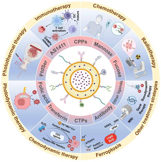
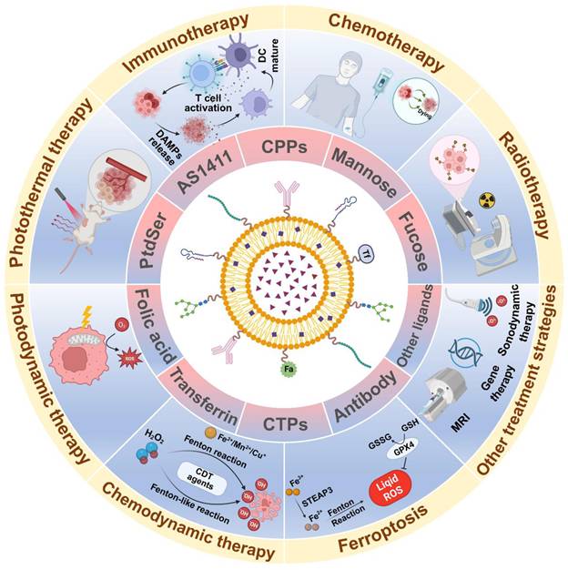
Herein, we summarize the recent developments in ligand-modified liposomal drug delivery systems (LLDDS) for multimodal cancer therapy (Figure 1). First, we outline the evolution of liposomal systems, from first-generation conventional liposomes to second-generation polyethylene glycol (PEG)-modified liposomes and, ultimately, third-generation ligand-modified liposomes. These three generations' benefits and drawbacks are contrasted, with special attention paid to how well ligand-modified liposomes work in comparison to regular liposomes. Next, the main strategies for conjugating targeting ligands to liposomes are introduced. More remarkably, we have systematically investigated integrated strategies for applying ligand-modified liposomal nanotechnology to combination cancer therapy, with a focus on liposomal formulation, targeted delivery, controlled drug release, and anticancer activity. Lastly, we go over the main obstacles and potential opportunities related to the practical use of LLDDS for multimodal combination cancer treatment. This study seeks to further the development of customized, selective, and multifunctional cancer therapeutics as well as contribute to a paradigm shift in precision nanomedicine by a thorough examination of several active targeting ligands and liposomal delivery methods.
Scheme of ligand-modified liposomal drug delivery systems for combination cancer therapy. Liposomal nanoplatforms functionalized with diverse ligands, including peptides such as CTPs and CPPs, glycans such as mannose and fucose, aptamers such as AS1411, antibodies, folic acid, and transferrin, can enhance tumor accumulation, promote receptor-mediated cellular uptake, and enable controlled drug delivery. By co-delivering therapeutic agents and functional components, these ligand-modified liposomes can integrate chemotherapy, immunotherapy, radiotherapy, photothermal therapy, photodynamic therapy, chemodynamic therapy, sonodynamic therapy, gene therapy, ferroptosis-based therapy, and imaging-guided interventions. The multifunctional ligand-modified liposomal drug delivery systems provide a versatile platform for precise and synergistic cancer treatment.
2. Materials and methods
We searched English databases between 2023 and 2026, including PubMed, Web of Science, and Google Scholar, then we screened relevant literature published in China and abroad. The databases were searched using the following terms: [‘‘ligand” OR ‘‘peptide” OR ‘‘aptamer” OR ‘‘glycan” AND ‘‘liposome”]. Liposomal drug delivery systems were retrieved in the database using the following terms: [‘‘liposome” AND ‘‘cancer”]. According to the situation of different databases, the subject words are comprehensively searched in combination with keywords, topics, abstracts, and free words to ensure the systematization and integrity of literature retrieval.
We searched all preclinical studies on the ligand-modified liposomal drug delivery systems for combination cancer therapy. To ensure the authenticity and systematicness of the results, we included relevant in vitro and in vivo studies involving cell and animal models.
3. The development process of liposomes
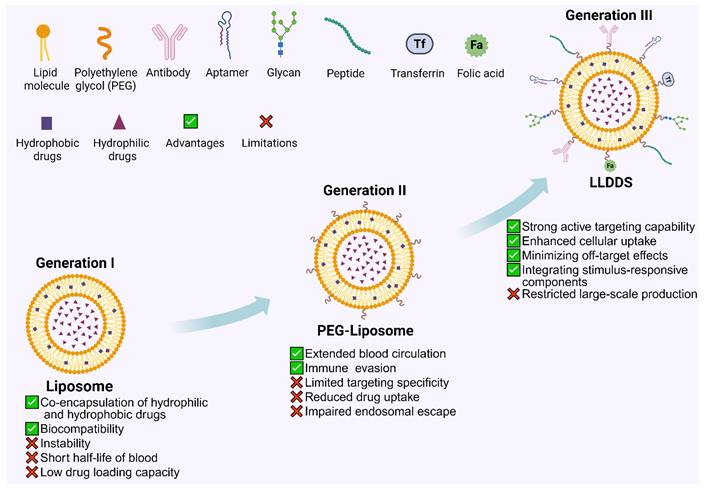
First-generation liposomes are those with simple lipid molecular structures. In 1965, British researchers Bangham and Standish discovered first-generation liposomes by dispersing phospholipids in water for electron microscopy observation. When phospholipids are dispersed in water, they naturally form multilayer vesicles, with each layer consisting of a lipid bilayer. The central region of the vesicles, as well as the spaces between layers, is filled with water, and the bilayer thickness is approximately 4 nm. In 1971, Rymen et al. from the UK utilized liposomes for drug delivery by encapsulating hydrophilic compounds in the aqueous core and binding hydrophobic compounds to the lipid bilayers. The simultaneous encapsulation of hydrophilic and hydrophobic medications, as well as their degradability and biocompatibility, were among the many benefits offered by first-generation liposomes. However, they also faced challenges, such as instability, low hydrophobic drug loading capacity, limited release of hydrophilic drugs, and a short blood half-life [26].
To correct the deficiencies of the first generation of liposomes, second-generation liposomal systems have been developed with improved in vivo stability and extended circulation time. Traditional liposomes are easily recognised, taken up and cleared by the mononuclear phagocyte system after intravenous injection, and thus have a limited therapeutic effect; therefore, these modified formulations have reduced rapid immune clearance and improved their prospects for clinical application. Blume and Klibanov modified the surface of liposomes with PEG in 1990 to address this problem and developed PEGylated liposomes. PEGylated doxorubicin liposomes (Doxil®) in 1995 were the first PEGylated nanomedicines approved by the US Food and Drug Administration and performed well. Doxil® was a precursor; now, several PEGylated liposomes, such as ThermoDox® and MM-302, have also advanced to clinical trials [27-28]. PEGylation alters the protein corona on the surface of liposomes to extend their circulation time in the blood and enhance drug delivery. PEG chains introduce steric hindrance at the surface of liposomes and, in turn, prevent aggregation caused by van der Waals forces. Thus, there will be no aggregation in the formulation. PEGylation can be added to liposomes to give them a "stealth" effect and thus prolong their circulation in the body. PEGylated liposomes take advantage of the EPR effect in tumours to prolong their circulation time and thus accumulate more drugs at the tumour site passively [29]. However, PEG modification has defects such as reduced drug uptake and impaired endosomal escape. Recently, some studies have shown that repeated administration of PEGylated liposomes induces the generation of anti-PEG antibodies. Antibodies bind to PEGylated liposomes, activate the complement system and increase the risk of allergic reactions [30].
Third-generation liposomes were developed to overcome the deficiencies of second-generation PEG liposomes, such as the protein corona on their surfaces that prevents cell entry. EPR effect is not beneficial to the therapeutic effect of nucleic acid, protein and peptide drugs that require good cell permeability. Given the above problems, many scholars have carried out relevant studies and developed third-generation liposomes. Liposomes are equipped with ligands, such as peptides, glycans, aptamers and antibodies, in a particular way. Due to the fluidity of lipids, surface-bound ligands have relatively more freedom in the lipid bilayer and are therefore better able to bind to receptors and pass through cells [31]. Ligand-modified liposomes improve the efficacy of drug delivery systems by selectively targeting and binding receptors overexpressed on tumor cell surfaces, while sparing healthy cells. Furthermore, minimizing off-target effects and integrating stimulus-responsive components are key advantages of third-generation liposomes. One of the major challenges in applying third-generation liposomes is scaling up. The absence of engineering advancements and accurate mathematical models to understand these processes hinders large-scale production (Figure 2).
The development process of liposomes: from first-generation conventional liposomes to second-generation PEG-modified liposomes and, ultimately, third-generation ligand-modified liposomal drug delivery systems (LLDDS). The advantages and limitations of each generation are shown in the diagram of the respective liposomes.
4. Ligand modification strategies of functionalized liposomes
In the construction of multifunctional liposomes based on ligand modification, two primary approaches are employed. The first approach involves directly mixing phospholipids with functional elements or active functional groups together with other phospholipid cofactors. This process enables the target moiety to be attached to the lipid. The second method is to functionalize the preformed liposomes with the desired target ligand on the liposome surface.
In both of the aforementioned methods for constructing functionalized liposomes, surface modification is achieved through the formation of covalent bonds. By creating strong chemical interactions, covalent bonding directly affixes functional components—like peptides, aptamers, or antibodies—to the liposome surface. Common covalent bonding methods include amide bond, hydrazone bond, and thioester bond. For example, aptamers or antibodies can be covalently attached to liposomes using these methods [32]. The first is that these strategies have high stability and, therefore, the functionalized liposomes are effective for a longer time. In addition, this way can control the concentration and distribution of functionalised liposomes more precisely to improve the efficiency of targeted drug delivery. Although covalent bonds are very stable, their synthesis involves numerous chemical reactions and relatively complex steps; thus, they are difficult to prepare in a lab. Due to the relatively complex synthesis process, the cost of raw materials and equipment is also high [33-34]. Non-covalent bonds are used to connect ligands with liposomes by means of physical and mechanical forces. Common non-covalent bonding modes are electrostatic interactions, hydrophobic interactions and simple adsorption. Cationic liposomes can bind negatively charged nucleic acids or peptides through electrostatic attraction, for example [35]. Non-covalent bonding is relatively simple, low in production cost and easy to realise; therefore, it is suitable for large-scale production. However, the bonded type is unstable in the presence of serum components, enzymes or pH changes in vivo and is therefore prone to degradation. These factors may cause ligand dissociation and thus reduce the drug's targeting and controlled-release effects. In addition, non-covalent bonds are relatively weak physical forces; thus, they may not effectively stabilize the liposome [36].
5. Ligand-modified liposomal drug delivery systems for cancer combination therapy
5.1. Peptide
Peptides are necessary active substances in the body, and they consist of oligomers of amino acids linked by amide bonds. Peptides have the advantages of high specificity, strong affinity and targeting capabilities. Peptides are also able to cross cell membranes and are responsive to the environment; thus, they have many applications in diagnostics and targeted drug delivery [37]. Research has shown that the biological activity of peptides is closely related to their conformation, such as α-helices or β-structures, which promote amphipathic behaviour and are necessary for their biological functions [38]. The two kinds of peptides are cell-penetrating peptides (CPPs) and cell-targeting peptides (CTPs). CPPs have up to 35 amino acids and are more biocompatible, more permeable to membranes, less toxic and less immunogenic than other cationic polymers. CPPs have shown promising results as modification ligands due to their excellent tissue penetration ability, and thus can achieve targeted drug delivery in a non-destructive manner. Polyarginine is a typical CPP that has been used to deliver therapeutic agents. R8 is an insulin delivery device and R7 is a cyclosporine A delivery device, for example [39, 40]. CTPs are short peptides that have a high affinity and specificity for cellular or tissue targets. CTPs can bind to specific molecules on the surface of target cells, such as receptors and glycoproteins, to deliver drugs, genes and other therapeutic substances into the target cells [41]. Peptides bind to cell-surface receptors, are taken up via endocytosis, and then both the peptide and its therapeutic payload enter the cell [42]. This strategy allows CTPs to improve the targeted transport and intracellular delivery of therapeutic agents, including drugs and genetic materials, while reducing off-target toxicity in normal or non-targeted cells [29, 43]. Peptides can be easily chemically synthesized, modified, and conjugated to liposomes through various methods. The most common chemical linkages include disulfide and thioester bonds, which covalently attach peptides to liposomes. Reactive functional groups in peptides, such as -COOH, -NH₂, and -SH, provide ideal sites for conjugation with liposomes, especially with DSPE-PEG-Mal/NHS [44, 45]. Interestingly, in tumor therapy, peptide ligands enhance tumor penetration and reduce the MPS-mediated clearance of liposomes. Research on using peptide-modified liposomes to deliver drugs to cancer cells is gaining increasing attention, particularly with the growing interest in cancer combination therapies.
5.1.1. Peptide-modified liposomes enabling chemo-immunotherapy synergy
Chemotherapy is a central modality in cancer treatment. It directly kills cancer cells and induces immunogenic cell death (ICD), playing a crucial role in enhancing therapeutic efficacy in multimodal treatment strategies. ICD is a specialized form of apoptosis that activates cellular immunity and restores anticancer immune responses [46]. During ICD, cancer cells release damage-associated molecular patterns (DAMPs), such as calreticulin (CRT), adenosine triphosphate (ATP), and high-mobility group box protein 1 (HMGB1) [47, 48]. DAMPs can bind to and promote the maturation of dendritic cells (DCs). DCs present antigens to cytotoxic T lymphocytes, activating T cell-mediated immune responses. Nevertheless, chemotherapeutic intervention may unintentionally induce an immunosuppressive tumor microenvironment, thereby weakening antitumor immune activity and reducing overall therapeutic effectiveness. Chemo-immunotherapy, which combines chemotherapy with immunotherapy, has emerged as a breakthrough strategy for targeting and eliminating malignant tumors, fundamentally transforming cancer treatment [49]. Significantly, peptide-functionalized liposomal drug delivery systems have significantly advanced chemo-immunotherapy by synergistically delivering both chemotherapeutic and immunotherapeutic agents, while also modulating the TME.
RGD is a well-known peptide sequence that targets tumors by binding to integrin receptors, namely αvβ3 and αvβ5 integrins, on the surface of tumor cells. These integrin receptors are frequently upregulated in malignant cells and tumor-associated vascular endothelial cells, which makes RGD peptides highly suitable targeting ligands for tumor-directed therapeutic delivery. RGD peptides not only promote endocytosis of tumor cells through integrin binding but also enhance the localization and delivery of anticancer drugs, thereby improving therapeutic efficacy [50]. In addition, cRGD peptides reduce the flexibility and instability inherent in free-chain peptides by cyclizing the RGD sequence, thereby enhancing their affinity and selectivity for integrin receptors. The cyclic structure enhances their stability in vivo, improving their performance in targeting tumor cells and tumor vasculature. cRGD peptides are widely used to target tumor cells and tumor-associated angiogenesis [51]. Wang et al. [52] proposed a strategy to degrade signal transducer and activator of transcription 3 (STAT3) using nano-integrated proteolysis-targeting chimeras (PROTACs). This approach efficiently reprogrammed hepatocellular carcinoma (HCC)-associated cancer stem cells (CSCs), suppressed the growth of CSCs, and stimulated anti-HCC immune responses. PROTAC technology is an innovative and promising therapeutic strategy that uses small molecules to induce ubiquitin-dependent protein degradation. PROTACs are developed by linking the target protein (POI) to ligands and E3 ligase ligands via an intermediate linker. inS3-TEG-VL-TK was incorporated into cRGD-modified cationic liposomes to enhance uptake by CSCs that overexpress integrin αvβ3, facilitating lysosomal escape and promoting interaction with cytoplasmic STAT3. The liposome (Lip@inS3-TEG-VL-TK) efficiently depleted endogenous STAT3 in CSCs, suppressed their stemness, induced anticancer immunity, and protected healthy cells, thereby reversing HCC progression. Furthermore, fluorescence imaging showed that the cRGD-modified liposome evaded hepatic and renal clearance. The cRGD-modified liposome also exhibited significantly higher tumor-specific accumulation than the non-RGD-modified liposome. This further supported the tumor-targeting efficacy of the liposomal PROTAC prodrug in vivo and validated the need for the cRGD-modified liposomal carrier. Relative to the control cohort, in which the mean number of metastatic nodules reached 21, treatment with Lip@inS3-TEG-VL-TK markedly inhibited pulmonary dissemination of HCC, lowering the average nodule burden to 1. This study presented a modular therapeutic strategy using peptide-modified liposomes to enhance the efficacy of HCC chemotherapy-immunotherapy combinations.
The advantages and limitations of each generation of liposomes.
| Each generation of liposomes | Advantages | Limitations |
|---|---|---|
| Generation I: Ordinary liposome | Co-encapsulation of hydrophilic and hydrophobic drugs; Biocompatibility | Instability; Short half-life of blood; Low drug loading capacity |
| Generation II: PEG-liposome | Extended blood circulation; Immune evasion | Limited targeting specificity; Reduced drug uptake; Impaired endosomal escape |
| Generation III: LLDDS | Strong active targeting capability; Enhanced cellular uptake; Minimizing off-target effects; Integrating stimulus-responsive components | Restricted large-scale production |
Previous studies have demonstrated that stimulation of the aryl hydrocarbon receptor (AhR) in splenic dendritic cells downregulates the production of key cytokines. These cytokines are critical for T helper cell polarization. A ligand with strong affinity and specificity for DCs was created by Zhang et al. [53], named synthetic peptide 65 (SP65), which was identified through phage display as a DC-targeting peptide. SP65 was conjugated to the neutral lipid dioleoylphosphatidylethanolamine (DOPE) via a linker to form SP65-DOPE. The phospholipid-peptide complex was incorporated into the surface of liposomes, forming SP65-lipo-CH. Uptake experiments showed that SP65-lipo-DiR treatment increased DiR intensity by 1.6-fold in the spleen and 1.8-fold in the kidneys compared with the non-SP65-modified group. Interestingly, SP65-lipo-CH significantly increased IL-12 production, activated NK cells and CD8+ T cells, and elevated interferon-γ (IFN-γ) levels. These changes led to effective anticancer activity against MC38 colorectal cancer and LLC lung cancer in mice. Similarly, Zhang et al. [54] designed a three-step artificial intelligence (AI) workflow to accelerate the design of dual-drug-loaded lipid carriers targeting CXCR4 for colorectal cancer treatment. Flexible docking and AlphaFold-based interface scoring identified a 9-mer fragment of SDF-1 (pSDF-1), known as the CXCR4-binding peptide (CXCR4BP). The CXCR4BP-modified liposome (with PTX located in the lipid shell) encapsulated berberine (BBR)-loaded functionalized mesoporous silica nanoparticles (FMSN), forming FMSN(BBR)-CXCR4BPL(PTX) for synergistic chemo-immunotherapy in colorectal cancer. The platform demonstrated high dual-drug encapsulation efficiency (78.8 ± 1.9% for BBR and 75.2 ± 2.4% for PTX), with sustained drug release over 72 hours. Concurrently, in vivo imaging confirmed that CXCR4BP conferred robust and selective targeting of CXCR4-positive CT26 colorectal cancer cells on the liposome. CXCR4BP modification also enhanced tumor accumulation without detectable uptake in non-target organs. In vivo anticancer studies showed that FMSN(BBR)-CXCR4BPL(PTX) caused significant tumor regression, reversed splenomegaly, and exhibited potent anti-proliferative, pro-apoptotic, and anti-angiogenic effects.
Moreover, octaarginine (R8) is a short peptide consisting of eight arginine residues. Due to its positive charge, the R8 peptide interacts with the negative charges on cell membranes, facilitating its transport across the membrane. R8 peptides are frequently used as CPPs to efficiently deliver bioactive molecules, such as small-molecule drugs, DNA, and RNA, into cells [55]. R8-modified liposomes (R8-Lip) were shown to enhance antigen presentation by MHC class I (MHC-I) in dendritic cells after antigen encapsulation. Nakamura and others [56] have shown that encapsulating polyinosinic-polycytidylic acid and ovalbumin (OVA) in R8-Lip significantly enhances their efficacy after intravenous injection. Treatment with the R8-Lip/PIC/OVA system increased the immune status of B16-OVA tumours and converted them from "cold" to "hot" tumours.
5.1.2. Peptide-modified liposomes enabling photo-chemotherapy synergy
Phototherapy has developed rapidly in recent years and now uses light of a particular wavelength to induce photochemical changes or heat damage in cancer cells and tissues. Among the various kinds of light, Photodynamic Therapy (PDT) and Photothermal Therapy (PTT) have been extensively studied and applied to date [57, 58]. Photodynamic therapy is the irradiation of a specified wavelength to activate photosensitizing agents that have accumulated in diseased tissues, generate cytotoxic reactive oxygen species (ROS), particularly singlet oxygen, and thus cause localised damage to the target tissues [59]. Photothermal therapy is a treatment modality in which absorbed light is converted into thermal energy. Upon photoexcitation, photothermal agents dissipate the absorbed energy through non-radiative relaxation as they return to the ground state, thereby producing heat and elevating the temperature within the local microenvironment. This localized hyperthermia can damage or eliminate tumor cells and pathogenic bacteria, while also inducing multiple biological alterations in cancer cells [60, 61]. Owing to their different modes of action, photodynamic therapy and photothermal therapy can serve as effective complements to conventional chemotherapy. At the cellular level, these light-based therapies may help reverse chemoresistance and restore therapeutic sensitivity by influencing dysregulated signaling networks. Within the tumor microenvironment, PDT and PTT can further improve tumor perfusion, increase vascular permeability, and remodel extracellular matrix barriers, thereby facilitating more efficient intratumoral drug accumulation and penetration. Furthermore, compared to chemotherapy-mediated systemic therapies, PDT and PTT offer the advantage of local specificity, providing better spatio-temporal control and reducing off-target toxicity [62, 63]. In addition to encouraging neutrophil recruitment and complement cascade activation, PDT and PTT may simultaneously increase the release of immune-modulatory cytokines including IL-6 and interleukin-4 (IL-4). Together, these responses establish an acute inflammatory milieu that supports the infiltration and activation of immune cells. Importantly, ICD triggered by PDT or PTT is associated with the liberation of DAMPs, which function as endogenous alarm signals to initiate and amplify antitumor immune responses [64, 65]. Combined chemotherapy and phototherapy can synergistically activate the immune system, enhancing therapeutic efficacy, particularly when combined with immunotherapies like immune checkpoint inhibitors. Integrating chemotherapy and phototherapy into a combined photo-chemotherapy strategy aims to overcome the limitations of monotherapy by leveraging the complementary effects of both approaches. The precision of phototherapy, the systemic action of chemotherapy, enhanced immune responses, and the ability to overcome drug resistance have generated growing interest in photo-chemotherapy.
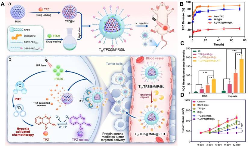
Recent studies have shown that when nanomaterials are introduced into biological systems or fluids, proteins in the bloodstream spontaneously adsorb onto nanoparticle surfaces, forming a “protein corona” (PC) [66]. While PC confers novel biological properties to nanoparticles, it impairs their functionality and targeting within cells. As a result, researchers are exploring the use of adsorbable proteins with active ligand properties to modify nanoparticle surfaces, forming endogenous PC in situ. This allows the ligand proteins to guide the nanoparticles to target sites. For example, Jin et al. [67] used the T10 peptide (sequence: CGGGHKYLRW), which has a high affinity for transferrin (Tf) and selectively binds Tf in the body. Transferrin (Tf) is abundant in the bloodstream, whereas its receptor, the transmembrane protein transferrin receptor (TfR), is frequently upregulated in diverse malignant cells. This expression pattern makes the Tf/TfR axis an attractive strategy for pre-PC tumor-targeted delivery. This study used T10 peptide-modified liposomes to create in situ Tf-PC-mediated liposomes carrying the hypoxia-sensitive chemotherapy drug tirapazamine (TPZ) and the photosensitizer indocyanine green (IR820) (Figure 3A). The water-soluble drug TPZ was encapsulated in mesoporous silica nanoparticles (MSNs) and coated with IR820-loaded liposomes (Figure 3B). Because of their superior biocompatibility, adaptable surface functionalization, and customizable structure and composition, MSNs constitute a significant class of biomedical nanomaterials [68]. Upon entering systemic circulation, the platform (T10/TPZ@M/IR@L) enabled T10 to bind specifically to plasma Tf, forming an in situ Tf liposome-PC complex. This approach demonstrated superior targeting efficacy over conventional ligand-modified targeting strategies. Simultaneously, upon exposure to near-infrared irradiation, IR820 activation promoted the infiltration of T10/TPZ@M/IR@L into deep breast tumor tissues. Crucially, under near-infrared irradiation, high ROS levels produced by IR820 directly damaged tumor cells and exacerbated tumor hypoxia (Figure 3C). Finally, TPZ was activated into cytotoxic metabolites in hypoxic tumor tissues, eliminating 4T1 tumor cells through the synergistic effects of photo-chemotherapy (Figure 3D). This strategy could aid the design of multifunctional liposome delivery systems for efficient active targeting and enhanced photo-chemotherapy in breast cancer.
Peptide-modified liposomal drug delivery systems for cancer photo-chemotherapy. (A): (a) Schematic illustration of design and preparation of the T10/TPZ@M/IR@L and (b) hypoxia-induced chemo-phototherapeutic effect under the mediation of Tf protein corona. (B) In vitro drug release of TPZ. (C) In vitro detection of reactive oxygen species/hypoxia in 4T1 cells treated with different formulations. (808 nm, 100 W/cm2, 5 min). (D) Tumor growth volume of the mice after different treatments. Adapted with permission from [67], Copyright 2024, American Chemical Society.
5.1.3. Peptide-modified liposomes enabling photo-immunotherapy synergy
In recent years, combining PDT with immunotherapy has attracted increasing attention as a promising strategy for cancer treatment. Numerous studies have shown that inducing ICD can address the issue of low immune responses in tumor immunotherapy. Compared with drug-induced ICD, PDT offers several advantages, including high selectivity, minimal side effects, precise spatiotemporal control, and the ability to overcome multidrug resistance. More notably, the clinical application of photodynamic therapy (PDT) in oncology has advanced substantially. Several photosensitizing agents approved by the Food and Drug Administration (FDA), including Photofrin and Verteporfin, have been introduced for the treatment of malignancies such as lung and esophageal cancers [69]. A cRGD-modified liposomal system including the photosensitiser pheophorbide A (Pa) and the anti-programmed cell death ligand 1 (PD-L1) antibody was created by Qin et al. [70]. Pa is a naturally occurring second-generation photosensitiser in the chlorophyll derivative family [71]. The photosensitizer has a high light-absorption cross-section and is highly phototoxic. These characteristics have made it a good candidate for PDT in various tumours [72]. cRGD-modified liposomes enhanced tumour targeting and improved the solubility and biocompatibility of Pa. The cRGD-PaNPs-αPD-L1 group exhibited a more favourable immune microenvironment in 4T1 tumours compared with the other groups. The system raised the activation rate of mature dendritic cells from 0.023% to 0.077% and the infiltration rate of cytotoxic T cells from 0.23% to 0.92%. Therefore, the two were combined in this study to improve the effect of PD-1/PD-L1 inhibition.
5.1.4. Peptide-modified liposomes enabling radio-immunotherapy synergy
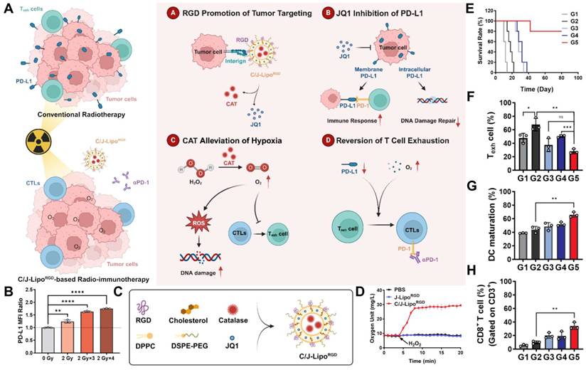
Radiation therapy (RT) is one of the three main kinds of cancer treatment that damages the DNA of cancer cells and stops them from dividing. Although widely used for the treatment of cancer, RT will inevitably induce changes in the TME and cause radiation resistance and immunosuppression. RT is now widely used to enhance the sensitivity and general efficacy of immunotherapy [73-74]. Based on the above evidence, RT directly kills malignant cells and, at the same time, remodels the immune environment in tumours by inducing immunogenic cell death, modifying anti-tumour immune responses, and triggering systemic abscopal effects. The efficacy of local RT combined with immunotherapeutic agents, particularly immune checkpoint inhibitors, has been validated in preclinical models and clinical trials [76]. Interestingly, Yue et al. [77] investigated radiation-induced changes in the TME. The study revealed that X-ray exposure elevated PD-L1 levels, aggravated intratumoral hypoxia, and promoted exhausted T-cell phenotypes, thereby weakening the therapeutic benefit of fractionated radiotherapy (Figure 4B). In response to these radiation-associated limitations, to accomplish tumor-selective accumulation and control the tumor microenvironment, the scientists created C/J-LipoRGD, a cRGD-functionalized liposomal nanoplatform co-loaded with catalase and the JQ1. (Figure 4A, C). Catalase converted excessive tumor H₂O₂ into oxygen and water, thereby alleviating hypoxia and improving local oxygen availability (Figure 4D). After cellular uptake, JQ1 suppressed PD-L1 expression, which inhibited PD-1/PD-L1-mediated immunosuppression and interfered with DNA damage repair. Flow cytometric profiling indicated that radiotherapy alone modestly increased lymph node dendritic cell maturation to 44.13%, whereas the triple regimen consisting of C/J-LipoRGD and αPD-1 markedly elevated this proportion to 70.10%. In parallel, treatment with C/J-LipoRGD increased intratumoral CD3⁺CD8⁺ T-cell infiltration and enhanced the expression of TNF-α and IFN-γ. Overall, C/J-LipoRGD-based radioimmunotherapy reshaped the TME by relieving hypoxia, suppressing PD-L1 signaling, reversing T-cell exhaustion, promoting DCs maturation, and strengthening CD8⁺ T-cell-mediated antitumor immunity (Figure 4E-H). These coordinated effects ultimately improved the therapeutic efficacy of radiotherapy combined with immune checkpoint blockade.
Peptide-functionalized liposomal platforms for cancer radioimmunotherapy. (A) Schematic illustration of C/J-LipoRGD-mediated tumor microenvironment remodeling for potentiating radioimmunotherapy. (B) Flow cytometric analysis of PD-L1 expression in B16-F10 cells 24 h after exposure to different doses of X-ray irradiation. (C) Diagram of cRGD-modified liposomes co-encapsulating JQ1 and catalase for tumor-targeted delivery. (D) Oxygen generation profiles of PBS, J-LipoRGD, and C/J-LipoRGD solutions following the addition of H₂O₂ at 4 min. (E) Survival curves of B16-F10 tumor-bearing mice subjected to various therapeutic regimens. (F) Flow cytometric detection and quantitative analysis of exhausted T cells, defined by PD-1 expression, within tumor tissues after different treatments. (G) Representative flow cytometry plots and corresponding quantification of mature dendritic cells in tumors following each treatment. (H) Flow cytometric assessment of tumor-infiltrating T cells and the proportion of CD8⁺ T cells among CD3⁺ T cells. Data are presented as mean ± SD (n = 3). *P < 0.05, **P < 0.01, ***P < 0.001. Adapted with permission from [77], Copyright 2025, Elsevier Ltd.
In summary, peptide-modified liposomal drug delivery systems enable targeted cancer therapy by enhancing intracellular uptake, improving tumor penetration, and reducing off-target effects. The integration of peptides with liposomes has shown promise in chemo-immunotherapy, photo-chemotherapy, and other combination therapies. However, challenges remain, including optimizing peptide stability and overcoming immune suppression within the TME.
5.2. Glycosylation
Tumor-associated alterations in cell-surface glycosylation are commonly manifested as the generation of truncated glycan structures, increased expression of highly branched N- and O-linked glycans, and dysregulated fucosylation or sialylation profiles [78]. Because malignant cells often have enhanced glucose uptake and abnormal expression of glucose transporters and sodium-dependent glucose cotransporters, these carbohydrate-related pathways have become attractive targets for tumor-selective therapy [79]. Glycosylation is a general kind of post-translational modification that adds carbohydrate chains to proteins and lipids to change their shape, stability and other biological functions. Changes occur in many areas during tumour development that alter the glycan pattern of cancer cells compared with those in normal tissues [80]. For example, tumour cells may have an increased amount of certain carbohydrate groups, such as Gal-β-D-mannose and N-acetyl-D-glucosamine, and a reduced quantity of other glycan structures [81, 82]. Based on the above, glycan-containing ligands have been gradually incorporated into drug delivery systems for glycosylation-directed targeting. Another therapeutic strategy is to inhibit glycosyltransferases or the glycosylation pathway in tumours that have been aberrantly activated. In particular, inhibition of O-GlcNAc transferase has been reported to suppress tumor growth and metastatic progression in several malignancies [83, 84]. Moreover, differences in the composition, density, and spatial distribution of glycans between malignant and healthy cells provide a molecular foundation for cancer-specific recognition and intervention. Therefore, exploiting tumor-associated glycosylation abnormalities represents a promising route for active targeting, and continued research in this area may support the development of more precise anticancer therapies [85, 86]. Liposomes are especially suitable for this purpose because their surfaces can be readily modified with defined carbohydrate ligands. By identifying tumor-associated biomarkers, these glycan-functionalized liposomal systems enhance targeted delivery and may find use in cancer imaging and diagnostics.
5.2.1. Glycosylated liposomes enabling photo-immunotherapy synergy
Mannose (Man) is a naturally occurring monosaccharide found in various plant sources, including citrus peels, peaches, and apples, as well as in biological materials such as ivory palm kernels and yeast [87]. Mannosyl groups are commonly used to modify anticancer drugs, such as platinum-based drugs, as well as drug carriers; proteins and chitosan are typical examples, and they show good potential in drug delivery systems. Maillard reaction provides a feasible strategy for preparing drug delivery platforms by coupling mannose with amino-group-containing materials, such as liposomes, chitosan and protein-based carriers [88]. Liposomes modified with mannose derivatives actively target hepatocellular carcinoma cells by binding to mannose receptors (MR) and have shown good results in targeted delivery [89]. Four members make up the extremely effective endogenous receptor system known as the mannose receptor family: MR (CD206), phospholipase A2 receptor (PLA2R), Endo180 (CD280), and DEC-205 (CD205) [90]. Its functions include clearing endogenous molecules, promoting antigen presentation, and regulating cell activation and trafficking [91, 92]. Additionally, MR is highly expressed in splenic and alveolar macrophages [93]. Accordingly, mannose-functionalized drug delivery platforms have attracted considerable interest as promising therapeutic carriers, owing to their favorable delivery performance, enhanced targeting potential, and relatively low toxicity. A recent landmark study demonstrated that trimannose conjugation markedly improved the pulmonary macrophage delivery of inhaled oligonucleotides, establishing this platform as the first mannosylated therapeutic candidate developed for COVID-19 [94].
Recent advances in cancer immunotherapy have increased interest in tumor vaccines. Tumor vaccines are characterized by durable immune memory and antigen-specific immune responses, and they have demonstrated promising therapeutic efficacy in clinical trials [95]. Among these, in situ vaccination generates endogenous antigens for autologous tumor cells in vivo, eliminating the need to identify and isolate tumor-associated antigens (TAAs), thereby eliciting a broad immune response [96, 97]. In PTT, near-infrared (NIR) light, with its strong tissue penetration, kills tumor cells and releases TAAs. As a result, localized PTT combined with immune activation from in situ vaccines achieves superior anticancer effects. Based on photothermal-immunotherapy, Li et al. [98] proposed a photothermal-triggered in situ vaccine consisting of acid-responsive liposome-coated polydopamine (PDA) nanoparticles, modified with Man and loaded with resiquimod (R848). First, acid-responsive liposomes (PMRL) were cleaved in the acidic TME at the tumor site, exposing PDA-Man@R848 nanoparticles. These nanoparticles not only mediated photothermal conversion but also induced ICD and promoted the release of TAAs. Meanwhile, the surface of PDA-Man@R848 nanoparticles was modified with Man. Relative to the PBS-treated cohort, administration of PMRL markedly enhanced T-cell accumulation within tumors, increasing CD4⁺ and CD8⁺ T-cell infiltration by 2.01- and 2.15-fold, respectively. Furthermore, ELISA analysis showed that the PMRL group had lower serum TGF-β levels and higher IFN-γ and TNF-α secretion than the other groups, indicating that PMRL induced a robust pro-inflammatory immune response. Man-modified PDA@R848 nanoparticles promoted the maturation of DCs, enhanced antigen cross-presentation, and strengthened anticancer adaptive immunity. In addition, the vaccine effectively inhibited distant tumor recurrence and lung metastasis in the 4T1 model by inducing long-term immune memory.
5.2.2. Glycosylated liposomes enabling chemo-immunotherapy synergy
Glucose serves as an essential cellular energy substrate, and its uptake is primarily mediated by glucose transporters (GLUTs), which are broadly expressed on the surface of most cell types. This results in the overexpression of GLUT-1 on tumor cell surfaces, sustaining their high glucose uptake. Consequently, GLUT-1 is considered a potential target for anticancer therapy. GLUT-1-specific carbohydrates, such as glucose, 2-deoxyglucose, and glucosamine analogues, can be conjugated to liposome drug delivery vehicles for transport via GLUT-1 [99]. Fu et al. [100] co-encapsulated 20(S)-protopanaxadiol (PPD) and cannabidiol (CBD) within n-dodecyl β-D-maltoside (Mal)-modified liposomes to evaluate their synergistic anticancer effect on breast cancer. The Mal surface contains two glucosyl residues, which mediate tumor-targeting functionality. Protopanaxadiol (PPD), a bioactive constituent originating from the traditional Chinese medicinal herb ginseng, exhibits diverse pharmacological activities and considerable therapeutic potential [101]. Cannabidiol (CBD), one of the major phytochemicals isolated from Cannabis sativa, has also been extensively investigated for applications in neurodegenerative disorders [102]. Notably, neither PPD nor CBD alone displays strong antitumor activity. However, when these two agents were co-loaded into maleimide-modified liposomes, the resulting combinational formulation produced a pronounced therapeutic effect, achieving an 82.2% tumor suppression rate.
5.2.3. Glycosylated liposomes enabling ferroptosis–immunotherapy synergy
Ferroptosis is a relatively new mode of cell death. Stockwell's laboratory in 2012 discovered a new kind of regulated cell death that occurs when iron-dependent accumulation of lethal membrane-localized lipid peroxides takes place [103, 104]. Recently, modulators of ferroptosis have been used to inhibit the development and spread of cancer, and thus have attracted much attention in etiology and therapy research [105, 106]. Therefore, ferroptosis is now regarded as a good target for cancer therapy, and in combination with other drugs, its effect can be enhanced to address problems such as the reduced efficacy of traditional treatments, drug resistance in tumours, and recurrence. Ferroptosis-immunotherapy can suppress cancer cells directly and boost the immune response via ICD to build a strong anti-cancer immunity [107]. Ferroptosis in the tumour microenvironment is regulated by complex cross-talk among malignant cells and immune cells [108]. TAAs and DAMPs are released by ferroptotic cells to promote DC maturation and T cell-mediated anti-tumor immunity. MHC-I recognition activates the JAK1/STAT1 pathway in tumour cells by CD8+ T cells, inhibits System Xc function, decreases GSH and GPX4, and thus increases susceptibility to ferroptosis [109, 110]. Furthermore, the ferroptosis pathway in immunosuppressive cell populations can be inhibited to reduce their tumour-promoting effects and alleviate immune suppression [111]. Not only do some ferroptosis-inducing agents cause ferroptotic death in M2-like macrophages, but they also promote their phenotypic switching to antitumor M1 macrophages. These M1-polarised macrophages can produce hydrogen peroxide and thus enhance Fenton chemistry in tumours, forming a positive feedback loop that further promotes ferroptosis [112]. Combine ferroptosis and immunotherapy to offer a new way of extended tumour suppression and improved treatment effect. Functionalized Drug Delivery Systems offer multiple advantages for this cooperative strategy. Glycosylated functionalized liposomes for tumour targeting through ligand-receptor interactions. At the same time, TME-responsive systems can achieve space-time-controlled drug release by using acidic pH, high GSH levels or enzyme overexpression [113].
For example, Gao et al. [114] reported a fucose-modified liposome that establishes a positive loop between ferroptotic therapy and immunotherapy. Fucose, a hexose sugar also known as 6-deoxy-L-galactose, is widely distributed in glycoproteins and glycolipids of living organisms. Liposomes are surface-modified with fucose, which binds to overexpressed mannose receptor C-type 1 (MRC1 or CD206) on tumor cell surfaces, mediating active targeting [115]. Interestingly, researchers engineered a lipid (DAPC) with phosphatidylcholine as its polar head and two arachidonic acid moieties as hydrophobic tails. This lipid responded to high ROS levels in the TME, inducing ferroptosis-specific peroxidation of DAPC and triggering the release of CpG ODNs. CpG ODN, as an immunostimulant, further promoted DC maturation and enhanced the effector function of CD8+ T cells. Additionally, IFN-γ released by activated CD8+ T cells promoted ferroptosis in cancer cells by inhibiting SLC7A11 and GSH biosynthesis. In the 4T1 tumor-bearing mouse model, this liposome (DAPC-Fuc/CpG) effectively suppressed tumor growth and demonstrated optimal therapeutic efficacy across all treatment groups. Overall, the DAPC-Fuc/CpG triple-action platform, integrating active targeting, ROS-responsive release, and selective lipid peroxidation, offered a promising strategy for coordinating ferroptosis-immunotherapy.
In summary, advances in glycobiology, glycan engineering, and glycomics have substantially accelerated the biomedical application of carbohydrate-based strategies. These sugar structures enable efficient active targeting of various tumor cells and can be used to deliver different types of drugs, genes, and therapeutic RNA molecules. Glycan-modified liposome delivery systems emphasize selective targeting of cancer cells via specific interactions with overexpressed glycan receptors.
5.3. Aptamer
In the early 1990s, two independent research groups made the co-discovery of aptamer technology and ushered in a new era of molecular recognition [116]. These synthetic oligonucleotides are called aptamers, and they are obtained by the technique of systematic evolution of ligands by exponential enrichment [117]. Aptamers are relatively small, functional single-stranded DNA or RNA oligonucleotides that can fold into specific three-dimensional shapes and bind to their targets with high affinity and specificity. Aptamers are taken up by tumour cells via clathrin-mediated endocytosis and micropinocytosis [118, 119]. Aptamers have a wide range of targets and can bind to ions, small molecules, peptides, proteins, cells and tissues. Both DNA and RNA aptamers have strong binding affinity for multiple targets, as shown in [120, 121]. Aptamers serve as specific ligands in the construction of target-delivery systems for anticancer nanomedicine frequently.
5.3.1. Aptamer-modified liposomes enabling photo-immunotherapy synergy
AS1411 aptamers are hydrophilic, guanosine-rich single-stranded DNA molecules. AS1411 has a length of 26 nucleotides and is a specific G-quadruplex. AS1411 has an interesting specific nucleotide sequence: 5'-GGTGGTGGTGGTGGT-3'. This peptide sequence can selectively recognise and bind to receptors expressed on the surface of cancer cells, thus having strong tumour-targeting ability [122]. AS1411 is a typical example of a receptor that binds to tumours, such as ribosomal protein S6, epithelial cell adhesion molecules, and nucleolin (a nucleic acid transporter). AS1411 aptamers have excellent specificity, can enhance receptor-mediated cellular uptake and are less prone to off-target toxicity. Significantly, the G-quadruplex structure of the AS1411 aptamer binds to DNA or RNA in cancer cells and thus inhibits their transcription and replication. Thus, inhibiting the growth of cancer cells and inducing apoptosis [123]. AS1411 can modify the function of T cells, natural killer (NK) cells and dendritic cells, etc., as shown in some studies. AS1411 can be combined with immune checkpoint inhibitors to boost the tumour-killing effect of the immune system and inhibit immune evasion [124]. AS1411 can be used in conjunction with a liposomal delivery system to fully realise its strong effects, and through multimodal therapy, it is now expected to serve as a high-precision and adaptable platform for breast cancer treatment.
Based on AS1411 aptamer modification, Li et al. [125] constructed a functionalized liposome loaded with Golgi-targeted carbon dots (CDs)-A-chain (RTA) conjugates and the photosensitizer pheophorbide a (PPa), enabling targeted synergistic chemotherapy and PDT. The liposome (AS1411/lip/PPa/C-R) preferentially accumulated at tumor sites due to the high affinity between nucleolin and the AS1411 aptamer. It was then internalized and transported to lysosomes. Under near-infrared laser irradiation, PPa generated large amounts of ROS for PDT. Concurrently, the CDs-RTA conjugate escaped from lysosomes and moved to the Golgi apparatus, where RTA dissociated from CDs. RTA was then transported retrogradely to the endoplasmic reticulum and ribosomes, where it irreversibly inhibited protein synthesis and induced cell death, thereby exerting potent chemotherapeutic effects. In conclusion, the bimodal synergistic targeted liposomal therapy system developed in this study achieved highly effective photodynamic-chemotherapy for breast cancer.
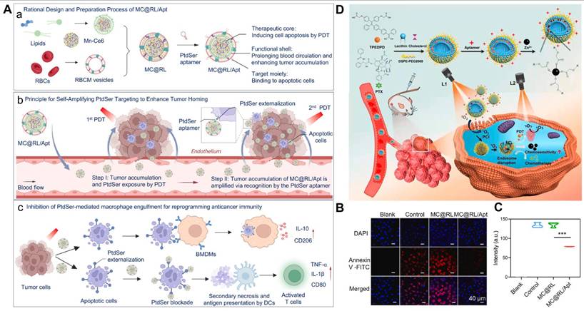
Similarly, Gao et al. [126] developed a prostate cancer-targeted liposomal system (PTX/PS-Zn@Lip-Apt) by co-loading PTX and Zn²⁺ and using aggregation-induced emission (AIE) to facilitate coordination between Zn²⁺ and AIE-associated carboxyl groups (Figure 5D). Notably, the investigators adopted a photochemical internalization (PCI) approach to achieve light-triggered, spatially controlled release of therapeutic cargos, including drugs and genetic materials. Studies showed that introducing the AS1411 aptamer mediated selective uptake by PC3 prostate cancer cells, while PCI enhanced endosomal escape efficiency. These findings were validated by tumor imaging, providing strong evidence for the efficacy of photodynamic-chemotherapy mediated by PTX/PS-Zn@Lip-Apt.
Aptamer-modified liposomal drug delivery systems for combination cancer therapy. (A) Diagrammatic representation of a self-amplifying PtdSer-targeted approach intended to improve tumor treatment and modify antitumor immunity by blocking efferocytosis and tumor homing. (a) An example of MC@RL/Apt' s logical creation and preparation. (b) When exposed to a 660 nm laser, MC@RL/Apt causes tumor cell death and encourages PtdSer externalization on the tumor cell membrane. This draws more MC@RL/Apt to the tumor location and creates a self-reinforcing accumulation effect. (c) By inhibiting macrophages from eliminating apoptotic cells and encouraging macrophage repolarization toward a pro-inflammatory M1 phenotype, MC@RL/Apt triggers potent anticancer immune responses. (B, C) Quantitative study of Annexin V-FITC staining in apoptotic CT26 cells after various treatments, along with representative confocal fluorescence pictures. The blank control was CT26 cells that had not been treated. Adapted with permission from [133], Copyright 2026, Elsevier Ltd. (D) Schematic representation of PTX/PS-Zn@Lip-Apt preparation and light-triggered therapeutic activation. The first irradiation phase (L1) was used to improve cellular internalisation by photochemical internalisation (PCI) after the accumulation of PTX/PS-Zn@Lip-Apt nanoparticles inside tumor tissues. Subsequently, a second light exposure (L2) was introduced to promote robust reactive oxygen species (ROS) generation for improved therapeutic efficacy. Adapted with permission from [126], Copyright 2025, Elsevier Ltd.
5.3.2. Aptamer-modified liposomes enabling chemo-immunotherapy synergy
Phosphatidylserine (PtdSer) is a negatively charged phospholipid consisting of a serine head group linked to a glycerophospholipid backbone. Under normal conditions, it resides primarily on the inner surface of cell membranes. In tumor cells, particularly in advanced cancer cells, PtdSer is frequently exposed on the outer leaflet of the cell membrane. The outward exposure of PtdSer on tumor cell membranes is a hallmark of tumor cell transformation and is closely linked to the invasive and metastatic properties of tumor cells [127-129]. Additionally, PtdSer is externalized to the outer leaflet of the plasma membrane during apoptosis, where it functions as an “eat-me” signal that promotes the recognition and engulfment of dying cells by macrophages and other professional phagocytes. Nevertheless, macrophages' efferocytosis of apoptotic cells might start immunosuppressive signalling, favoring their polarisation toward an anti-inflammatory M2-like phenotype and encouraging the production of TGF-β and interleukin-10 (IL-10) [130-132]. Together, these processes limit effective antitumor immune activation and help create an immunosuppressive tumor microenvironment.
Based on this, Ren et al. [133] developed a PtdSer aptamer-functionalized, red blood cell membrane-camouflaged Mn-Ce6 nanocomplex, termed MC@RL/Apt, to facilitate tumor homing and suppress efferocytosis, thereby reshaping antitumor immunity (Figure 5A). By utilizing erythrocyte membrane camouflage, this platform achieved extended systemic circulation. Upon 660 nm laser irradiation, Mn-Ce6 induced apoptotic death in tumor cells and enhanced PtdSer externalization, which further promoted progressive “snowball-like” tumor accumulation through specific recognition by PtdSer aptamers (Figure 5B, C). Simultaneously, PtdSer blocked the interaction between PtdSer and the Tim-4 receptor on the macrophage surface, thereby inhibiting phagocytosis. This increased M1-type marker expression (iNOS, TNF-α) threefold while decreasing M2-type markers (Arg-1, IL-10) by 60%. The process reprogrammed macrophages into the pro-inflammatory M1 type and activated antigen presentation in dendritic cells. The “killing two birds with one stone” strategy resulted in MC@RL/Apt accumulation at the tumor site being 1.46 times that of the non-functionalized group. In vivo experiments showed that these functionalized liposomes reduced tumor volume by 72% in the CT26 tumor model, increased CD8+ T cell infiltration fourfold, and induced a systemic anticancer immune response. This study pioneered the integration of PtdSer’s “targeting” and “immunomodulatory” functions, overcoming the limitations of traditional nanomedicines’ “passive accumulation.” The self-amplifying design addressed tumor heterogeneity challenges, while remodeling the immune microenvironment laid the foundation for combining chemotherapy with immunotherapy.
In conclusion, aptamers offer several advantages: simple synthesis, structural versatility, high chemical stability, strong tissue penetration, and low immunogenicity. In preclinical studies and recent clinical applications, aptamers have shown promise as ligands for targeted treatment of tumors.
5.4. Other ligand modifications
5.4.1. Folic acid
Folic acid is a water-soluble vitamin found in foods like leafy green vegetables, legumes, nuts, and certain animal livers [134]. It has significant potential as a targeting ligand in drug delivery systems due to its high binding affinity for folate receptors (FRs) [135, 136]. FRs are cysteine-rich cell surface glycoproteins present in three isoforms: FR-α, FR-β, and FR-γ. Among these, FR-α is the most studied isoform, showing low expression in normal cells but being overexpressed in malignancies like lung, colon, breast, and ovarian cancers [137]. Research shows that FR density increases with cancer stage and grade [138-140]. Therefore, FR-α can serve as a target for designing active cancer-targeting therapies. For instance, folate can be covalently attached to liposomes.
Extensive research shows that enhanced immunotherapy efficacy requires sufficient infiltration by NK cells and CD8+ T cells. Interleukin-15 (IL-15), a stimulatory cytokine, enhances the proliferation and activation of natural killer (NK) and CD8+ T cells by efficiently binding to the IL-15 receptor [141]. Liu et al. [142] designed FA-PEG-modified liposomes for the separate delivery of plasmid IL-15 (pIL-15) and gemcitabine (GEM) (FPCL@pIL-15 + FPGL). Both FPCL@pIL-15 and FPGL exhibited symmetrical spherical structures with particle sizes of 150 nm and 120 nm, respectively, demonstrating ideal penetration and accumulation in tumor tissues. After tumor cell-specific uptake of FPCL@pIL-15, the encapsulated pIL-15 escaped from endosomal vesicles into the cytoplasm, facilitating transfection and expression and thereby significantly promoting the proliferation and activation of NK cells and T cells. Concurrently, FPGL upregulated NKG2DLs, mediating the NKG2D/NKG2DL axis to enhance NK cell recognition and reduce immune escape. It also promoted CD8+ T cell activation via the ICD effect. Activated NK and CD8+ T cells delivered potent cytotoxic effects against cancer cells by increasing granzyme B, IFN-γ, and TNF-α expression. Crucially, FPCL@pIL-15 and FPGL-mediated chemo-immunotherapy demonstrated promising antitumor efficacy in a mouse 4T1 tumor model.
Light-responsive liposomal platforms have been engineered to enable spatiotemporally controlled release of drugs in response to external light stimulation. Chitgupi and colleagues [143] developed FA-modified liposomes co-loaded with light-sensitive porphyrin-phospholipid (PoP) and doxorubicin (Dox) for ovarian cancer therapy. In vitro co-incubation experiments showed that FA-conjugated liposomes had superior uptake rates compared to non-FA-modified liposomes when incubated with human KB cancer cells overexpressing FRs. Interestingly, Dox accumulation under phototherapy was 4-6 times higher than that achieved with chemotherapy alone. Furthermore, in vivo xenograft mouse experiments showed delayed tumor growth and improved survival rates in the functionalized liposome-treated group compared to other groups.
In short, folate-conjugated liposomes are an effective strategy for combination cancer therapy. These modified liposomes selectively target tumor cells, enhance intracellular delivery, and improve stability, biocompatibility, the ability to overcome drug resistance, and pharmacokinetic properties.
5.4.2. Transferrin
TfR is upregulated on tumor cell surfaces as a result of the unchecked growth and multiplication of cancer cells, which need much more iron than normal cells, according to several studies. TfR is widely expressed in most cell types and is involved in iron uptake. However, various tumor cells and brain capillary endothelial cells overexpress TfR at levels at least 100 times higher than in normal cells [144, 145]. Therefore, TfR is an attractive target for developing cancer therapy ligands. Researchers have explored using Tf as the natural ligand for TfR, enabling drug delivery to target cells via Tf-TfR receptor-mediated endocytosis. This approach represents an emerging strategy for targeted cancer therapy [146]. Tf is a glycoprotein primarily found in the bloodstream, with the main function of transporting iron ions (Fe³⁺) to cells throughout the body. Compared to traditional antibodies or other ligands, as a naturally occurring biomolecule, Tf is less likely to elicit an immune response and has minimal immunogenicity. This makes Tf-modified drug delivery systems safer for practical use, reducing the risk of immune rejection. In addition, transferrin-modified drug delivery systems offer other advantages, including reduced systemic drug distribution, higher drug concentrations at the target site, and enhanced synergistic toxicity against tumor cells [147, 148].
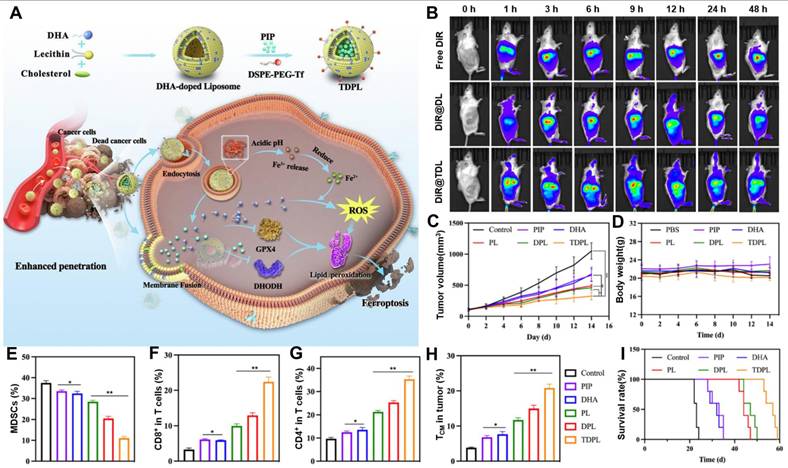
Huang et al. [149] developed Tf-modified liposomes (TDPL) doped with the unsaturated fatty acid docosahexaenoic acid (DHA) as a lipid peroxidation inducer and loaded with piperine (PIP) to enable effective ferroptosis-chemo-immunotherapy (Figure 6A). In this platform, Tf served a dual role, acting as both a ligand targeting TfR and a Fe3+ ionophore. Triggered by the low pH in the lysosome, Tf-bound Fe3+ was released and reduced to Fe2+, which subsequently catalyzed the oxidation of unsaturated lipids in addition to participating in the Fenton reaction (Figure 6B). Meanwhile, DHA incorporated into the lipid bilayer fuses with the cell membrane, inactivating GPX4 and inducing lipid peroxidation. In addition, piperine (PIP), an alkaloid found primarily in Piper longum, exerts potent anticancer effects by inducing cell cycle arrest and apoptosis, suppressing signaling protein expression, and targeting multiple cancer-related signaling pathways. In vitro experiments showed that this liposome reduced GPX4 and DHODH levels, effectively overcoming the GPX4-mediated ferroptosis defense pathway. Concurrently, piperine (PIP) downregulated DHODH expression, thereby further promoting lipid peroxidation. In vivo studies using 4T1 tumor-bearing mice demonstrated that the TDPL-treated group achieved the most pronounced antitumor efficacy and the longest survival duration among all experimental groups (Figure 6C, D, I). Notably, analysis of tumor-draining lymph nodes revealed that TDPL markedly increased dendritic cell maturation to 42.7%, which was substantially higher than that observed in the other treatment groups. These findings suggested that TDPL effectively induced immunogenic cell death and promoted dendritic cell activation in vivo (Figure 6E-H). Overall, this work provides a useful reference for integrating polyunsaturated fatty acids with natural bioactive compounds in multimodal cancer therapy.
Transferrin-modified liposomal drug delivery systems for cancer ferroptosis-immunotherapy. (A) Schematic diagram of the preparation of transferrin-modified liposomes (TDPL) and therapeutic mechanism. (B) In vivo biodistribution of free DiR, DHA-containing DiR-loaded liposomes (DiR@DL), and transferrin-functionalized DHA-containing DiR-loaded liposomes (DiR@TDL) in 4T1 tumor-bearing mice following intravenous administration. (C) Mean tumor volume changes in mice receiving different treatments, including control, PIP, DHA, PL, DPL, and TDPL groups (n = 5). (D) Body weight variations of mice throughout the treatment period (n = 5). The proportions of (E) CD8⁺ T cells among total T cells, (F) CD4⁺ T cells among total T cells, (G) myeloid-derived suppressor cells (MDSCs), and (H) central memory T cells (TCM) (n = 3) indicate the in vivo immune regulation after different therapeutic treatments. (I) Kaplan-Meier survival analysis of five mice given various treatments. *P < 0.05, **P < 0.01, ***P < 0.001. Adapted with permission from [149], Copyright 2025, Elsevier Ltd.
5.4.3. Nanobody
In the design of functionalized liposomes, nanobodies have emerged as a novel class of ligand molecule. They are derived from the variable domains of naturally occurring heavy-chain-only antibodies in camelids and fish [150-153]. Specifically, nanobodies are particularly useful as they are roughly 10 times smaller (12-15 kDa) than conventional full-length antibodies, contributing to better tumor-penetrating characteristics [154]. Nanobodies are highly stable under acidic pH conditions and at high temperatures, allowing them to withstand the harsh tumor microenvironment. These properties make them particularly suitable for modification, including radiolabeling and chemical conjugation to fluorescent dyes, liposomes, photosensitizers, and immunomodulatory molecules [155]. More importantly, the simple structure and easy production of nanobodies enable diverse molecular engineering strategies. Consequently, nanobodies can be engineered in multivalent forms to enhance stability and affinity, or fused with Fc domains to confer effector functions.
Bouma and colleagues [156] developed a novel vaccine formulation by conjugating CD169- or DC-SIGN-specific nanobodies to liposomes. This design enabled high-affinity targeting and promoted selective uptake by antigen-presenting cells (APCs). In vivo and in vitro studies showed that both nanobody-conjugated liposomes exhibited increased uptake compared to control liposomes or those bearing the native ligands for CD169 and DC-SIGN. This enhanced uptake increased T cell activation by human APCs and stimulated naive T cell activation in mouse models. Table 2 summarizes common ligands for functionalized liposome modification, corresponding receptors, and typical target cells.
Common ligands for functionalized liposome modification, corresponding receptors, and typical target cells.
| Ligand type | Ligand name | Receptor name | Typical target cells |
|---|---|---|---|
| Peptide | cRGD | integrin αvβ3 | Glioblastoma (U87MG), Breast cancer (MDA-MB-231) |
| iRGD | integrin αvβ3/αvβ5, NRP-1 | Pancreatic cancer (PANC-1) | |
| LyP-1 | p32 | Triple negative breast cancer (4T1) | |
| RVG29 | nAChR | Glioma (C6) | |
| SP94 | GRP78 | Liver cancer (HepG2) | |
| GE11 | EGFR | Hepatocellular carcinoma (SMMC-7721) | |
| T7 (HAIYPRH) | TfR1/CD71 | Lung cancer (A549) | |
| T10 | TfR | Colorectal cancer (HCT-116) | |
| Octaarginine (R8) | syndecan-4 | Broad-spectrum tumor cells (HeLa, A549) | |
| Glycans | Mannose | CD206, MRC1 | Dendritic cell, Tumor associated macrophages |
| Dextran sulfate | SRA, MSR1, CD204 | Tumor associated macrophages | |
| Fucose | MRC1/CD206 | Dendritic cell, Tumor associated macrophages | |
| Chondroitin sulfate | CD44 | Triple negative breast cancer (4T1) | |
| Hyaluronic acid | CD44 | Lung cancer (A549), Ovarian cancer (SKOV-3) | |
| Galactose | ASGPR | Liver cancer (HepG2) | |
| GalNAc | ASGPR, ASGR1 | Hepatocytes | |
| Aptamer | AS1411 | Nucleolin | Breast cancer (MCF-7), Pancreatic cancer (MIA PaCa-2), Glioblastoma (U87MG) |
| PtdSer | TIM-4 | Macrophage, Dendritic cell | |
| A10 | PSMA | Prostate cancer (LNCaP) | |
| sgc8 | PTK7 | Leukemia (CCRF-CEM) | |
| EpCAM aptamer | EpCAM | Colorectal cancer (CT-26) | |
| Others | Folic acid | FRα, FOLR1 | Ovarian cancer (SKOV-3), Cervical cancer (KB) |
| Transferrin | TfR1, CD71 | Glioblastoma (U87MG), Breast cancer (MCF-7), Leukemia (K562) | |
| Anti-DC-SIGN nanobody | DC-SIGN/CD209 | Immature/monocyte-derived dendritic cells, Certain macrophages | |
| Anti-CD169 nanobody | CD169, Siglec-1 | CD169 positive antigen-presenting cells, Macrophage |
5.5. Multiple ligand modifications
Single-target-modified liposomes have reached a relatively mature stage of development, but their further application faces limitations. Single-target modifications targeting cancer cell receptors often cause non-selective toxicity, as normal cells also express these receptors. To address this issue, researchers have explored multi-target modification strategies for liposomes to improve the targeting precision of liposomal drug delivery systems.
5.5.1. Glycosylation-Peptide
Dual-targeted liposomal drug delivery systems based on glycosylation and peptide co-modification for cancer chemo-immunotherapy combinations have recently gained attention in research. LyP-1 (CGNKRTR) is a peptide that binds to PI3K-receptor complexes on the surface of cancer cells and has high selectivity and affinity. A peptide can help a drug enter the tumour cells by binding to receptors on the surface of the tumour cells and thus increase the therapeutic effect of the drug. LyP-1 is expected to target cancer cells and promote the delivery of drugs to the tumour site [157]. Chondroitin sulfate (CS) is an anionic polysaccharide in the sulfated glycosaminoglycan family derived from animal cartilage. CS has the above anti-cancer, antioxidant, anti-inflammatory and immunomodulatory properties and can be used to treat various diseases [158, 159]. CS also has the attributes of biocompatibility, degradability, mucosal adhesion and hydrophilicity, and has been widely used in biomedicine [160]. CS has carboxyl, hydroxyl and amino functional groups that can be modified with hydrophobic components, therapeutic agents or probes to develop various drug delivery systems flexibly [161]. CS has a high affinity for the CD44 receptor and is thus taken up by the cell via receptor-mediated endocytosis. Cluster of differentiation 44 (CD44) is a cell surface glycoprotein overexpressed in various solid tumors, including pancreatic, breast, ovarian, brain, and lung cancers [162]. CD44 is primarily involved in cell proliferation, cell-to-cell interaction, cellular migration, and immune response generation. Thus, CS serves as an effective ligand targeting the CD44 receptor. Luo et al. [25] designed a dual-ligand-modified liposomal system functionalized with the LyP-1 peptide and CS and co-loaded with PTX and the STAT3 inhibitor cryptotanshinone to achieve combined chemotherapy and immunotherapy for triple-negative breast cancer (TNBC) (Figure 7A). After intravenous injection, the liposome accumulated in tumor tissue through p32/CD44 dual-receptor-mediated active targeting and was subsequently internalized by 4T1 tumor cells (Figure 7B, C). As shown in Figure 7D-F, treatment of 4T1 cells with CS/LyP-1-P increased CRT expression, HMGB1 release, and ATP secretion. These results showed that CS/LyP-1-P effectively activated antitumor immunity in vivo by inducing ICD by PTX-based chemotherapy. Concurrently, cryptotanshinone downregulated STAT3 expression, reduced the secretion of immunosuppressive factors, decreased the accumulation of immunosuppressive cells, and reversed the immunosuppressive tumor microenvironment (Figure 7G). Importantly, in the 4T1 tumor-bearing mouse model, CS/LyP-1-PC Lip exhibited significant antitumor effects and effectively inhibited lung metastasis (Figure 7H). This work established a multifunctional liposomal drug delivery platform that modulated the TME, offering a potential strategy for chemo-immunotherapy in TNBC. Similarly, Yuba and colleagues [163] incorporated pH-responsive polysaccharide derivatives and a glycopeptide containing multiple mannose residues (Man9GlcNAc2-Asn) into liposomes to balance cell uptake via lectin-mediated mechanisms with antigen delivery capabilities in the cytoplasm. Man9GlcNAc2-Asn served as a ligand for antigen-presenting cell lectins (e.g., DC-SIGN and DC-SIGNR), significantly enhancing DC uptake of liposomes and improving cross-presentation efficiency. Compared to unmodified liposomes, co-modified liposomes bearing both glycopeptide and pH-responsive polysaccharide moieties cooperatively promoted cellular association via lectin binding and scavenger receptor recognition. Concordant in vitro and in vivo results confirmed the strong adjuvant activity of this system, as reflected by increased populations of splenic dendritic cells, M1-polarized macrophages, and effector T cells. In addition, Haas and colleagues [164] investigated the immunostimulatory effects of liposomal vaccines modified with the sugar chain LeY and synthetic SLP peptides. Results showed that LeY modification enhanced the targeting and uptake of liposomes by DCs, while LeY-modified liposomes exhibited potent antitumor effects in the B16-OVA tumor model. Furthermore, binding of synthetic SLP peptides to α-glycosylcholine (α-GC) promoted activation of invariant natural killer (iNK) cells and enhanced CD4+ T cell responses.
Peptide/glycans dual-modified liposomal drug delivery systems for cancer chemo-immunotherapy. (A) Schematic illustration of the preparation of the dual-modified LyP-1/chondroitin sulfate liposomal system (CS/LyP-1-PC Lip), together with its intracellular trafficking behavior and proposed therapeutic mechanism. The CS/LyP-1-PC Lip formulation enhances chemoimmunotherapy against triple-negative breast cancer (TNBC) by inducing tumor immunogenicity and suppressing STAT3 signaling. (B) Confocal laser scanning microscopy (CLSM) images showing the lysosomal escape behavior of CS/LyP-1-C6 Lip in 4T1 cells at 1, 4, and 8 h. (C) Ex vivo fluorescence images of major organs, including the heart (H), liver (Li), spleen (S), lung (Lu), and kidney (K), as well as tumor tissue (T), at 8 and 24 h after injection. (D) Quantitative evaluation of the expression of calreticulin (CRT) in 4T1 cells. (E) HMGB1 release and (F) ATP secretion after targeted liposomal formulation treatment (n = 3). (G) Western blot examination, STAT3 and phosphorylated STAT3 (p-STAT3) expression. (H) Antimetastatic activity in a metastatic tumor model, as assessed by the total number of pulmonary metastatic nodules after different treatments (n = 5). *P < 0.05, **P < 0.01, ***P < 0.001. Adapted with permission from [25], Copyright 2023, Wiley-VCH GmbH.
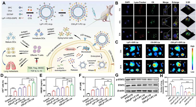
Attractively, multifunctional liposomal drug delivery platforms based on cyclodextrin and matrix metalloproteinase-2 (MMP-2)-responsive peptides can enable effective cancer photo-immunotherapy. Cyclodextrin (CD) has been used in drug delivery since the 1950s. CDs possess a mildly hydrophobic central cavity that can encapsulate lipophilic drugs or drug molecules. The formation of CD complexes enhances drug solubility, improves chemical stability, and reduces irritation [165, 166]. With advancements in biotechnological manufacturing, CD has achieved large-scale production and increased interest in the pharmaceutical industry. Over the past decade, CD has been primarily used in drug delivery systems to enhance the solubility and stability of small-molecule drugs [167]. Owing to these properties, CD-based drug delivery systems have demonstrated improved bioavailability and therapeutic efficacy in numerous clinical studies. Interestingly, β-cyclodextrin (β-CD) is a cyclodextrin with an alpha-(1->4) linkage and contains seven D-glucopyranose units [168, 169]. Some clinical trials have investigated the tumor-targeting properties of β-CD. For example, a CD dimer synthesized via click chemistry connected hydrophobic and hydrophilic portions, resulting in a self-assembled, noncovalently bonded micellar nanostructure. The researchers validated the fabrication strategy for the drug-loaded nanocarrier and demonstrated its potential for tumor-targeted therapy through endocytosis-related studies [170]. More importantly, β-CD-based liposomal drug delivery systems can achieve precise tumor targeting and effective cancer combination therapy. Although PDT combined with immunotherapy has been extensively studied, research has primarily focused on inducing ICD to activate CTLs. In contrast, leveraging the synergistic effects of PDT and NK cell immunotherapy, Liu et al. [171] designed a TME/light dual-responsive liposome system (MLRN) to achieve Ce6-mediated PDT combined with NKG2D-enhanced immunotherapy using the therapeutic agent SB-3CT (Figure 8A). The system (Ce6&SB-3CT@MLRNs) comprised two components: SB-3CT-loaded β-CD and Ce6-loaded nanoliposomes, connected via an MMP-2-responsive peptide (Figure 8B). SB-3CT is a potent and selective competitive inhibitor of MMP-2 and MMP-9, with significant anticancer activity. In the tumor microenvironment, abundant MMP-2 triggered β-CD cleavage and subsequent SB-3CT release (Figure 8C, D). After the nanoplatform accumulated within melanoma tissues, the liberated SB-3CT inhibited MMP-2 activity, thereby reducing the shedding of soluble NKG2D ligands and increasing their retention on the tumor cell surface. Simultaneously, Ce6-loaded liposomes were activated by 660 nm laser irradiation, inducing apoptotic death of tumor cells. Through this spatiotemporally coordinated photoimmunotherapeutic strategy, the liposomal system markedly promoted cytotoxicity against A375 melanoma cells, reaching 83.31%, suppressed tumor progression, with a tumor proliferation rate of only 1.13% relative to the saline control, and increased NK cell abundance among tumor-infiltrating lymphocytes (Figure 8E).
Peptide/glycans dual-modified liposomal drug delivery systems for cancer photo-immunotherapy. (A) (a) Schematic representation of the synthetic route for the preparation of Ce6&SB-3CT@MLRNs. (b) Illustration of the dual-responsive drug release behavior and the underlying mechanism by which Ce6&SB-3CT@MLRNs enhance photodynamic immunotherapy in tumors. (B) Transmission electron microscopy (TEM) images of different MLRN formulations, including blank MLRNs, Ce6@MLRNs loaded with Ce6 alone, SB-3CT@MLRNs loaded with SB-3CT alone, and Ce6&SB-3CT@MLRNs co-loaded with both Ce6 and SB-3CT. (C, D) Quantitative analysis of the mean positive area percentages of MMP-2 and MICA. *P < 0.05, ***P < 0.001. (E) Photographs of the tumor tissues of mice from different treatment groups. Adapted with permission from [171], Copyright 2023, American Chemical Society.
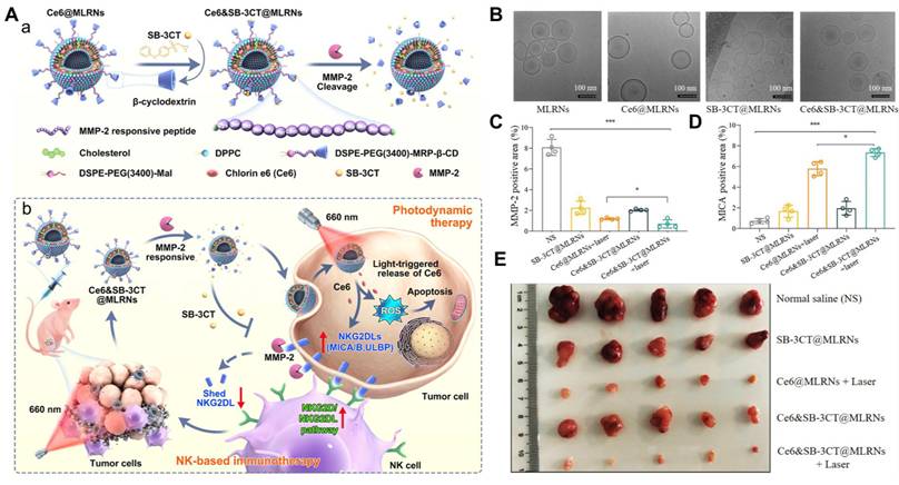
CDT is a novel therapeutic strategy that selectively kills tumor cells by activating TME-specific Fenton (or Fenton-like) reactions to generate hydroxyl radicals (·OH). CDT primarily relies on nanocatalysts to exploit the elevated H₂O₂ levels within the tumor microenvironment, converting them into highly toxic reactive oxygen species. This process amplifies oxidative stress in malignant cells, causes severe intracellular damage, and ultimately promotes tumor cell death [172-174]. Compared to traditional therapies, CDT requires no external excitation and exhibits higher specificity. Notably, CDT can be combined with chemotherapy and magnetic resonance imaging (MRI) to enhance tumor-killing efficacy. Manganese dioxide (MnO₂), a typical CDT agent, reacts with endogenous H₂O₂ in the tumor microenvironment to generate oxygen. The oxygen released from subsequent reactions alleviates the hypoxic TME, attenuates HIF-1α expression, and ultimately reverses multidrug-resistant (MDR) tumors [175, 176]. In addition, MnO₂ can be reduced to Mn²⁺ ions at lower pH values in the acidic TME, making it suitable for MRI [177, 178]. Therefore, drug delivery systems integrating CDT, chemotherapy, and MRI are extensively researched. For example, Liang et al. [179] engineered a dual-ligand-functionalized liposomal platform incorporating NAG/R8 modification and stimuli-responsive dePEGylation. This system was co-loaded with MnO₂ and paclitaxel to enable MRI-guided synergistic chemotherapy and chemodynamic therapy for lung cancer. The active targeting molecule N-acetyl-d-glucosamine (NAG), a monosaccharide derivative of glucose, simultaneously targets GLUT1 and lectin receptors. The synergistic interaction of PEG, NAG, and R8 enhanced endocytosis. The multistage liposome (C-NAG-R8-PTXL/MnO₂-lip) demonstrated superior performance under hypoxic conditions and effectively reversed hypoxia-induced chemoresistance. Concurrently, O₂-induced liposome disruption significantly promoted PTX release. The reaction between H₂O₂ and MnO₂ generated highly toxic •OH radicals, which acted synergistically with PTX to exert anti-NSCLC effects. Furthermore, in vivo experiments showed that C-NAG-R8-PTXL/MnO₂-lip exhibited outstanding T1-weighted imaging performance.
5.5.2. Dual aptamer
Based on two types of DNA aptamers (anti-CD44 and anti-PD-L1), Kim and colleagues [180] a nanosized cationic liposomal system, Aptm [DOX/IDO1], loaded with DOX and IDO1 siRNA for targeted chemoimmunotherapy and TME modulation. This nanoliposome effectively delivered DOX and IDO1 siRNA via aptamer-mediated endocytosis, successfully binding to breast cancer cells overexpressing CD44 and PD-L1. Among these, DOX was employed as the ICD inducer to kill cancer cells and initiate an anticancer immune response by activating CTLs. Meanwhile, IDO1 siRNA was combined to achieve a synergistic effect by inhibiting DOX-induced IDO1 overexpression. Aptm [DOX/IDO1] promoted the ICD response while reversing the immunosuppressive TME and reducing IDO1 expression in 4T1 tumor-bearing mice, thereby diminishing tumor volume and achieving a highly synergistic antitumor effect. More intriguingly, compared to the saline group, the lungs of Aptm [DOX/IDO1]-treated mice showed no metastatic tumors, exhibiting tissue characteristics similar to normal lungs. These findings indicated that this liposome eliminated breast cancer cells through targeted drug delivery while suppressing tumor metastasis. Collectively, this study developed a tumor-specific multithreaded chemo-immunomodulatory liposomal nanomedicine that enhances chemo-immunotherapy for breast cancer. Table 3 summarizes various ligand-modified liposomal drug delivery systems for combination cancer therapy.
Ligand-modified liposomal drug delivery systems for combination cancer therapy.
| Ligand type | Delivery platform | Cargo | Model cancer cell | Combination therapy strategy | Refs. |
|---|---|---|---|---|---|
| Peptide | FMSN(BBR)-CXCR4BPL(PTX) | BBR, PTX | Mice colon cancer CT-26 cells | Chemotherapy, Immunotherapy | [54] |
| T10/TPZ@M/IR@L | TPZ, IR820 | Mice breast cancer 4T1 cells | Chemotherapy, PDT | [67] | |
| PoIC/OVA-R8L | OVA | Mice T-cell lymphoma cells | Immunotherapy, PDT | [56] | |
| cRGD-PaNPs-αPD-L1 | Pa, αPD-L1 | Mice breast cancer 4T1 cells | Immunotherapy, PDT | [70] | |
| SP65-lipo-CH | CH223191 | Mice colon cancer MC38 cells | Chemotherapy, Immunotherapy | [53] | |
| C/J-LipoRGD | JQ1, CAT | Mice skin melanoma B16F10 cells | Radiotherapy, Immunotherapy | [77] | |
| Lip@inS3-TEG-VL-TK | PROTACs | Mice liver cancer Hepa 1-6-Luc cells | Chemotherapy, Immunotherapy | [52] | |
| Glycans | PMRL | R848 | Mice breast cancer 4T1 cells | Immunotherapy, PTT | [98] |
| GMCP-Lip | CBD, PPD | Mice breast cancer 4T1 cells | Chemotherapy, Immunotherapy | [100] | |
| DAPC-Fuc/CpG | CpG ODNs | Mice breast cancer 4T1 cells | Ferroptosis, Immunotherapy | [114] | |
| Aptamer | MC@RL/Apt | Mn-Ce6 | Mice colon cancer CT-26 cells, Mice skin melanoma B16F10 cells | Immunotherapy, PDT | [133] |
| AS1411/lip/PPa/C-R | CDs-RTA conjugates, PPa | Human breast cancer MCF-7 cells | Chemotherapy, PDT | [125] | |
| PTX/PS-Zn@Lip-Apt | PTX, AIE PSs, Zn2+ | Human prostate cancer PC3 cells | Chemotherapy, PDT | [126] | |
| Folic acid | FPGL, FPCL@pIL-15 | pIL-15, GEM | Mice breast cancer 4T1 cells | Chemotherapy, Immunotherapy | [142] |
| Dox-FA-PoP | DOX | Human oral epidermoid carcinoma KB cells | Chemotherapy, PDT | [143] | |
| Transferrin | TDPL | DHA, PIP | Mice breast cancer 4T1 cells | Ferroptosis, Immunotherapy | [149] |
| Nanobody | Nbs-Lip | OVA | Mice breast cancer 4T1 cells | Immunotherapy, PDT | [156] |
| Peptide/Glycans | C-NAG-R8-PTXL/MnO2-lip | PTX, MnO2 | Human non-small cell lung cancer A549 cells | Chemotherapy, CDT, MRI | [179] |
| Ce6&SB-3CT@MLRNs | SB-3CT, Ce6 | Human melanoma A375 cells | Immunotherapy, PDT | [171] | |
| SLP-αGC-LeY | αGC | Mice melanoma B16 cells | Chemotherapy, Immunotherapy | [164] | |
| SBA-pH-Lip | SBA | Mice T-cell lymphoma cells | Immunotherapy, PTT | [163] | |
| CD44/PD-L1 Aptamer | Aptm [DOX/IDO1] | DOX, IDO1 siRNA | Human breast cancer MDA-MB-231 cells, Mice breast cancer 4T1 cells | Chemotherapy, Immunotherapy | [180] |
6. Challenges and perspectives
This review systematically summarizes recent advances in ligand-modified liposomal drug delivery systems (LLDDS) for combination cancer therapy. LLDDS creates a potent platform for active targeted drug administration by deftly fusing the inherent benefits of liposomes with the high specificity of ligands for tumor-associated indicators. These liposomes allow for more effective drug accumulation at tumor locations while lowering off-target toxicity, in contrast to traditional passive targeting techniques that mainly depend on the EPR effect. Many studies on various kinds of cancer cells in vitro and in vivo have shown that LLDDs can increase the therapeutic effect of many drugs, such as traditional chemotherapy and new nucleic acid drugs. The above devices can address the deficiencies of conventional chemotherapy and prolong the time needed for treatment. Ligand modification can also be used to enhance the multi-functionality of liposomes as a drug delivery system. Many problems still need to be solved before the therapeutic use of ligand-modified liposomes can be realised, although some progress has been made.
6.1. The shortcomings of ligand-functionalized liposome technology and overcoming strategies
6.1.1. The stability of ligand-functionalized liposomes
First, the addition of functional ligands may reduce the physicochemical stability of liposomes. The above structural modifications may promote phase separation or aggregation in a physiological environment and thus increase the risk of early drug leakage, off-target release and damage to normal tissues. Therefore, to design and optimise ligand-modified liposomal formulations rationally, it is necessary to understand how the surface ligands interact with encapsulated cargo and lipid bilayers. To improve the stability of liposomes, the concentration, kind and spatial distribution of ligands need to be controlled precisely to achieve a uniform arrangement in the lipid bilayer [181]. Other factors will also change the circulation time, biodistribution, tumour-targeting efficiency and clearance of these particles, such as particle size and surface charge [182]. Among them, one of the problems in developing active targeting nanoparticles is how to set the optimal density for surface ligands on the nanoparticles [183]. Although a large number of ligands is expected to bind to the target cells with high affinity, an extremely high density of ligands can also have many negative effects [184]. The above reasons lead to a larger nanoparticle size, an increased risk of opsonisation and excessive depletion of receptors on the surface of target cells. Finally, the individual liposomes compete for cellular uptake and thus have reduced targeting efficiency [185]. In addition, based on experimental analysis and theoretical calculations, molecular dynamics simulations, differential scanning calorimetry, fluorescence microscopy, etc., can be used to investigate the mechanism of action for the design and optimisation of liposomal carriers to improve their overall performance.
6.1.2. Regulation of protein corona
In addition, studies have shown that ligand modification may lead to the formation of a protein corona (PC) [186]. Upon entering the bloodstream, modified liposomes interact with plasma proteins, leading to the formation of a 50-80 nm thick PC layer on their surface. Compared with passively targeted liposomes, the surface molecules and ligands on actively targeted liposomes may promote greater plasma protein binding. The PC influences the circulation time, retention, zeta potential, drug release behavior, and cellular uptake of liposomal delivery systems. More importantly, the PC may also mask ligand-binding sites, thereby reducing targeting efficiency [187]. Currently, two main strategies are used to minimize PC formation. The first approach is to use PEG-modified liposomes. The second approach is to use diphosphate polymers as materials for constructing liposomal formulations. A key feature of these materials is their electrostatic interaction with water molecules, which enables the formation of a hydration layer and thereby reduces nonspecific protein adsorption. Interestingly, AI technologies, including machine learning (ML) and deep learning (DL), have shown considerable potential in early cancer diagnosis and the systematic design of nanoplatforms [188]. Simultaneously, based on published studies, AI can be used to predict the possible composition of the PC and its biological effects in vivo [189, 190].
6.1.3. Integration of stimulus-responsive strategies
Currently, most ligand-modified liposomes still rely on passive drug release, which limits their ability to achieve precise and controlled drug delivery in vivo. Passive drug release can lead to premature drug leakage, insufficient therapeutic concentrations at target sites, and toxicity in normal tissues, thereby compromising both efficacy and safety. Endogenous stimulus-responsive strategies can take advantage of tumor-associated factors, such as elevated glutathione levels, overexpressed enzymes, and acidic pH [191]. Representative examples include pH-responsive liposomes that undergo structural transformation in acidic microenvironments, as well as enzyme-cleavable linker systems that enable payload release upon exposure to specific proteases. However, these strategies remain insufficiently explored. When designing ligand-modified stimulus-responsive liposomes, exterior stimuli including light, heat, magnetic fields, and ultrasound may be taken into account in addition to internal stimuli.
6.2. LLDDS design driven by multiligand modification and imaging technologies
To increase the binding avidity of ligands to their corresponding receptors on target cells, multiple targeting ligands are required to bind to multiple targets on the same cell simultaneously. Single-ligand-functionalised liposomes are often limited by slow receptor turnover and recycling kinetics, thus reducing their efficiency of cellular uptake. Dual-ligand-modified liposomal systems can bind to two different receptor populations simultaneously and thus enhance receptor-mediated endocytosis and improve intracellular delivery. With the continuous development of research in this area, future designs of ligand-modified actively targeted liposomes will increasingly be dual- or even multi-ligand modified. In addition, the design of imaging-technology-driven ligand-modified liposomal drug delivery systems can be realised via various means, such as magnetic resonance imaging (MRI)-guided systems, fluorescence imaging, positron emission tomography (PET)-guided systems, photothermal therapy (PTT)-guided systems, etc., for real-time monitoring of drug uptake and distribution in personalised medicine [192, 193].
6.3. Clinical translation
In addition to verifying the excellent anti-tumour effect of ligand-modified liposomal systems, clinical application also requires the development of large-scale, reproducible and economical manufacturing platforms. Functionalized liposomes with specific ligand designs for quality control are a more difficult problem than traditional liposomes. Although some ligands, such as aptamers, have shown safety in early human trials [194], additional steps are still required before they can be included in complex liposomal nanoplatforms. These are the processes of nonclinical safety, clinical comparability, chemistry, production and controls. The above reasons make it more challenging to ensure the consistency of properties for nanomedicines. The U.S.A. At present, the FDA requires assessments of human pharmacokinetics, toxicology and pharmacodynamics for liposomal products and has requested strict control over key quality attributes. Functionalised liposomes are becoming more complex, and their production costs are also rising. For instance, in chemical synthesis of functional lipids and formulation of surface-modification reagents, production costs have risen, quality control has been complicated, and batch-to-batch variability may have occurred; therefore, regulatory approval has become more difficult. To solve the problems above, reformulate and adjust the proportion of basic lipids and functional molecules for better cost performance. Cost-effective manufacturing may also be promoted by using synthetic phospholipids, such as DSPC, DPPC and DOPC, and a modular formulation strategy with simplified surface-functionalisation methods. Build a scalable and highly reproducible production platform for microfluidic-based fabrication and continuous extrusion technology to increase manufacturing efficiency, improve batch-to-batch consistency, and promote broad clinical applications.
7. Summary
In short, although ligand-modified liposomal drug delivery systems (LLDDS) are still in the exploration stage, their excellent active targeting ability, compatibility with new therapeutic modes and high biocompatibility make them a promising platform for clinical application. With the continuous progress of target discovery, formulation optimisation, biosafety assessment and large-scale production, LLDDS are expected to show good results.
Abbreviations
LLDDS: ligand-modified liposomal drug delivery systems; CAR-T: chimeric antigen receptor T-cell; ICB: immune checkpoint blockade; TDDS: targeted drug delivery systems; EPR: enhanced permeability and retention; NPs: nanoparticles; PTT: photothermal therapy; PDT: photodynamic therapy; CDT: chemodynamic therapy; PEG: polyethylene glycol; MPS: mononuclear phagocyte system; Doxil®: doxorubicin liposome; CPPs: cell-penetrating peptides; CTPs: cell-targeting peptides; ICD: immunogenic cell death; DAMPs: damage-associated molecular patterns; CRT: calreticulin; ATP: adenosine triphosphate; HMGB1: high-mobility group box protein 1; DCs: dendritic cells; TME: tumor microenvironment; STAT3: signal transducer and activator of transcription 3; PROTACs: proteolysis-targeting chimeras; HCC: hepatocellular carcinoma; CSCs: cancer stem cells; AhR: aryl hydrocarbon receptor; IL-12: interleukin-12; IL-6: interleukin-6; IL-10: interleukin-10; IL-15: interleukin-15; TGF-β: transforming growth factor-β; SP65: synthetic peptide 65; DOPE: dioleoylphosphatidylethanolamine; IFN-γ: interferon-γ; TNF-α: tumor necrosis factor-α; AI: artificial intelligence; BBR: berberine; FMSN: functionalized mesoporous silica nanoparticles; PTX: paclitaxel; OVA: ovalbumin; ROS: reactive oxygen species; IL-4: interleukin-4; PC: protein corona; Tf: transferrin; TPZ: tirapazamine; MSNs: mesoporous silica nanoparticles; FDA: Food and Drug Administration; Pa: pheophorbide A; PD-L1: programmed cell death ligand 1; RT: radiotherapy; Man: mannose; MR: mannose receptor; TAAs: tumor-associated antigens; NIR: near-infrared; PDA: polydopamine; GLUT: glucose transporters; AIE: aggregation-induced emission; PCI: photochemical internalization; PtdSer: phosphatidylserine; FRs: folate receptors; GEM: gemcitabine; NKG2DLs: natural killer group 2 member D ligands; Dox: doxorubicin; PIP: piperine; APCs: antigen-presenting cells; CS: chondroitin sulfate; CD44: cluster of differentiation 44; TNBC: triple-negative breast cancer; α-GC: α-glycosylcholine; iNK: invariant natural killer; MMP-2: matrix metalloproteinase-2; β-CD: β-cyclodextrin; MRI: magnetic resonance imaging; MDR: multidrug-resistant; NSCLC: non-small-cell lung cancer; ML: machine learning; DL: deep learning.
Acknowledgements
This work was supported by the National Natural Science Foundation of China (82204633, 82304880), the Natural Science Foundation of Sichuan Province (2024NSFSC2112), the Young Elite Scientists Sponsorship Program by China Association of Chinese Medicine (2024-QNRC2-B28), and the Talent Research Promotion Plan of “Xinglin Scholar” Discipline in Chengdu University of Traditional Chinese Medicine (QJRC2024021).
Authorship contribution statement
Xuehong Zhang: Writing – original draft, Writing – review & editing, Conceptualization, Investigation. Yingjie Jiang: Writing – original draft, Writing – review & editing, Conceptualization, Investigation. Xing Duan: Writing – original draft, Investigation, Visualization. Fuchun Li: Software, Investigation. Xiaozhuo Chen: Software, Investigation. Aikepaer Aikedai: Methodology, Investigation. Wanting Pan: Methodology, Investigation. Liangjie Ren: Software, Methodology. Yufei Su: Software, Methodology. Chengqi Li: Software, Methodology. Zhaoshuo Gao: Visualization, Methodology. Shengcai Liao: Visualization, Methodology. Qiang Zhang: Writing – review & editing, Funding acquisition. Zhenyu Zhu: Writing – review & editing, Supervision. Kaipei Luo: Writing – review & editing, Resources, Funding acquisition, Supervision, Validation, Conceptualization. Yingji Wang: Writing – review & editing, Project administration, Resources, Supervision, Validation, Conceptualization.
Competing Interests
The authors have declared that no competing interest exists.
References
1. Siegel RL, Kratzer TB, Wagle NS, Sung H, Jemal A. Cancer statistics, 2026. CA Cancer J Clin. 2026;76:e70043
2. Bot A, Scharenberg A, Friedman K, Guey L, Hofmeister R, Andorko JI. et al. In vivo chimeric antigen receptor (CAR)-T cell therapy. Nat Rev Drug Discov. 2026;25:116-137
3. Konen JM, Wu H, Gibbons DL. Immune checkpoint blockade resistance in lung cancer: emerging mechanisms and therapeutic opportunities. Trends Pharmacol Sci. 2024;45:520-536
4. Molinaro M, Skrodzki D, Pan D. Chemoradiotherapy and nanomedicine: drug mechanisms and delivery systems. Wiley Interdiscip Rev Nanomed Nanobiotechnol. 2024;16:e1984
5. Gu Y, Yang R, Zhang Y, Guo M, Takehiro K, Zhan M. et al. Molecular mechanisms and therapeutic strategies in overcoming chemotherapy resistance in cancer. Mol Biomed. 2025;6:2
6. Zheng X, Song X, Zhu G, Pan D, Li H, Hu J. et al. Nanomedicine combats drug resistance in lung cancer. Adv Mater. 2024;36:e2308977
7. Jiang Y, Hu Z, Wei Y, Su Y, Yang Y, Aikedai A. et al. Biomimetic cell membrane-mediated nanodelivery platform based on natural products: emerging strategies for cancer combination therapy. Biomaterials. 2026;328:123908
8. Yin S, Chen Z, Chen D, Yan D. Strategies targeting PD-L1 expression and associated opportunities for cancer combination therapy. Theranostics. 2023;13:1520-1544
9. Sun B, Li R, Ji N, Liu H, Wang H, Chen C. et al. Brain-targeting drug delivery systems: the state of the art in treatment of glioblastoma. Mater Today Bio. 2025;30:101443
10. Baker AG, Fruk L. Reimagining drug nanocarriers: clinical realities and smarter strategies for targeted drug delivery. Mater Horiz. 2025;12:6423-6427
11. Lahooti B, Akwii RG, Zahra FT, Sajib MS, Lamprou M, Alobaida A. et al. Targeting endothelial permeability in the EPR effect. J Control Release. 2023;361:212-235
12. Matsumura Y, Maeda H. A new concept for macromolecular therapeutics in cancer chemotherapy: mechanism of tumoritropic accumulation of proteins and the antitumor agent smancs. Cancer Res. 1986;46:6387-6392
13. Forgham H, Chang Y, Wang Y, Zhu J, Liu L, Biggs H. et al. The evolution of nanomedicine: the rise of next-generation nanomaterials in cancer nanomedicine. Sci Adv. 2025;11:eadx1576
14. Liu Y, Xu D, Cheng J, Fei Z, Dong H, Li Y. et al. Self-reinforcing nanomedicine orchestrates EPR effect and neutrophil hitchhiking for spatiotemporal accumulation in solid tumors. J Nanobiotechnology. 2026;24:14
15. Tong F, Wang Y, Gao H. Progress and challenges in the translation of cancer nanomedicines. Curr Opin Biotechnol. 2024;85:103045
16. Li X, Zou J, He Z, Sun Y, Song X, He W. The interaction between particles and vascular endothelium in blood flow. Adv Drug Deliv Rev. 2024;207:115216
17. Belyaev IB, Griaznova OY, Yaremenko AV, Deyev SM, Zelepukin IV. Beyond the EPR effect: intravital microscopy analysis of nanoparticle drug delivery to tumors. Adv Drug Deliv Rev. 2025;219:115550
18. Xu W, Yang S, Lu L, Xu Q, Wu S, Zhou J. et al. Influence of lung cancer model characteristics on tumor targeting behavior of nanodrugs. J Control Release. 2023;354:538-553
19. Jana D, Han Z, Huang X, Wadhwa A, Raveendran A, Ebeid K. et al. Enhanced prostate-specific membrane antigen targeting by precision control of DNA scaffolded nanoparticle ligand presentation. ACS Nano. 2024;18:16674-16683
20. Shi P, Cheng Z, Zhao K, Chen Y, Zhang A, Gan W. et al. Active targeting schemes for nano-drug delivery systems in osteosarcoma therapeutics. J Nanobiotechnology. 2023;21:103
21. Cheng Z, Huang H, Yin M, Liu H. Applications of liposomes and lipid nanoparticles in cancer therapy: current advances and prospects. Exp Hematol Oncol. 2025;14:11
22. Wang C, Lan X, Zhu L, Wang Y, Gao X, Li J. et al. Construction strategy of functionalized liposomes and multidimensional application. Small. 2024;20:e2309031
23. Patel D, Solanki J, Kher MM, Azagury A. A review: surface engineering of lipid-based drug delivery systems. Small. 2024;20:e2401990
24. Lei L, Dai W, Man J, Hu H, Jin Q, Zhang B. et al. Lonidamine liposomes to enhance photodynamic and photothermal therapy of hepatocellular carcinoma by inhibiting glycolysis. J Nanobiotechnology. 2023;21:482
25. Luo K, Yang L, Yan C, Zhao Y, Li Q, Liu X. et al. A dual-targeting liposome enhances triple-negative breast cancer chemoimmunotherapy through inducing immunogenic cell death and inhibiting STAT3 activation. Small. 2023;19:e2302834
26. Nakmode D, Bhavana V, Thakor P, Madan J, Singh PK, Singh SB. et al. Fundamental aspects of lipid-based excipients in lipid-based product development. Pharmaceutics. 2022;14:831
27. Nambiar NR, Gaur S, Ramachandran G, Pandey RS, Sabitha M, Nath LR. et al. Remote loading in liposome: a review of current strategies and recent developments. J Liposome Res. 2024;34:658-670
28. Dymek M, Sikora E. Liposomes as biocompatible and smart delivery systems - the current state. Adv Colloid Interface Sci. 2022;309:102757
29. He Z, Luo Y, Duan Z, Su B, Zeng W, Guo Y. et al. IRF5 siRNA nanoimmunotherapy: restoring macrophage efferocytosis in atherosclerosis. Circulation. 2025;152:1564-1581
30. Wang H, Lin S, Wu X, Jiang K, Lu H, Zhan C. Interplay between liposomes and IgM: principles, challenges, and opportunities. Adv Sci (Weinh). 2023;10:e2301777
31. Qiao X, Wang XL, Huang X. Cholesterol-mediated functionalization of liposomes for artificial cell design. Trends Chem. 2024;6:596-611
32. Hasanbegloo K, Banihashem S, Faraji Dizaji B, Bybordi S, Farrokh-Eslamlou N, Abadi PG. et al. Paclitaxel-loaded liposome-incorporated chitosan (core)/poly(ε-caprolactone)/chitosan (shell) nanofibers for the treatment of breast cancer. Int J Biol Macromol. 2023;230:123380
33. Dhara M, Al Hoque A, Sen R, Dutta D, Mukherjee B, Paul B. et al. Phosphorothioated amino-AS1411 aptamer functionalized stealth nanoliposome accelerates bio-therapeutic threshold of apigenin in neoplastic rat liver: a mechanistic approach. J Nanobiotechnology. 2023;21:28
34. Liu X, Meng L, Wang Z, Yu Z, Zhang C, Liu L. et al. Novel construction of multifunctional photo-responsive and nucleic acid-triggered doxorubicin-releasing liposomes for cancer therapy. Eur J Med Chem. 2023;250:115207
35. Chen X, Yang H, Li C, Hu W, Cui H, Lin L. Enhancing the targeting performance and prolonging the antibacterial effects of clove essential oil liposomes to Campylobacter jejuni by antibody modification. Food Res Int. 2023;167:112736
36. Simón M, Jørgensen JT, Norregaard K, Henriksen JR, Clergeaud G, Andresen TL. et al. Neoadjuvant gold nanoshell-based photothermal therapy combined with liposomal doxorubicin in a mouse model of colorectal cancer. Int J Nanomedicine. 2023;18:829-841
37. Hao Y, Song K, Tan X, Ren L, Guo X, Zhou C. et al. Reactive oxygen species-responsive polypeptide drug delivery system targeted activated hepatic stellate cells to ameliorate liver fibrosis. ACS Nano. 2022;16:20739-20757
38. Nam SH, Park J, Koo H. Recent advances in selective and targeted drug/gene delivery systems using cell-penetrating peptides. Arch Pharm Res. 2023;46:18-34
39. Wu J, Roesger S, Jones N, Hu CJ, Li SD. Cell-penetrating peptides for transmucosal delivery of proteins. J Control Release. 2024;366:864-878
40. Zorko M, Jones S, Langel Ü. Cell-penetrating peptides in protein mimicry and cancer therapeutics. Adv Drug Deliv Rev. 2022;180:114044
41. He Z, Chen W, Hu K, Luo Y, Zeng W, He X. et al. Resolvin D1 delivery to lesional macrophages using antioxidative black phosphorus nanosheets for atherosclerosis treatment. Nat Nanotechnol. 2024;19:1386-1398
42. He Z, Luo Y, Yang S, Shi H, Huang YC, Zhou Z. et al. Reprogramming lesional macrophage homeostasis via interferon regulatory factor 5 targeted siRNA nanoimmunotherapy for atherosclerosis. ACS Nano. 2026;20:8350-8371
43. Heh E, Allen J, Ramirez F, Lovasz D, Fernandez L, Hogg T. et al. Peptide drug conjugates and their role in cancer therapy. Int J Mol Sci. 2023;24:829
44. Hu C, Song Y, Zhang Y, He S, Liu X, Yang X. et al. Sequential delivery of PD-1/PD-L1 blockade peptide and IDO inhibitor for immunosuppressive microenvironment remodeling via an MMP-2 responsive dual-targeting liposome. Acta Pharm Sin B. 2023;13:2176-2187
45. Bai Z, Yang Y, Cui Z, Liang W, Zhang X, Zhang Z. et al. Double-targeted liposomes coated with matrix metallopeptidase-2-responsive polypeptide nanogel for chemotherapy and enhanced immunotherapy against cervical cancer. Mater Today Bio. 2025;30:101412
46. Yan C, Zhao Y, Liu X, Jiang Y, Li Q, Yang L. et al. Self-delivery nanobooster to enhance immunogenic cell death for cancer chemoimmunotherapy. ACS Appl Mater Interfaces. 2024;16:33169-33181
47. Krysko DV, Garg AD, Kaczmarek A, Krysko O, Agostinis P, Vandenabeele P. Immunogenic cell death and DAMPs in cancer therapy. Nat Rev Cancer. 2012;12:860-875
48. Obeid M, Tesniere A, Ghiringhelli F, Fimia GM, Apetoh L, Perfettini JL. et al. Calreticulin exposure dictates the immunogenicity of cancer cell death. Nat Med. 2007;13:54-61
49. Wang J, Li L, Xu ZP. Enhancing cancer chemo-immunotherapy: innovative approaches for overcoming immunosuppression by functional nanomaterials. Small Methods. 2024;8:e2301005
50. Song Y, Cui L, Liu Z, Tang Z, Chen X. Multivalent RGD peptide-mediated nanochimera for lysosomal degradation of PDL1 protein. Nano Lett. 2025;25:4078-4086
51. Benyettou F, Khair M, Prakasam T, Varghese S, Matouk Z, Alkaabi M. et al. cRGD-peptide modified covalent organic frameworks for precision chemotherapy in triple-negative breast cancer. ACS Appl Mater Interfaces. 2024;16:56676-56695
52. Wang X, Zhao Y, Li X, Zhang Q, He J, Liu Y. et al. Liposomal STAT3-degrading PROTAC prodrugs promote anti-hepatocellular carcinoma immunity via chemically reprogramming cancer stem cells. Nano Lett. 2024;24:4858-4868
53. Zhang CG, Yeh CY, Hsu SY, Prakash M, Abarientos AB, Chiang-Hsieh HM. et al. Dendritic cell-targeted liposomes for cancer immunotherapy via inhibition of aryl hydrocarbon receptor. J Nanobiotechnology. 2025;23:683
54. Jang Y, Babu A, Chahal S, Vasukutty A, Moon JJ, Park IK. et al. AI-guided design of a CXCR4-targeted core-shell nanocarrier for co-delivery of berberine/paclitaxel in cancer therapy. J Nanobiotechnology. 2025;23:773
55. Zhang S, Tamura A, Yui N. Cothreading of unmodified and monoazidated β-cyclodextrins in polyrotaxanes for orthogonal modification of cell-penetrating peptides via click chemistry. ACS Appl Polym Mater. 2022;4:3866-3876
56. Nakamura T, Haloho SEE, Harashima H. Intravenous liposomal vaccine enhances CTL generation, but not until antigen presentation. J Control Release. 2022;343:1-12
57. Overchuk M, Weersink RA, Wilson BC, Zheng G. Photodynamic and photothermal therapies: synergy opportunities for nanomedicine. ACS Nano. 2023;17:7979-8003
58. Cai Y, Chai T, Nguyen W, Liu J, Xiao E, Ran X. et al. Phototherapy in cancer treatment: strategies and challenges. Signal Transduct Target Ther. 2025;10:115
59. Du Y, Zhao J, Li S, Yuan H. Application of photodynamic activation of prodrugs combined with phototherapy in tumor treatment. Mol Cancer. 2025;24:200
60. Ding Q, Chen S, Hua S, Yoo J, Yoon C, Li Z. et al. Photoactivated nanovaccines. Chem Soc Rev. 2025;54:9807-9848
61. Wang Y, Ma K, Kang M, Yan D, Niu N, Yan S. et al. A new era of cancer phototherapy: mechanisms and applications. Chem Soc Rev. 2024;53:12014-12042
62. Du P, Wei Y, Liang Y, An R, Liu S, Lei P. et al. Near-infrared-responsive rare earth nanoparticles for optical imaging and wireless phototherapy. Adv Sci (Weinh). 2024;11:e2305308
63. Ding Q, Chen H, Zhang Y, Yang J, Li M, He Q. et al. Innovative integration of nanomedicines and phototherapy to modulate autophagy for enhanced tumor eradication. J Control Release. 2025;377:855-879
64. Cai W, Sun T, Qiu C, Sheng H, Chen R, Xie C. et al. Stable triangle: nanomedicine-based synergistic application of phototherapy and immunotherapy for tumor treatment. J Nanobiotechnology. 2024;22:635
65. Li D, Liu S, Ma Y, Liu S, Liu Y, Ding J. Biomaterials that induce immunogenic cell death. Small Methods. 2023;7:e2300204
66. Hajipour MJ, Safavi-Sohi R, Sharifi S, Mahmoud N, Ashkarran AA, Voke E. et al. An overview of nanoparticle protein corona literature. Small. 2023;19:e2301838
67. Jin M, Wu H, Jin W, Zeng B, Liu Y, Wang N. et al. Transferrin protein corona-targeted codelivery of tirapazamine and IR820 facilitates efficient PDT-induced hypoxic chemotherapy on 4T1 breast cancer. ACS Appl Mater Interfaces. 2025;17:1892-1910
68. Xu B, Li S, Shi R, Liu H. Multifunctional mesoporous silica nanoparticles for biomedical applications. Signal Transduct Target Ther. 2023;8:435
69. Zhao R, Li S, Zhao J, Yao C. Advancements in nano-delivery systems for photodynamic and photothermal therapy to induce immunogenic cell death in tumor immunotherapy. Int J Nanomedicine. 2025;20:8221-8248
70. Tong Q, Xu J, Wu A, Zhang C, Yang A, Zhang S. et al. Pheophorbide A-mediated photodynamic therapy potentiates checkpoint blockade therapy of tumor with low PD-L1 expression. Pharmaceutics. 2022;14:2513
71. Lu X, He Z, Xiao X, Wei X, Song X, Zhang S. Natural antioxidant-based nanodrug for atherosclerosis treatment. Small. 2023;19:e2303459
72. Guo Y, Zhang Q, Zhu Q, Gao J, Zhu X, Yu H. et al. Copackaging photosensitizer and PD-L1 siRNA in a nucleic acid nanogel for synergistic cancer photoimmunotherapy. Sci Adv. 2022;8:eabn2941
73. Pointer KB, Pitroda SP, Weichselbaum RR. Radiotherapy and immunotherapy: open questions and future strategies. Trends Cancer. 2022;8:9-20
74. Jin Y, Jiang J, Mao W, Bai M, Chen Q, Zhu J. Treatment strategies and molecular mechanism of radiotherapy combined with immunotherapy in colorectal cancer. Cancer Lett. 2024;591:216858
75. Wu Y, Yi M, Niu M, Zhou B, Mei Q, Wu K. Beyond success: unveiling the hidden potential of radiotherapy and immunotherapy in solid tumors. Cancer Commun (Lond). 2024;44:739-760
76. Galluzzi L, Aryankalayil MJ, Coleman CN, Formenti SC. Emerging evidence for adapting radiotherapy to immunotherapy. Nat Rev Clin Oncol. 2023;20:543-557
77. Liu Y, Zhang Y, Yang X, Lang S, Zhu Y, Song J. et al. Reprogramming of radiation-deteriorated TME by liposomal nanomedicine to potentiate radio-immunotherapy. J Control Release. 2025;383:113792
78. Thomas D, Rathinavel AK, Radhakrishnan P. Altered glycosylation in cancer: a promising target for biomarkers and therapeutics. Biochim Biophys Acta Rev Cancer. 2021;1875:188464
79. Komaniecka N, Maroszek S, Drozdzik M, Oswald S, Drozdzik M. Transporter proteins as therapeutic drug targets-with a focus on SGLT2 inhibitors. Int J Mol Sci. 2024;25:6926
80. Wang Y, Chen H. Protein glycosylation alterations in hepatocellular carcinoma: function and clinical implications. Oncogene. 2023;42:1970-1979
81. Lin Y, Lubman DM. The role of N-glycosylation in cancer. Acta Pharm Sin B. 2024;14:1098-1110
82. Yue J, Huang R, Lan Z, Xiao B, Luo Z. Abnormal glycosylation in glioma: related changes in biology, biomarkers and targeted therapy. Biomark Res. 2023;11:54
83. He X, Hu X, Wen G, Wang Z, Lin W. O-GlcNAcylation in cancer development and immunotherapy. Cancer Lett. 2023;566:216258
84. Cheng SS, Mody AC, Woo CM. Opportunities for therapeutic modulation of O-GlcNAc. Chem Rev. 2024;124:12918-13019
85. Zare I, Zirak Hassan Kiadeh S, Varol A, Ören Varol T, Varol M, Sezen S. et al. Glycosylated nanoplatforms: from glycosylation strategies to implications and opportunities for cancer theranostics. J Control Release. 2024;371:158-178
86. Khan H, Mirzaei HR, Amiri A, Kupeli Akkol E, Ashhad Halimi SM, Mirzaei H. Glyco-nanoparticles: new drug delivery systems in cancer therapy. Semin Cancer Biol. 2021;69:24-42
87. Jin H, Liu X, Liu HX. Biological function, regulatory mechanism, and clinical application of mannose in cancer. Biochim Biophys Acta Rev Cancer. 2023;1878:188970
88. Ju H, Liu Y, Gong J, Gong PX, Wang ZX, Wu YC. et al. Revolutionizing cancer treatment: harnessing the power of terrestrial microbial polysaccharides. Int J Biol Macromol. 2024;274:133171
89. Nahar UJ, Toth I, Skwarczynski M. Mannose in vaccine delivery. J Control Release. 2022;351:284-300
90. Paurević M, Šrajer Gajdošik M, Ribić R. Mannose ligands for mannose receptor targeting. Int J Mol Sci. 2024;25:1370
91. Colaço M, Cruz MT, Almeida LP, Borges O. Mannose and lactobionic acid in nasal vaccination: enhancing antigen delivery via C-type lectin receptors. Pharmaceutics. 2024;16:1308
92. Cummings RD. The mannose receptor ligands and the macrophage glycome. Curr Opin Struct Biol. 2022;75:102394
93. Rojekar S, Gholap AD, Togre N, Bhoj P, Haeck C, Hatvate N. et al. Current status of mannose receptor-targeted drug delivery for improved anti-HIV therapy. J Control Release. 2024;372:494-521
94. Beck C, Ramanujam D, Vaccarello P, Widenmeyer F, Feuerherd M, Cheng CC. et al. Trimannose-coupled antimiR-21 for macrophage-targeted inhalation treatment of acute inflammatory lung damage. Nat Commun. 2023;14:4564
95. Pail O, Lin MJ, Anagnostou T, Brown BD, Brody JD. Cancer vaccines and the future of immunotherapy. Lancet. 2025;406:189-202
96. Katsikis PD, Ishii KJ, Schliehe C. Challenges in developing personalized neoantigen cancer vaccines. Nat Rev Immunol. 2024;24:213-227
97. Gong N, Alameh MG, El-Mayta R, Xue L, Weissman D, Mitchell MJ. Enhancing in situ cancer vaccines using delivery technologies. Nat Rev Drug Discov. 2024;23:607-625
98. Li Y, Luo Y, Hou L, Huang Z, Wang Y, Zhou S. Antigen-capturing dendritic-cell-targeting nanoparticles for enhanced tumor immunotherapy based on photothermal-therapy-induced in situ vaccination. Adv Healthc Mater. 2023;12:e2202871
99. Yu LY, Shueng PW, Chiu HC, Yen YW, Kuo TY, Li CR. et al. Glucose transporter 1-mediated transcytosis of glucosamine-labeled liposomal ceramide targets hypoxia niches and cancer stem cells to enhance therapeutic efficacy. ACS Nano. 2023;17:13158-13175
100. Fu J, Zhang K, Lu L, Li M, Han M, Guo Y. et al. Improved therapeutic efficacy of CBD with good tolerance in the treatment of breast cancer through nanoencapsulation and in combination with 20(S)-protopanaxadiol (PPD). Pharmaceutics. 2022;14:1533
101. Zhou Y, Wang Z, Ren S, Li W. Mechanism of action of protopanaxadiol ginsenosides on hepatocellular carcinoma and network pharmacological analysis. Chin Herb Med. 2024;16:548-557
102. Tihăuan BM, Onisei T, Slootweg W, Gună D, Iliescu C, Chifiriuc MC. Cannabidiol-A friend or a foe? Eur J Pharm Sci. 2025;208:107036
103. Wu Y, Li H, Yue K, Jing C, Duan Y. Ferroptosis in cancer: metabolism, mechanisms and therapeutic prospects. Mol Cancer. 2025;24:303
104. Ubellacker JM, Dixon SJ. Prospects for ferroptosis therapies in cancer. Nat Cancer. 2025;6:1326-1336
105. Zhou Q, Meng Y, Li D, Yao L, Le J, Liu Y. et al. Ferroptosis in cancer: from molecular mechanisms to therapeutic strategies. Signal Transduct Target Ther. 2024;9:55
106. Caforio M, Iacovelli S, Locatelli F, Folgiero V. Inducing ferroptosis to improve cancer therapy: a promising tool for enhancing immunotherapy. J Exp Clin Cancer Res. 2025;45:10
107. Wen S, Yuan X, Shi H, Luo Y, Wang S, He Z. et al. Nanohydrogel-mediated delivery of Resolvin D1 for periodontal bone regeneration via immuno-osteogenic crosstalk modulation. Chem Eng J. 2025;256:170187
108. Yuan X, Wen S, Shi H, Luo Y, Wang S, He Z. et al. Multifunctional inorganic-nanosheets-based nanohydrogel for periodontal bone regeneration via antibacterial and anti-ferroptotic immunomodulation. ACS Appl Mater Interfaces. 2025;17:64149-64167
109. Mao Z, Hu Y, Zhao Y, Zhang X, Guo L, Wang X. et al. The mutual regulatory role of ferroptosis and immunotherapy in anti-tumor therapy. Apoptosis. 2024;29:1291-1308
110. Gong D, Chen M, Wang Y, Shi J, Hou Y. Role of ferroptosis on tumor progression and immunotherapy. Cell Death Discov. 2022;8:427
111. Zhao L, Zhou X, Xie F, Zhang L, Yan H, Huang J. et al. Ferroptosis in cancer and cancer immunotherapy. Cancer Commun (Lond). 2022;42:88-116
112. Zhang X, Tang B, Luo J, Yang Y, Weng Q, Fang S. et al. Cuproptosis, ferroptosis and PANoptosis in tumor immune microenvironment remodeling and immunotherapy: culprits or new hope. Mol Cancer. 2024;23:255
113. Wei X, Jiang Y, Chenwu F, Li Z, Wan J, Li Z. et al. Synergistic ferroptosis-immunotherapy nanoplatforms: multidimensional engineering for tumor microenvironment remodeling and therapeutic optimization. Nanomicro Lett. 2025;18:56
114. Gao Z, Zhang J, Hou Y, Lu J, Liang J, Gao Y. et al. Boosting the synergism between cancer ferroptosis and immunotherapy via targeted stimuli-responsive liposomes. Biomaterials. 2024;305:122442
115. Wu H, Owen CD, Juge N. Structure and function of microbial α-l-fucosidases: a mini review. Essays Biochem. 2023;67:399-414
116. Yang W, Ran C, Lian X, Wang Z, Du Z, Bing T. et al. Aptamer-based targeted drug delivery and disease therapy in preclinical and clinical applications. Adv Drug Deliv Rev. 2025;226:115680
117. Wong KY, Wong MS, Lee JH, Liu J. From cell-SELEX to tissue-SELEX for targeted drug delivery and aptamer nanomedicine. Adv Drug Deliv Rev. 2025;224:115646
118. He S, Du Y, Tao H, Duan H. Advances in aptamer-mediated targeted delivery system for cancer treatment. Int J Biol Macromol. 2023;238:124173
119. Coppola G, Cennamo F, Ciccone G, Ibba ML, Di Ruscio A, Di Vito A. et al. Aptamer-based applications in delivering cancer gene therapies and beyond: state of the art and the missing links to clinical translation. Adv Drug Deliv Rev. 2025;224:115639
120. Jabbari A, Sameiyan E, Yaghoobi E, Ramezani M, Alibolandi M, Abnous K. et al. Aptamer-based targeted delivery systems for cancer treatment using DNA origami and DNA nanostructures. Int J Pharm. 2023;646:123448
121. Liu B, Liu J, Hu X, Xiang W, Hou W, Li C. et al. Recent advances in aptamer-based therapeutic strategies for targeting cancer stem cells. Mater Today Bio. 2023;19:100605
122. Tong X, Ga L, Ai J, Wang Y. Progress in cancer drug delivery based on AS1411 oriented nanomaterials. J Nanobiotechnology. 2022;20:57
123. Thongchot S, Aksonnam K, Thuwajit P, Yenchitsomanus PT, Thuwajit C. Nucleolin-based targeting strategies in cancer treatment: focus on cancer immunotherapy (Review). Int J Mol Med. 2023;52:81
124. Verma Y, Khan I, Dongsar TT, Alsayari A, Wahab S, Sahebkar A. et al. AS1411 aptamer based nanomaterials: a novel approach for breast cancer therapy. Int J Biol Macromol. 2025;318:145125
125. Li C, Xie J, Men C, Liu L, Huang C, Li C. et al. Precise delivery of ricin A-chain and photosensitizer by aptamer-functionalized liposome for targeted chemo-photodynamic synergistic therapy. ACS Mater Lett. 2024;6:2050-2058
126. Gao L, Tang Z, Xiao D, Chen X, Zhu Y. Prostate cancer-targeting liposome loaded with Zinc ion-coordinated photosensitizer for enhanced chemo-photodynamic therapy. Pharmaceutics. 2025;17:448
127. Sakuragi T, Nagata S. Regulation of phospholipid distribution in the lipid bilayer by flippases and scramblases. Nat Rev Mol Cell Biol. 2023;24:576-596
128. Chung WY, Ahuja M, McNally BA, Leibow SR, Ohman HKE, Movahed Abtahi A. et al. PtdSer as a signaling lipid determined by privileged localization of ORP5 and ORP8 at ER/PM junctional foci to determine PM and ER PtdSer/PI(4)P ratio and cell function. Proc Natl Acad Sci U S A. 2023;120:e2301410120
129. Zhuang J, Zhang Y, Shu H, Zhang S, Zhao W, Ward N. et al. Phosphatidylserine in the nervous system: cytoplasmic regulator of the AKT and PKC signaling pathways and extracellular "eat-me" signal in microglial phagocytosis. Mol Neurobiol. 2023;60:1050-1066
130. Wu Y, Wang C, Yan Y, Hao Y, Liu B, Dong Z. et al. Efferocytosis nanoinhibitors to promote secondary necrosis and potentiate the immunogenicity of conventional cancer therapies for improved therapeutic benefits. ACS Nano. 2023;17:18089-18102
131. Cheng M, Chen S, Li K, Wang G, Xiong G, Ling R. et al. CD276-dependent efferocytosis by tumor-associated macrophages promotes immune evasion in bladder cancer. Nat Commun. 2024;15:2818
132. Lin J, Xu A, Jin J, Zhang M, Lou J, Qian C. et al. MerTK-mediated efferocytosis promotes immune tolerance and tumor progression in osteosarcoma through enhancing M2 polarization and PD-L1 expression. Oncoimmunology. 2022;11:2024941
133. Ren Y, Fan Q, Yao X, Zhang J, Wen K, Qu X. et al. A phosphatidylserine-targeted self-amplifying nanosystem improves tumor accumulation and enables efficient tumor therapy by modulating anticancer immunity. Biomaterials. 2026;324:123526
134. Luo Y, He Z, Guo M, Wang X, Jin Z, Sun M. et al. Targeted tilianin lipid nanoparticles for the treatment of atherosclerosis through remodeling lesional macrophage phenotype. Nano Res. 2025;18:94907144
135. Ebrahimnejad P, Sodagar Taleghani A, Asare-Addo K, Nokhodchi A. An updated review of folate-functionalized nanocarriers: a promising ligand in cancer. Drug Discov Today. 2022;27:471-489
136. Moharil P, Wan Z, Pardeshi A, Li J, Huang H, Luo Z. et al. Engineering a folic acid-decorated ultrasmall gemcitabine nanocarrier for breast cancer therapy: dual targeting of tumor cells and tumor-associated macrophages. Acta Pharm Sin B. 2022;12:1148-1162
137. Guo M, He Z, Jin Z, Huang L, Yuan J, Qin S. et al. Oral nanoparticles containing naringenin suppress atherosclerotic progression by targeting delivery to plaque macrophages. Nano Res. 2023;16:925-937
138. Kesharwani P, Halwai K, Jha SK, Al Mughram MH, Almujri SS, Almalki WH. et al. Folate-engineered chitosan nanoparticles: next-generation anticancer nanocarriers. Mol Cancer. 2024;23:244
139. Ibrahim MAI, Othman R, Chee CF, Ahmad Fisol F. Evaluation of folate-functionalized nanoparticle drug delivery systems-effectiveness and concerns. Biomedicines. 2023;11:2080
140. Moghimipour E, Handali S. Functionalized liposomes as a potential drug delivery systems for colon cancer treatment: a systematic review. Int J Biol Macromol. 2024;269:132023
141. Ma S, Caligiuri MA, Yu J. Harnessing IL-15 signaling to potentiate NK cell-mediated cancer immunotherapy. Trends Immunol. 2022;43:833-847
142. Liu J, Han Y, Zhao M, Wang L, Hu H, Chen D. Unlocking the power of immunotherapy: combinatorial delivery of plasmid IL-15 and gemcitabine to synergistically remodeling the tumor microenvironment. Int J Pharm. 2024;655:124027
143. Chitgupi U, Qin Y, Ghosh S, Quinn B, Carter K, He X. et al. Folate-targeted nanoliposomal chemophototherapy. Pharmaceutics. 2023;15:2385
144. Song MS, Bustos AH, Bastue L, Mikutavicius J, Swiderski P, Clemens KJ. et al. New transferrin receptor-targeting conjugate effectively delivers DNA to mouse brain. Angew Chem Int Ed Engl. 2025;64:e202500247
145. Li C, Zhou L, Yin X. Pathophysiological aspects of transferrin-A potential nano-based drug delivery signaling molecule in therapeutic target for varied diseases. Front Pharmacol. 2024;15:1342181
146. Guo Q, Qian C, Wang X, Qian ZM. Transferrin receptors. Exp Mol Med. 2025;57:724-732
147. Kim H, Villareal LB, Liu Z, Haneef M, Falcon DM, Martin DR. et al. Transferrin receptor-mediated iron uptake promotes colon tumorigenesis. Adv Sci (Weinh). 2023;10:e2207693
148. Mojarad-Jabali S, Mahdinloo S, Farshbaf M, Sarfraz M, Fatahi Y, Atyabi F. et al. Transferrin receptor-mediated liposomal drug delivery: recent trends in targeted therapy of cancer. Expert Opin Drug Deliv. 2022;19:685-705
149. Wong KH, Wang Y, Wang X, Yin Y, Feng K, Chen M. Unsaturated fatty acid-doped liposomes deliver piperine to deactivate defensive mechanism for ferroptosis in cancer therapy. J Control Release. 2025;382:113656
150. Liu L, Tu B, Sun Y, Liao L, Lu X, Liu E. et al. Nanobody-based drug delivery systems for cancer therapy. J Control Release. 2025;381:113562
151. Feng Q, Ma X, Cheng K, Liu G, Li Y, Yue Y. et al. Engineered bacterial outer membrane vesicles as controllable two-way adaptors to activate macrophage phagocytosis for improved tumor immunotherapy. Adv Mater. 2022;34:e2206200
152. Jiang S, Lv X, Ouyang Z, Chi H, Zeng Y, Wang Y. et al. Programmable circular multivalent nanobody-targeting chimeras (mNbTACs) for multireceptor-mediated protein degradation and targeted drug delivery. Angew Chem Int Ed Engl. 2024;63:e202407986
153. Noh K, Yi S, Kim H, Lee J, Kim S, Yoo W. et al. Targeting CD155 in lung adenocarcinoma: A5 nanobody-based therapeutics for precision treatment and enhanced drug delivery. Signal Transduct Target Ther. 2025;10:218
154. Sanjanwala D, Patravale V. Aptamers and nanobodies as alternatives to antibodies for ligand-targeted drug delivery in cancer. Drug Discov Today. 2023;28:103550
155. Maksymova L, Pilger YA, Nuhn L, Van Ginderachter JA. Nanobodies targeting the tumor microenvironment and their formulation as nanomedicines. Mol Cancer. 2025;24:65
156. Bouma RG, Nijen Twilhaar MK, Brink HJ, Affandi AJ, Mesquita BS, Olesek K. et al. Nanobody-liposomes as novel cancer vaccine platform to efficiently stimulate T cell immunity. Int J Pharm. 2024;660:124254
157. Zhang Y, Ouyang Z, Zhan M, Yang R, Gao Y, Li L. et al. An intelligent vascular disrupting dendritic nanodevice incorporating copper sulfide nanoparticles for immune modulation-mediated combination tumor therapy. Small. 2023;19:e2301914
158. Sharma R, Kuche K, Thakor P, Bhavana V, Srivastava S, Mehra NK. et al. Chondroitin Sulfate: emerging biomaterial for biopharmaceutical purpose and tissue engineering. Carbohydr Polym. 2022;286:119305
159. Vijayakumar S, González-Sánchez ZI, Divya M, Amanullah M, Durán-Lara EF, Li M. Efficacy of chondroitin sulfate as an emerging biomaterial for cancer-targeted drug delivery: a short review. Int J Biol Macromol. 2024;283:137704
160. Rajesh A, Sajeev D, Kumaar NR, Rangasamy J, Nair SC. Chondroitin sulfate: from bioactive molecule to versatile drug delivery system for advancing regenerative medicine. Int J Biol Macromol. 2025;311:143746
161. Peng C, Zheng A, Wang L, Shen Y, Peng C, Yu J. et al. Advances in chondroitin sulfate-based nanoplatforms for biomedical applications. Int J Nanomedicine. 2025;20:9857-9881
162. Kesharwani P, Chadar R, Sheikh A, Rizg WY, Safhi AY. CD44-targeted nanocarrier for cancer therapy. Front Pharmacol. 2021;12:800481
163. Yuba E, Gupta RK. Preparation of glycopeptide-modified pH-sensitive liposomes for promoting antigen cross-presentation and induction of antigen-specific cellular immunity. Biomater Sci. 2024;12:1490-1501
164. de Haas AM, Stolk DA, Schetters STT, Goossens-Kruijssen L, Keuning E, Ambrosini M. et al. Vaccination with DC-SIGN-targeting αGC liposomes leads to tumor control, irrespective of suboptimally activated T-cells. Pharmaceutics. 2024;16:581
165. Singh P, Mahar R. Cyclodextrin in drug delivery: exploring scaffolds, properties, and cutting-edge applications. Int J Pharm. 2024;662:124485
166. Liu H, Guo S, Wei S, Liu J, Tian B. Pharmacokinetics and pharmacodynamics of cyclodextrin-based oral drug delivery formulations for disease therapy. Carbohydr Polym. 2024;329:121763
167. Soh WWM, Li J. Cyclodextrin-based pseudocopolymers and their biomedical applications for drug and gene delivery. Small. 2025;21:e01304
168. Yadav S, Bukke SPN, Prajapati S, Ansari I, Tomar PS, Jai R. et al. Advances in cancer therapy: a critical review of β-cyclodextrins conjugated nanoparticle delivery systems as molecularly targeted therapies. Int J Pharm. 2025;682:125937
169. Jiang Y, Yan C, Li M, Chen S, Chen Z, Yang L. et al. Delivery of natural products via polysaccharide-based nanocarriers for cancer therapy: a review on recent advances and future challenges. Int J Biol Macromol. 2024;278:135072
170. Kali G, Haddadzadegan S, Bernkop-Schnürch A. Cyclodextrins and derivatives in drug delivery: new developments, relevant clinical trials, and advanced products. Carbohydr Polym. 2024;324:121500
171. Liu H, Lei D, Li J, Xin J, Zhang L, Fu L. et al. MMP-2 inhibitor-mediated tumor microenvironment regulation using a sequentially released bio-nanosystem for enhanced cancer photo-immunotherapy. ACS Appl Mater Interfaces. 2022;14:41834-41850
172. Gao H, Cao Z, Liu H, Chen L, Bai Y, Wu Q. et al. Multifunctional nanomedicines-enabled chemodynamic-synergized multimodal tumor therapy via Fenton and Fenton-like reactions. Theranostics. 2023;13:1974-2014
173. Kong S, Zhang J, Ding B, He C, Zhang X. Nanoplatform-based synergistic cancer immuno-chemodynamic therapy. Int J Pharm. 2024;667:124956
174. Zhao P, Li H, Bu W. A forward vision for chemodynamic therapy: issues and opportunities. Angew Chem Int Ed Engl. 2023;62:e202210415
175. Kang H, Chen L, Li Q, Chen H, Zhang L. Dual-oxygenation/dual-Fenton synergistic photothermal/chemodynamic/starvation therapy for tumor treatment. ACS Appl Mater Interfaces. 2023;15:15129-15139
176. Gao Y, Ouyang Z, Shen S, Yu H, Jia B, Wang H. et al. Manganese dioxide-entrapping dendrimers co-deliver protein and nucleotide for magnetic resonance imaging-guided chemodynamic/starvation/immune therapy of tumors. ACS Nano. 2023;17:23889-23902
177. Chen W, Hu F, Gao Q, Zheng C, Bai Q, Liu J. et al. Tumor acidification and GSH depletion by bimetallic composite nanoparticles for enhanced chemodynamic therapy of TNBC. J Nanobiotechnology. 2024;22:98
178. Wang L, Guo H, Zhang W, Li X, Su Z, Huang X. Injectable hydrogels for Fenton-like Mn(2+)/Fe(2+) delivery with enhanced chemodynamic therapy prevent osteosarcoma recurrence and promote wound healing after excision surgery. Mater Today Bio. 2024;29:101297
179. Liang Y, Wang PY, Li YJ, Liu ZY, Wang RR, Sun GB. et al. Multistage O(2)-producing liposome for MRI-guided synergistic chemodynamic/chemotherapy to reverse cancer multidrug resistance. Int J Pharm. 2023;631:122488
180. Kim M, Lee JS, Kim W, Lee JH, Jun BH, Kim KS. et al. Aptamer-conjugated nano-liposome for immunogenic chemotherapy with reversal of immunosuppression. J Control Release. 2022;348:893-910
181. Lu X, Yang L, Ma L, Wu J, Zhang H, Wang Y. Liposome stability: multifactorial regulation and optimization strategies in in vivo delivery. J Liposome Res. 2025;35:607-618
182. Xu X, Tian F, Pan Y, Zhang T, Deng L, Jiang H. et al. Emerging mechanistic insights into liposomal stability: full process management from production and storage to food application. Chem Eng J. 2025;505:159552
183. Chen C, Zhou Y, Chen C, Zhu S, Yan X. Quantification of available ligand density on the surface of targeted liposomal nanomedicines at the single-particle level. ACS Nano. 2022;16:6886-6897
184. Wu YL, Lee K, Diloknawarit B, Odom TW. Ligand separation on nanoconstructs affects targeting selectivity to protein dimers on cell membranes. Nano Lett. 2024;24:519-524
185. Adjei-Sowah E, Rangasami V, Loiselle AE, Benoit DSW. Optimizing ligand valency to maximize tendon accumulation of peptide-targeted nanoparticles. ACS Appl Mater Interfaces. 2024;16:68864-68876
186. Björgvinsdóttir UJ, Larsen JB, Bak M, Andresen TL, Münter R. Targeting antibodies dissociate from drug delivery liposomes during blood circulation. J Control Release. 2025;379:982-992
187. Guo F, Luo S, Wang L, Wang M, Wu F, Wang Y. et al. Protein corona, influence on drug delivery system and its improvement strategy: a review. Int J Biol Macromol. 2024;256:128513
188. Zhao H, Xu J, Gao X, Zhu J, He Z. Artificial intelligence in the rational design of lipid nanoparticles for mRNA therapeutics. Innov Drug Discov. 2026;1:100006
189. Eugster R, Orsi M, Buttitta G, Serafini N, Tiboni M, Casettari L. et al. Leveraging machine learning to streamline the development of liposomal drug delivery systems. J Control Release. 2024;376:1025-1038
190. Ban Z, Yuan P, Yu F, Peng T, Zhou Q, Hu X. Machine learning predicts the functional composition of the protein corona and the cellular recognition of nanoparticles. Proc Natl Acad Sci U S A. 2020;117:10492-10499
191. Li Q, Liu X, Yan C, Zhao B, Zhao Y, Yang L. et al. Polysaccharide-based stimulus-responsive nanomedicines for combination cancer immunotherapy. Small. 2023;19:e2206211
192. Sofias AM, Guo B, Xu J, Lammers T. Image-guided drug delivery: biomedical and imaging advances. Adv Drug Deliv Rev. 2024;206:115187
193. Zeng W, Fan Y, Wang C, Yang S, Xu J, Wang C. et al. Restoring immune homeostasis in atherosclerotic plaques via inorganic violet phosphorus nano-immunotherapy. Cell Rep Med. 2026;7:102528
194. Giordano FA, Layer JP, Leonardelli S, Friker LL, Turiello R, Corvino D. et al. L-RNA aptamer-based CXCL12 inhibition combined with radiotherapy in newly-diagnosed glioblastoma: dose escalation of the phase I/II GLORIA trial. Nat Commun. 2024;15:4210
Author contact
Corresponding authors: Q. Zhang, E-mail address: zhangqiangedu.cn; Z. Zhu, zhu.zhenyu.48kkyoto-u.ac.jp; K. Luo, Email: luokaipeiedu.cn; Y. Wang, Email: wangyingjiuestc.edu.cn.