Impact Factor




Theranostics 2026; 16(14):7985-8016. doi:10.7150/thno.137940 This issue Cite
Review
The challenges and strategies for navigating in vivo barriers to therapeutic bacterial delivery
Haoda Yu1,†, Jieyu Liu1,†, Shuxin Zhang1, Bing Wang1,  , Kui Luo1,2, , Xian Jiang1,
1. Department of Dermatology, Department of Radiology, Institution of Radiology and Medical Imaging, Huaxi MR Research Center (HMRRC), Department of Critical Care Medicine, West China Hospital, Sichuan University, Chengdu, Sichuan 610041, China.
2. Functional and Molecular Imaging Key Laboratory of Sichuan Province, NHC Key Laboratory of Transplant Engineering and Immunology, Research Unit of Psychoradiology, Chinese Academy of Medical Sciences, Chengdu 610041, China.
† These authors contributed equally to this work.
Received 2026-5-18; Accepted 2026-6-25; Published 2026-7-13
Abstract
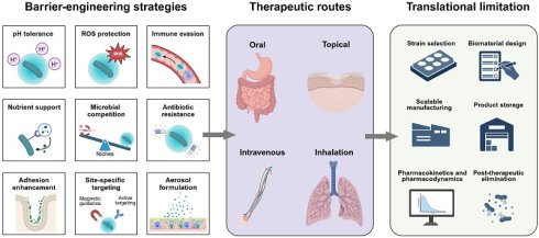
Bacteria have emerged as promising living therapeutics for the treatment of infections, inflammatory disorders, metabolic diseases, and neoplastic diseases. However, their clinical efficacy is often compromised by in vivo barriers that reduce their viability and impair their function after administration. Rational engineering can help them sustain their viability under a harsh microenvironment and enhance their targetability as well as retention, ultimately improving their functional performance in vivo. This review starts with a concise synopsis of current status in live bacterial therapy, with particular emphasis on the major biological barriers to their in vivo application. Engineering strategies for harnessing bacterial intrinsic characteristics and overcoming their specific barriers are discussed. Representative examples are then presented to illustrate how engineered live bacteria overcome these biological barriers and achieve therapeutic applications via oral, topical, intravenous, and inhalational administration. Finally, we identify the key challenges associated with translating laboratory advances into reliable and safe clinical therapeutics.
Keywords: bacterial therapy, drug delivery, therapeutics, biological barriers, probiotics
1. Introduction
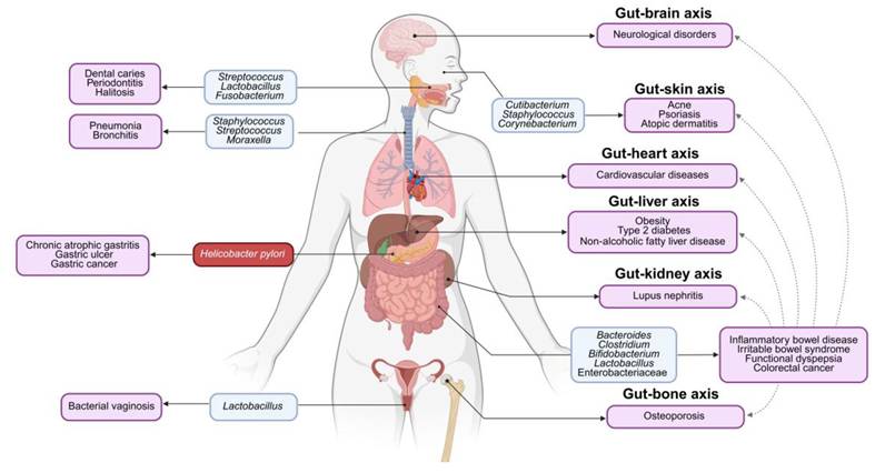
An adult human body hosts estimated 38 trillion bacteria, exceeding 30 trillion human cells within the body. Increasing evidence has seen microbial dysbiosis as a driving force to pathogenesis of metabolic disorders, inflammation, cancer, and infections [1] (Figure 1). The therapeutic use of bacteria can be traced back to over a century ago, when Tissier isolated Bifidobacterium from the feces of infants and observed its association with reduced incidences of diarrhea in 1889. In this context, bacteria have been widely investigated as promising living therapeutics for disease treatment because they can produce bioactive metabolites and modulate immunity [2]. Moreover, bacteria can be used as drug delivery systems to hypoxic lesions, such as tumor tissues [3, 4]. To note, bacterial therapeutics are used as a broad term in this review, including probiotic bacteria, commensal bacteria, genetically engineered bacteria and attenuated bacteria.
Schematic diagram for representative examples of bacteria and their associated diseases. Notably, the gut microbiota can induce various syndromes in distant organs via the gut-organ axes, such as the gut-brain, gut-skin, gut-heart, gut-liver, gut-kidney or gut-bone axis. Created in https://BioRender.com.
Despite their encouraging therapeutic promise, the efficacy of exogenously administered bacteria is significantly impacted by multiple biological barriers encountered during delivery and after administration. Oral bacteria must survive gastric acidity, oxidative stress, intestinal peristalsis, competition from resident microbiota, and, in some cases, concurrent antibiotic exposure [5]. Topically delivered bacteria are exposed to local hostile physicochemical conditions [6]. Intravenously administered bacteria are subjected to complement activation, phagocytic clearance, and insufficient accumulation at target lesions [7], and inhaled bacteria must withstand formulation-associated stress and evade mucociliary clearance. Collectively, these route-dependent biological barriers impose major constraints on the in vivo performance of therapeutic bacteria, thereby motivating the development of engineering strategies to improve their viability, persistence, targeting, and therapeutic efficacy [8].
Engineering bacteria provides a potent avenue for enhancing the therapeutic efficacy by improving bacterial tolerance to hostile in vivo environments and endowing them with protective, targeting, and therapeutic functions beyond their native capacity. The integrated engineering approaches hold promises for addressing multiple barriers encountered by bacteria during the courses of their delivery and therapeutic action. However, effective bacterial engineering relies on the successful incorporation of functional moieties onto bacterial cells, as well as the mechanistic action of the bacterial chassis. The structural, physicochemical, and biosynthetic attributes of bacteria offer a set of engineerable interfaces, and these interfaces can be selectively exploited for precise moieties coupling. More importantly, these engineerable interfaces allow rational alignment of exogenous functions with the native biological activities of bacterial cells, achieving context-specific therapeutic synergy and enhancing therapeutic efficacy.
In this review, we provide an overview of engineering strategies to improve bacterial delivery efficiency and enhance therapeutic efficacy. Importantly, we elaborate on the rationale behind employing these strategies to overcome the in vivo biological barriers that impact bacterial performance. This review initially introduces bacterial structural and biological features. By harnessing specific structural, physicochemical, or biological characteristics of bacteria, engineering strategies can be systematically classified according to the specific bacterial characteristics they exploit. Major categories include surface engineering based on bacterial physicochemical properties, covalent or dynamic surface functionalization enabled by exposed reactive groups, biosynthesis-expanded surface engineering that introduces non-native modification handles through endogenous bacterial processes, and strategies involving intracellular cargo loading or bulk encapsulation. We delve into functional tailoring of these strategies to address the significant biological barriers that bacteria encounter during oral, topical, intravenous, and inhalational delivery, such as physicochemical stress, inadequate adhesion, nutritional constraints, microbial competition, immune and antibiotic clearance, and insufficient site-specific targeting. We then survey therapeutic application examples of engineered bacteria via different administration routes. Finally, we discuss principal translational challenges and propose future directions for the development of next-generation bacterial therapeutics.
2. Foundations for bacterial therapeutic engineering
2.1 Biological foundations of bacterial therapeutics
Resident bacteria in the human body confer health benefits through multiple mechanisms. The development of bacterial therapeutics starts with the selection of appropriate bacterial chassis from the resident bacterial community to confer therapeutic benefits. Because different bacterial species vary in their colonization niches, oxygen preferences, and tissue-tropic behaviors, the selection of bacterial species should be aligned with the target organ or the disease microenvironment to achieve effective and feasible bacterial therapy. For example, in the context of inflammatory bowel disease (IBD) characterized with dysbiotic gut microbiota, probiotic and commensal bacteria, such as Lactobacillus, Bifidobacterium, Escherichia coli Nissle 1917 (EcN), and Clostridium, contribute to disease resolution through four distinct mechanisms [9]. First, they engage in direct competition with pathogens for the nutrients available and adhesion sites on the intestinal mucosa [10]. Second, bacteria suppress pathogen proliferation via producing a range of biologically active metabolites including bacteriocins, antimicrobial peptides (AMPs), and lactic acid. Third, they maintain the integrity of the intestinal epithelium by upregulating the expression of tight junction proteins, stimulating mucus secretion by intestinal epithelial cells, and promoting the secretion of short-chain fatty acids (SCFAs). Finally, bacteria can help resolve inflammation and neutralize reactive oxygen species (ROS) through inducing the differentiation of regulatory T cells.
The skin is colonized by diverse microbial communities. Strikingly, mutually beneficial bacteria play a critical role in training the skin immune system to resist the invasion of pathogenic substances. For instance, Staphylococcus epidermidis offers protection to the host against skin infections through activating CD8+ T cells and exciting keratinocytes to discharge AMPs [11]. In addition, Staphylococcal lipoteichoic acid can maintain the integrity of epithelium, and facilitate tissue repair through alleviating inflammation by blocking the Toll-like receptor 2 (TLR2)-associated pathway [12]. In conclusion, skin microbiota plays an indispensable role in maintaining epithelial homeostasis and overall skin health.
The tumor microenvironment (TME) is an immunosuppressive milieu characterized by hypoxia, abundant immunosuppressive metabolites, and deprived nutrients, which collectively diminish the efficacy of immunotherapy [13-17]. Recent studies have revealed that gut microbiota can stimulate host immunity and act in synergy with cancer immunotherapy for anticancer treatment. For instance, Lactobacillus reuteri and Lactobacillus plantarum have been shown to activate neutrophils and natural killer (NK) cells, thereby promoting pro-inflammatory cytokine production within the TME and inhibiting tumor progression [18]. Furthermore, dendritic cells (DCs) can be activated upon recognizing bacterial components, such as lipopolysaccharide and peptidoglycan, subsequently recruiting effector T cells to eliminate tumors [19]. Notably, the abundance of Bifidobacterium and Enterococcus in the feces of melanoma patients is positively correlated with the therapeutic response to anti-PD-L1 antibodies [20]. However, intratumoral bacteria are found to interact with immune cells and promote tumor progression [21]. For example, Fusobacterium nucleatum residing in colorectal cancer can protect tumor cells from the elimination by cytotoxic NK cells via various means [22]. Therefore, bacteria can be administered to regulate the population of intratumoral immune cells and activate their anti-tumor immune responses, offering new insights into cancer immunotherapy.
Although bacteria exhibit multiple therapeutic mechanisms, their effective delivery to target sites is often hindered by a myriad of biological barriers. In this section, we summarize the application of bacteria for a variety of indications, with a particular focus on those native or genetically engineered bacteria that have entered clinical trial phases (Table 1). We then introduce bacterial delivery challenges including oral, topical, intravenous and inhalational administration.
Probiotic treatment in clinical trials
| Routes | Probiotics | Indications | Phase | ID numbers |
|---|---|---|---|---|
| Oral | B. breve | Crohn’s disease | Not applicable | NCT04842149 |
| B. longum | Not applicable | NCT00305409 | ||
| L. acidophilus; L. rhamnosus | Not applicable | NCT00374374 | ||
| L. acidophilus; L. rhamnosus | Ulcerative colitis | Not applicable | NCT00374725 | |
| L. plantarum | Phase 2/3 | NCT01193894 | ||
| EcN | Phase 4 | NCT01772615 | ||
| P. freudenreichii | Not applicable | NCT02488954 | ||
| Engineered L. lactis to secret IL-10 | Phase 2 | NCT00729872 | ||
| Lactobacillus; Streptococcus; Bifidobacterium | Necrotizing enterocolitis | Not applicable | NCT06118801 | |
| L. reuteri | Gingivitis | Not applicable | NCT06234839 | |
| L. reuteri | Periodontitis | Not applicable | NCT04478643 | |
| L. rhamnosus; B. longum | Dental caries | Not applicable | NCT03078179 | |
| S. salivarius | Halitosis | Not applicable | NCT07384130 | |
| B. lactis | Respiratory tract infections | Not applicable | NCT04122495 | |
| L. rhamnosus | Anxiety | Not applicable | NCT03427515 | |
| B. bifidum | Attention-deficit/Hyperactivity disorder | Phase 3 | NCT04958460 | |
| Lactobacillus | Epilepsy | Phase 2 | NCT05539287 | |
| Bifidobacterium; Lactobacillus; Enterococcus | Parkinson's disease | Phase 4 | NCT04871464 | |
| L. paracasei; L. plantarum; L. rhamnosus; L. helveticus; B. breve | Alzheimer's disease | Phase 1 | NCT06181513 | |
| Lactobacillus | Breast cancer | Not applicable | NCT03290651 | |
| Bifidobacterium; Lactobacillus; Lactococcus | Atherosclerosis | Not applicable | NCT03100162 | |
| L. salivarius; B. breve | Atopic dermatitis | Phase 3 | NCT01500941 | |
| L. acidophilus; B. lactis | Acne | Phase 4 | NCT05216289 | |
| L. rhamnosus | Vaginal dysbiosis | Not applicable | NCT05796921 | |
| L. rhamnosus; L. reuteri | Bacterial vaginosis | Phase 4 | NCT03894813 | |
| Lactobacillus | Polycystic ovary syndrome | Phase 2 | NCT04029805 | |
| Engineered EcN to convert oxalate to formate | Hyperoxaluria | Phase 1 | NCT04629170 | |
| Engineered EcN to reduce methionine levels | Homocystinuria | Phase 1 | NCT05462132 | |
| Engineered EcN to express PAL and LAAD | Phenylketonuria | Phase 1/2 | NCT03516487 | |
| Engineered L. lactis to secrete proinsulin and IL-10 | Type 1 diabetes | Phase 1/2 | NCT03751007 | |
| Engineered EcN to convert ammonia to L-arginine | Hyperammonemia | Phase 1/2 | NCT03447730 | |
| Topical | Engineered L. lactis to secrete human TFF1 | Oral mucositis | Phase 2 | NCT03234465 |
| B. subtilis; B. clausii | Vaginitis | Not applicable | NCT06165354 | |
| Lactobacilli | Bacterial vaginosis | Not applicable | NCT00752193 | |
| L. reuteri | Wound healing | Not applicable | NCT03210779 | |
| Engineered L. lactis to secret FGF-2, IL-4 and CSF-1 | Phase 2 | NCT06111183 | ||
| Engineered L. reuteri to secret CXCL12 | Phase 2 | NCT05608187 | ||
| Inhalational | L. lactis | Chronic rhinosinusitis | NCT04048174 | |
| L. sakei | NCT05427695 | |||
| L. casei; L. rhamnosus | NCT03587545 | |||
| L. lactis | COVID-19 | NCT04458519 | ||
| IT injection | Engineered EcN to express c-di-AMP | Solid Tumors and Lymphoma | Phase 1 | NCT04167137 |
| IV injection | Engineered B. longum to express IL-12 | Solid Tumors | Phase 1 | NCT04025307 |
L. acidophilus: Lactobacillus acidophilus; L. rhamnosus: Lactobacillus rhamnosus; L. plantarum: Lactobacillus plantarum; L. reuteri: Lactobacillus reuteri; L. salivarius: Lactobacillus salivarius; Lactobacilli, Lactobacillus species; L. lactis: Lactococcus lactis; L. sakei: Lactobacillus sakei; L. casei: Lactobacillus casei; B. breve: Bifidobacterium breve; B. longum: Bifidobacterium longum; B. lactis: Bifidobacterium lactis; B. subtilis: Bacillus subtilis; B. clausii: Bacillus clausii; EcN: Escherichia coli Nissle 1917; P. freudenreichii: Propionibacterium freudenreichii; S. salivarius: Streptococcus salivarius; PAL: phenylalanine ammonia lyase; LAAD: L-amino acid deaminase; TFF1: trefoil factor 1; FGF-2: fibroblast growth factor 2; IL-4: interleukin-4; CSF-1: colony-stimulating factor 1; CXCL12: C-X-C motif chemokine ligand 12; c-di-AMP: cyclic di-adenosine monophosphate; IL-10: interleukin-10; IL-12: interleukin-12.
2.2 Administration route-specific barriers
Oral administration is the most conventional and widely accepted route for bacterial therapy. When bacteria enter the body, they are exposed to a series of harsh gastrointestinal microenvironments. Gastric fluid is the first crucial biological barrier. Gastric acid can significantly reduce the survival rate of bacteria through breaking the integrity of microbial cell membranes and inducing protein denaturation [23]. After the bacteria survive through the stomach, they enter an alkaline environment of the small intestine. Bile salts and digestive enzymes serve as the second barrier. Bile salts can penetrate the phospholipid double layer of the bacterial cell membrane, leading to the damage of cell membranes and the disturbance of the mechanisms of DNA repair, ultimately reducing bacterial survival and preventing bacterial growth [24]. It is noted that bacteria must achieve context-dependent optimal colonization within the gut to bring sustained therapeutic benefits. Unfortunately, the administered bacteria may be expelled by the originally resident microorganism group, because these bacteria compete for nutritional metabolites as well as attachment sites at the intestine site with resident bacteria. In addition, they may not colonize successfully at the intestine site due to rapid intestine movements [25]. It is worth noting that the intestinal micro-environment of colitis displays the characteristics of excessive oxidative stress and inflammatory reaction, which can inhibit the colonization, survival, and growth of bacteria [26]. Once bacteria successfully establish themselves in the intestine tract and proliferate to an adequate quantity, they can commence their therapeutic function.
Bacterial therapy can be topically applied to treat skin and mucous membrane diseases by the restoration of the microbial population balance at these sites. Nevertheless, physical and biological barriers may directly impact the restoration process, thus diminishing their therapeutic efficacy [27]. Physical and chemical properties at these sites, including local pH, water moisture, and surface chemistry, may reduce bacterial viability and impair their metabolic activity [6]. In addition, the resident microbial groups in the host prevent the applied bacteria from occupying ecological niches by competing for nutrients and attachment positions [28]. For example, these resident bacteria may form thick harmful biofilms, and these biofilms act as an extra physical obstacle to block the approach of bacteria to the surface of the skin tissue, obstructing adherence and inhabitation of topically applied bacteria [1]. Furthermore, the host inflammation reaction adds one more layer of complexity. The reaction may lead to the formation of a micro-environment rich in cytokines and ROS, which can damage bacterial membranes and weaken the survival of bacteria [29]. The restoration process by topical application of bacteria may be significantly impeded when antibiotics are used for infection control. The exposure to antimicrobial drugs can kill pathogenic bacteria at these sites; however, this may eliminate beneficial resident microbial groups as well as bacteria, thus reducing the likelihood of prolonged and steady settling of bacteria after the treatment [30].
Intravenous route of bacterial therapy presents an advantage of bypassing gastrointestinal barriers. After administration, they directly enter the circulatory system, thus enhancing their delivery efficiency. However, this route is impeded by host immune clearance. The mononuclear phagocyte system serves as the primary mechanism to eliminate circulating foreign bacteria [31]. Besides, the activated complement system can directly lyse bacteria through the formation of the membrane attack complexes [32]. Since systemic administration of billions of bacterial spores inevitably leads to septicaemia [7], it is crucial to ensure the intravenously administered bacteria can target lesion sites and maintain their biological function on these sites.
The respiratory tract is directly exposed to an external environment, and it displays a branching architecture with regionally distinct deposition patterns, creating formulation and translational challenges for local delivery of living bacteria. To reach the targeted pulmonary region, inhaled bacteria must be delivered as aerosols, and the aerodynamic particle size of aerosols is critical for their efficient deposition because of variations in the airway caliber and airflow throughout the bronchial tree [33]. The bacterial viability and function must be maintained during preparation and administration of aerosols. Nebulization of liquid formulations may alter the bacterial population [34], and the preparation of dry powders introduces dehydration and processing stresses that may lead to a reduction in bacterial viability [35]. After delivery of aerosols, clearance through mucus or mucociliary structures and alveolar phagocytes may diminish bacterial persistence in the targeted region and compromise their therapeutic activity [36]. Moreover, chronic lung diseases are often accompanied with tissue injury, sustained inflammation and local microbial dysbiosis [37]. Therefore, interactions between administered bacteria and this altered pulmonary microenvironment may provoke unintended inflammatory responses, and extensive safety evaluation should be performed during clinical translation. Furthermore, excessive ROS in the diseased pulmonary microenvironment may impair the bioactivity of administered probiotic bacteria, thereby reducing their therapeutic efficacy [38].
Collectively, unsatisfied therapeutic efficacy of exogenous bacteria may be attributed to their low viability as well as their susceptibility to ecological stress due to physicochemical/biological barriers at the target site. Therefore, these microorganisms before administration can be engineered to improve overall fitness and stress tolerance, ultimately enhancing their therapeutic performance [39].
3. Engineering bacteria to overcome in vivo barriers
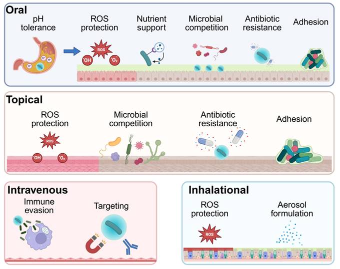
The curative effect of administered bacteria on reshaping the microbe community and treating a broad spectrum of diseases has been extensively documented. However, bacteria which are administered without engineering manipulation often have poor survival in vivo, because they face harsh microenvironmental conditions, including acidic pH, excess ROS, poor adhesion, and weak niche competition with existing microbial groups on the disease site. For solving these shortcomings, many kinds of surface engineering methods have been studied to raise the survival ability and biological usability of treatment bacteria when compared with their not modified original types (Figure 2). Accordingly, this section highlights the major engineering approaches designed to overcome the challenges encountered by administered bacteria. The ability of engineered bacterial therapeutics to overcome in vivo biological barriers and achieve enhanced therapeutic efficacy predominantly depends on the materials integrated with bacterial cells. These materials can protect bacterial viability under harsh physiological conditions, improve resistance to immune clearance and external stresses, and enhance bacterial adhesion and targeting capabilities. As a result, material engineering plays a critical role in improving the in vivo performance of bacterial therapeutics.
Representative route-specific engineering strategies of therapeutic bacteria. Therapeutic bacteria encounter distinct biological barriers depending on the route of administration. During oral delivery, engineered coatings or encapsulation systems can protect bacteria from gastric acidity, reactive oxygen species (ROS), nutrient limitation, microbial competition, antibiotics, and weak mucosal adhesion, thereby improving survival and intestinal retention. For topical delivery, bacteria must tolerate oxidative stress, compete with resident or pathogenic microorganisms, survive after local antibiotic exposure, and adhere to skin or mucosal surfaces. Intravenously administered bacteria face rapid immune clearance and immune-evasive modification, or active targeting strategies can be used to improve circulation persistence and lesion accumulation. For inhalational delivery, bacteria should be formulated into aerosols, in the format of respirable droplets or dry powder particles, for effective deposition in the bronchial and small airway regions. In addition, ROS-scavenging biomaterials can protect bacteria from oxidative stress in inflamed lung tissues and help maintain their therapeutic activity. Created in https://BioRender.com.
3.1 pH shielding
The pH value in an environment plays a crucial role in the survival and function of live bacterial therapeutics. Exposure of them to unfavorable pH can damage their membranes, disrupt their metabolic activity, and reduce their viability, thereby substantially diminishing the proportion of administered bacteria that retain their functional efficacy at the target site. This challenge is especially associated with the oral delivery route. In this route, a strongly acidic gastric environment represents a major barrier. A crucial aspect in bacterial engineering is safeguarding them against acid-induced damage.
An array of coatings have been designed to shield bacteria from the gastric environment and dissolve upon entry into the intestine, including Eudragit L100-55, a clinically used polymer coating [40, 41], chitosan/sodium alginate coatings [42], two-dimensional nanosheets [43], and polyelectrolyte composite coatings [44]. Notably, a polyelectrolyte complex formed via electrostatic and hydrogen-bonding interactions between oxidized starch and polyethylenimine enhanced the survival of EcN by approximately 40-fold in a simulated gastric fluid and 74-fold in an intestinal fluid [25]. Encapsulation of bacteria within biomaterial matrices, such as colloids and hydrogels, is another effective strategy to shield them from the gastric fluid [45, 46]. Of particular interest are positively charged colloids composed of tannic acid and poly-β-cyclodextrin, which electrostatically encapsulated bacteria and formed a thick protective shell. Up to 19% of the bacteria inside the shell remained viable in a simulated gastric fluid, and the value is 7,500 times higher than that of Eudragit L100-55 [46] (Figure 3A-B). Apart from physically shielding bacteria from gastric acid, protons can be obtained during engineering bacteria shielding can also seize protons to provide acidic protection. For example, EcN was coated with carboxymethylated β-glucan (mGN), conferring a 1720-fold survival rate in a simulated gastric fluid compared to the bare counterpartner, which may be attributed to the capture of H⁺ by -COO- of gastric acids [47]. However, simulated gastric and intestinal fluids cannot recapitulate the complexity in the gastrointestinal environment, including mucus diffusion, peristalsis, bile and enzyme dynamics, microbiota competition, dietary effects, or disease-related changes in the gut. Therefore, improved survival rates in the simulated fluids should be viewed as the evidence to support stress tolerance, rather than in vivo colonization or therapeutic efficacy.
Representative examples of pH-protective engineering strategies. (A) Schematic of colloidal encapsulation of bacteria to enhance their gastric acid resistance during oral delivery. (B) Survival of NTcEcN, naked EcN, and L100EcN after exposure to a simulated gastric fluid for 2 h. Adapted with permission from [46], copyright 2023, American Chemical Society. (C) Diagram for preparing theranostic CaFeMn@EcN via in situ sequential mineralization. The outer CaCO3 layer neutralizes gastric acids to protect the activities of the encapsulated bacteria. (D) Photographs of culture plates, (E-F) transmission electron microscopy (TEM) images and the survival rate of EcN, FeMn@EcN, and CaFeMn@EcN after 30-min exposure to a simulated gastric fluid. Adapted with permission from [48], copyright 2025, The American Association for the Advacement of Science.
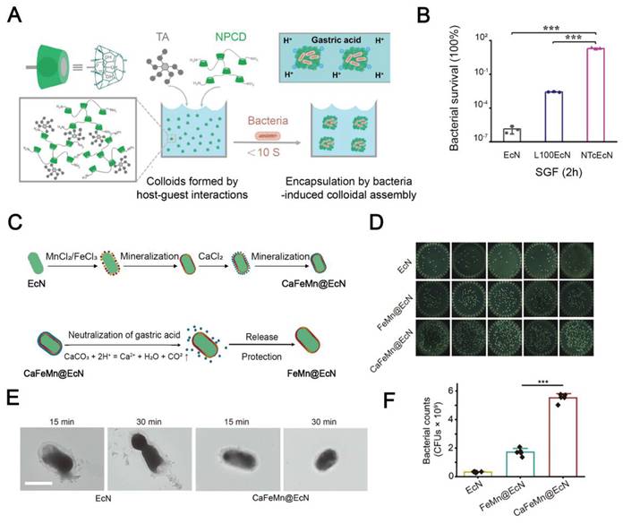
Mineralization offers an alternative acid-protective approach by neutralizing excess acids in a gastric environment. A mineral layer composed of MnO2 and Fe2O3 nanoparticles was first deposited on the bacterial surface, and the inner layer served as a substrate for the sequential growth of an outer CaCO3 shell. The CaCO3 shell neutralized gastric acids and preserved the bacterial viability [48] (Figure 3C-F). Collaboratively, engineered bacteria with acid resistance may survive in a harsh stomach environment and colonize the intestinal tract.
3.2 ROS protection
Oxidative stress is a well-established feature in inflammatory diseases, such as IBD and chronic wounds. Excessive ROS contributes to epithelial injury, inflammatory amplification and impaired tissue repair. This redox-hostile milieu compromises the viability and bioactivity of bacteria at diseased sites, thereby diminishing their colonization and reducing their therapeutic persistence [49, 50]. Therefore, it is a valuable therapeutic strategy to engineer bacteria with the capacity for withstanding oxidative stress. The widely used ROS-scavenging strategies can be roughly divided into direct ROS consumption and catalytic ROS scavenging [51].
One approach for direct ROS consumption is to coat bacteria with ROS-responsive organic materials. For example, Zhang et al. decorated EcN with hyaluronic acid-poly(propylene sulfide) nanoparticles. Poly(propylene sulfide) acted as a ROS-consuming component because its sulfur atoms can be readily oxidized by ROS [52] (Figure 4A-B). This design enabled direct ROS scavenging and protected the bacteria against oxidative damage in an inflammatory intestinal environment. Another example for direct ROS consumption is the employment of lipid coatings loaded with antioxidant compounds, such as pterostilbene or astaxanthin. These compounds react with ROS to mitigate oxidative damage and improve bacterial persistence in the presence of a high level of ROS [53, 54].
Representative examples of ROS-scavenging engineering strategies. (A) Diagram for preparing HPN-NE-EcN and its ROS-scavenging activity by oxidizing sulfur atoms to form sulfoxides and then sulfones. (B) The normalized proliferation rate of EcN after culture in the LB medium containing H2O2 with or without HPN treatment. Adapted with permission from [52], copyright 2022, The American Association for the Advancement of Science. (C) Schematic illustration of preparing antioxidative engineered EcN-Fh. (D) The ROS-scavenging activity of EcN-Fh@F in vitro. Adapted with permission from [57], copyright 2024, American Chemical Society.
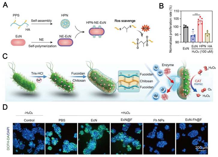
Nanozymes have attracted considerable interest in engineering bacteria because their catalytic nature endows them with sustained ROS-scavenging capability. Mechanistically, SOD-like nanozymes catalyze the dismutation of superoxide anions into H2O2 and O2, whereas CAT-like nanozymes decompose H2O2 into H2O and O2 [55]. Diverse nanozyme systems, including metal-based nanozymes, carbon-based nanozymes, and single-atom nanozymes, have been developed for bacterial engineering to eliminate excess ROS at inflammatory sites and generate a habitable microenvironment for bacterial colonization [56-60]. For example, ferrihydrite nanoparticles were anchored onto EcN to construct EcN-Fh, which exhibited CAT-like activity. The CAT-like nanozymes reduced the H2O2 content by 80% within 12 h [57] (Figure 4C-E). This activity was attributed to the Fe³⁺-rich oxyhydroxide interface of ferrihydrite, where surface iron-associated hydroxyl groups and adjacent Fe sites cooperatively activated H2O2 and promoted its decomposition into H2O and O2 [61]. More recently, hybrid coatings have been developed by integrating direct ROS consumption with catalytic ROS elimination. Liang et al. reported a biocompatible polydopamine-based nanozyme shell. The catechol groups on the shell enabled direct radical scavenging and cerium oxide provided CAT-like activity. The resulting nanozyme shell for probiotics (EcN@DMCe) rapidly decomposed H2O2 into O2 at a H2O2 concentration of 10 mM and oxygen generation reached an equilibrium state within 10 min [62], as the defects in the crystal lattice and oxygen vacancy-coupled Ce3+/Ce4+ cycling facilitated H2O2 adsorption and decomposition into H2O and O2 [63].
3.3 Adhesion enhancement
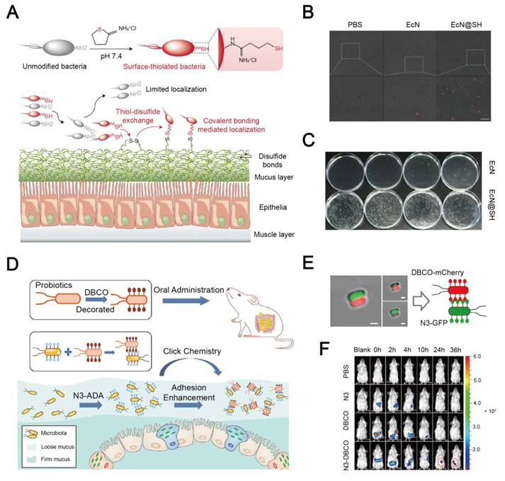
Effective adhesion and prolonged retention at the target site are critical for bacterial efficacy. Competitively occupying the target site by administered bacteria can prevent their clearance, sustain the abundance of the local bacterial population, and promote intimate host–microbe interactions. Securing spatial niches at the target site also supports pathogen exclusion, thereby prolonging the therapeutic activity of bacteria [64, 65]. However, disease-associated mucosal damage, together with persistent peristalsis and luminal washout, could markedly accelerate bacterial clearance. To address these challenging issues, multiple strategies have been developed to enhance the adhesion and colonization of engineered bacteria [65-68]. For instance, conversion of primary amino groups on the bacterial surface into free thiols enables the formation of covalent bonds between bacteria and disulfide-rich mucin through catalyst-free thiol-disulfide exchange [67]. The number of engineered bacteria colonizing jejunal mucus was 171.3 times higher than native bacteria (Figure 5A-C). Tannic acid-incorporated coatings can also improve intestinal mucosal retention by 2.5-fold through hydrogen-bond-mediated interactions and scavenge excessive ROS under pathological conditions [66]. In addition to direct mucus binding, bacterial colonization can be directed toward specific local niches by exploiting interactions with the bacteria that are established at the target site. For example, incorporation of azido-modified D-alanine into peptidoglycan of gut-resident bacteria facilitates bioorthogonal anchoring of DBCO-modified Clostridium butyricum, thereby extending the adhesion and colonization time at the target intestinal site to 48 h [68] (Figure 5D-F).
Representative examples of adhesion-enhancing engineering strategies. (A) Schematic illustration of preparing surface-thiolated bacteria that can be chemically bound to poly(disulfide)s-abundant mucin via catalyst-free dynamic thiol-disulfide exchange reaction. (B) Representative confocal laser scanning microscopy (CLSM) images of the mouse mucus layer. (Scale bar: 10 µm). (C) Typical digital images of agar plates containing EcN collected from mouse jejunal mucus. Adapted with permission from [67], copyright 2022, Springer Nature. (D) Schematic illustration of colonization enhancement for delivered bacteria through biorthogonal conjugation with gut inhabitants via click chemistry. (E) CLSM images of bacterial adhesion between N3-GFP and DBCO-mCherry. (Scale bar: 1 µm). (F) Representative in vivo imaging system (IVIS) images for evaluating bacterial retention in the gastrointestinal tract of mice. Adapted with permission from [68], copyright 2022, American Chemical Society.
3.4 Nutrient support
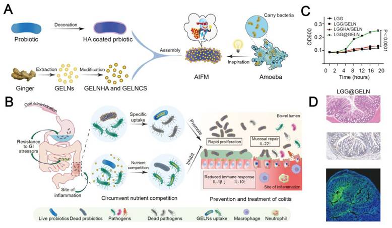
Bacteria, as living therapeutics, require adequate nutritional support to sustain their viability, metabolic activity, and functional persistence after administration. Natural polysaccharides have been widely used in developing protective coatings for bacteria. Meanwhile, they are often employed as bacterial carbon sources, such as inulin, β-glucan, and sodium alginate [47, 69, 70]. However, carbon sources alone are insufficient to meet the holistic metabolic demands for the proliferation of bacteria. Ginger-derived exosomes enriched with polysaccharides, nucleic acids, proteins, and lipids have been assembled onto bacteria as versatile nutrient depots [71] (Figure 6). Inspired by the food-carrying strategy of ameba, Pan et al. prebound ginger-derived exosome-like nanoparticles (GELNs) to Lacticaseibacillus rhamnosus GG (LGG). Due to the close contact between LGG and GELNs, LGG can exclusively uptake GELNs despite the existence of other intestinal microorganisms. This nutrient-providing strategy has been demonstrated to elevate the LGG proliferation rate approximately 1.5 times under nutrient-deprived, competitive conditions. Nevertheless, supplementation of rich nutrients may support the growth of opportunistic pathogens. In this context, prebiotics, such as inulin, galacto-oligosaccharides, and xylo-oligosaccharides [2], which are preferentially utilized by beneficial microbes and confer measurable health benefits, can be employed for engineering bacteria. This rational approach can selectively enhance the competition capacity and fitness of engineered bacteria. Besides, nutrient-rich systems may inadvertently support opportunistic pathogens, and close contact between probiotic bacteria and the nutrient depot can help inhibit growth of opportunistic pathogens.
Representative examples of nutrient-supplying engineering strategies. (A) Schematic illustration of the fabrication of nutrient-carrying microecologics (AIFM). (B) Diagram for enhanced survival, proliferation, and colonization of bacteria through specific uptake of prebound nutrients from AIFM. (C) The proliferation of L.reuteri in a simulated nutrient-deprived intestinal fluid without the presence of pathogens. (D) The therapeutic efficacy of LGG@GELN in dextran sulfate sodium (DSS)-induced colitis. Adapted with permission from [71], copyright 2025, Springer Nature.
3.5 Microbial competition
In therapeutic settings, bacterial colonization is frequently constrained by ecological competition from microorganisms residing in the local microenvironment [72, 73]. Exogenous strains must compete for nutrients, attachment sites, and host-derived resources. Meanwhile, disease-associated niches may be favorite for the expansion of pathobionts or opportunistic pathogens [74, 75]. Strategies for relieving this dual competitive pressure for administered bacteria have been developed, including equipping exogenous bacteria with the capacity for selective support of their survival and persistence and restricting competing microbes, thereby improving their therapeutic performance [39, 76, 77]. Xu et al. constructed an EPS-based hydrogel to create a local selective advantage for bacteria while suppressing competing pathogens. EPS-M76 in the hydrogel promoted the proliferation and metabolic activity of Lactobacillus paracasei TYM202, while lactic acid and acetic acid are released from the system to suppress the growth of pathogenic bacteria, thereby conferring a direct competitive advantage to the bacterial cargo [78]. Oxygen regulation is another engineering route. Functionalized chlorella and Bacillus subtilis were encapsulated in a living microecological hydrogel. Sustained oxygen generation from chlorella reshaped the local oxygen distribution that favored bacterial proliferation while suppressing pathogenic bacteria. Therefore, differential oxygen responsiveness between beneficial and pathogenic microorganisms could be exploited for engineering other bacteria [79]. Furthermore, competition can be shifted by creating an environment that selectively eliminates pathogens while preserving the bacterial payload. Wei et al. integrated a hydrogel containing peroxidase-like nanozymes with Lactobacillus. During hydrogel degradation in an infected vaginal microenvironment, Lactobacillus restored an acidic niche and provided H2O2, which was converted by the nanozymes into fungicidal ROS, thereby achieving selective killing of Candida albicans. Therefore, this approach preserved the bacterial viability and reshaped the vaginal microbiota toward a healthier state [80].
3.6 Antibiotic resistance
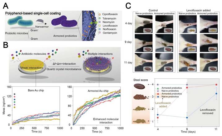
When bacteria are applied to infection-associated indications, such as severe wounds and gastrointestinal infections, antibiotic therapy is often used to rapidly suppress pathogen overgrowth. Although antibiotics remain indispensable in these settings, their non-specific bactericidal activity can disrupt commensal microbiota and inadvertently impair administered bacteria, thereby diminishing their colonization and compromising their therapeutic efficacy [81]. Therefore, it is essential to protect bacteria from antibiotic-induced collateral damage when live biotherapeutics are deployed in conjunction with conventional anti-infective treatment. Current engineering strategies are designed to localize and inactivate antibiotics at the bacterial interface [82-85] (Figure 7). For example, a coating for bacteria was prepared from a biocompatible metal-phenolic network derived from tannic acid and Fe3+, which adsorbed and inactivated diverse antibiotic molecules through interfacial molecular interactions, thereby protecting engineered bacteria from collateral damage [83]. This nano-armoring strategy reduced the detrimental impact of antibiotic therapy on exogenous bacteria and facilitated their subsequent colonization and proliferation.
Representative examples of antibiotic-shielding engineering strategies. (A) Schematic illustration of a nano-armor that protects bacteria from a wide range of antibiotics with different molecular structures and properties. (B) Diagram for multiple interactions between polyphenol moieties and antibiotic molecules. (C) The severity of antibiotic-associated diarrhea (AAD) and dysfunction of the gastrointestinal tract assessed from the fecal samples and the stool scores. Adapted with permission from [82], copyright 2022, Springer Nature.
3.7 Immune evasion
A significant impediment to the efficacy of bacterial therapy is rapid immune recognition and subsequent clearance of the administered bacteria, particularly, after systemic delivery such as intravenous injection. Rich conserved microbial molecular components, including lipopolysaccharides, peptidoglycan, flagellin, and CpG-rich DNA, are displayed on the bacterial surface. These components are detected by pattern-recognition receptors (PRRs), which promptly initiates inflammatory signaling, complement activation, and phagocytic elimination [86, 87]. Therefore, immune surveillance can markedly compromise bacterial persistence and reduce their accumulation at target sites, thereby diminishing their therapeutic efficacy. For instance, intravenously administered Salmonella Typhimurium VNP20009 displayed unsatisfactory antitumor efficacy, even at the maximum-tolerated dose, which was supported by insufficient tumor accumulation at the tumor site [88]. External surface engineering has become an important strategy to improve the performance of live bacterial therapeutics by masking pathogen-associated molecular patterns or camouflaging bacteria as host cells for immune evasion.
To minimize the recognition of bacterial surface antigens by immune cells in the body, an artificial shell can be directly coated on the bacterial surface. An inducible capsular polysaccharide system reported in a recent study helped the bacteria temporarily evade immune clearance and enhanced systemic delivery efficiency [89]. In a FeTA-coated EcN system, an iron–tannic acid coating mitigated both complement-mediated and phagocyte-mediated bacterial killing, leading to a prolonged circulation time and a high tumor colonization level after intravenous injection [90]. In another study, a tannic acid-based compact layer in conjunction with an albumin coating was used to mitigate endotoxin exposure, improving bacterial biocompatibility and reducing immune elimination [91]. (Figure 8A-D) In addition, a polymeric stealth coating onto engineered Lactobacillus acidophilus protected the bacteria from macrophage recognition and phagocytosis, prolonging bacterial survival during the delivery process [92].
Representative examples of immune-evasive engineering strategies. (A-B) Schematic illustration of developing BSA-TA-E. coli that exhibits extended retention and enhanced biocompatibility in vivo. (C-D) Fluorescence images and flow cytometry plots confirm a significantly lower proportion of BSA-TA-E. coli is engulfed by macrophages compared to uncoated E. coli. Adapted with permission from [91], copyright 2025, Elsevier. (E) The process of preparing cell-membrane-coated bacteria (CMCB). (F-G) A bar chart and tumor images of the luminescence signal intensity in 4T1 tumor-bearing mice post-injection of EcN or CMCB. Adapted with permission from [93], copyright 2019, Springer Nature.
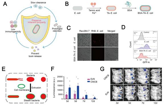
An alternative approach is to cloak bacteria with host cell membranes to present host-derived interfacial features during circulation. Camouflaging EcN by wrapping with red blood cell membrane lead to a 42-fold increase in tumor accumulation and a 14-fold prolongation in blood persistence compared with uncoated bacteria following intravenous injection. The encouraging results can be attributed to the membrane-derived anti-phagocytic properties, such as CD47-associated self-recognition signals [93] (Figure 8E-G). Collectively, these studies support that bacteria are rapidly eliminated by immune cells in the body because their native surfaces are exposed to complement proteins, phagocytes and innate immune receptors during systemic circulation. Bacterial surface engineering can improve their in vivo delivery efficiency and bacterial persistence by mitigating their early immune clearance.
3.8 Site-specific targeting and responsive release
The accumulation of sufficient bacteria at lesional sites is crucial for efficacious bacterial therapy. Therefore, site-specific targeting has become a critical attribute for bacteria. Targeting moieties can be introduced onto the surface of engineered bacteria to enhance their active targeting of specific cells or molecular signatures within inflamed or tumor tissues. In addition, external physical guidance strategies can be employed to direct engineered bacteria to diseased sites and improve their localized accumulation.
Diseased tissues often exhibit distinct physicochemical features, including gradients in pH, ROS, and glutathione (GSH), which can be exploited to achieve stimuli-responsive, site-specific release of engineered bacteria. Eudragit L100-55 is a commonly used pH-responsive polymer coating. The carboxyl groups in Eudragit L100-55 remain protonated under an acidic condition, thereby maintaining the integrity of the polymer during gastrointestinal transit. However, these groups become ionized at an intestinal pH, leading to polymer dissolution and localized release of bacteria [40, 94-96]. To enable bacterial selective release at inflammatory lesions, a hyaluronic acid-based thiolated thioketal hydrogel was degraded to locally release Lactobacillus reuteri in response to excess ROS in the inflamed colon [97]. In addition, a disulfide-rich self-assembling peptide coating on Salmonella underwent disassembly to facilitate local bacterial release in response to an elevated GSH level in a reductive TME [98].
Engineered bacteria can actively target disease sites via recognizing overexpressed cell surface-exposed and microenvironment-associated signal cues. The cues on the host cell surface are activated in response to pathological conditions, including aberrant cellular activation, epithelial injury, and membrane perturbation, leading to the upregulation of surface molecules that are accessible for microbial recognition. By decorating bacterial surfaces with appropriate targeting ligands, these interfacial changes can result in enhanced adhesion and retention at lesional sites. For instance, a fucoidan-based protective coating on EcN promoted bacterial localization to diseased mucosa through fucoidan-mediated binding to P-selectin, an endothelial adhesion molecule, as well as electrostatic adhesion to damaged epithelial surfaces. In vivo imaging results confirmed a 2.42-fold increase in the bacterial signal in the inflamed colon after administration of engineered EcN compared with bare EcN [57]. Functionalization of the EcN surface with Annexin A5 was performed. The resulting engineered EcN recognized phosphatidylserine exposed on injured or apoptotic epithelial cells, thereby extending their lesional enrichment time up to 120 h [99]. In addition, cell surface-directed localization was achieved by a hyaluronic acid-modified bacterial delivery system, and this system improved bacterial retention at CD44-enriched pathological sites [100, 101]. Furthermore, active tumor targeting can be realized by conjugation of antibodies or aptamers onto the surface of engineered bacteria. These antibodies or aptamers can selectively recognize overexpressed proteins on tumor cells through ligand–receptor interactions [102-105]. For instance, Geng et al. developed tumor-targeting engineered bacteria by conjugating AS1411, an aptamer, to Salmonella Typhimurium VNP20009 through amide condensation. The engineered bacteria exhibited a 4-fold increase in tumor accumulation at 60 h after intravenous administration, compared with their unmodified counterpartner [105].
Pathological lesions are often characterized by aberrant intercellular signaling and tissue remodeling, leading to localized accumulation of diffusible bioactive factors and extracellular enzymes. These soluble mediators can be exploited by engineered bacteria as cues for direct targeting or lesion responsive activation. For example, EcN with a surface IL-6 aptamer exhibited a 197.2-fold increase in the residence time than bare EcN in an inflamed colon tissue [106].
Spatiotemporal manipulation strategies have been developed to guide engineered bacteria to diseased sites by external physical stimuli, including a magnetic field, ultrasound, and light irradiation [107-111]. For example, the Magnetococcus marinus strain MC-1 was guided toward solid tumors under an external magnetic field and then intrinsically migrated along an oxygen gradient toward hypoxic, necrotic tumor regions. Experimental data indicated that 55% of MC-1 cells successfully penetrated deep into hypoxic regions of colorectal cancers [107]. A magnetically engineered bacterial strain was explored for localized anti-CD47 antibody release in another study [108]. In this system, engineered bacteria carrying Fe3O4@lipid nanocomposites were guided to colon tumors via an external magnetic field. At the tumor site, the magnetic signals of Fe3O4 nanoparticles were converted into thermal signals. This magnetothermal effect subsequently induced bacterial lysis, leading to the release of therapeutic anti-CD47 antibodies, thereby enabling precise tumor immunotherapy.
3.9 Aerosol formulation
The pulmonary deposition level of inhaled bacteria is predominantly governed by the aerodynamic size of the bacteria-containing droplets or powder particles, rather than the size of individual bacterial cells. According to a coupled airway and mucus flow model, aerosols larger than 5 μm deposit primarily in the upper airways because of inertial impaction, whereas aerosols smaller than 1 μm may remain suspended in the air flow and they are then exhaled. By contrast, the deep lung delivery efficacy for aerosols is highest at the 1-5 μm size range [33]. Accordingly, inhaled bacteria formulations for distal lung diseases should be prepared within a respirable aerodynamic size window. In alignment with this principle, Nicola et al. developed a spray dried Lactobacillus live biotherapeutic formulation at a mass median aerodynamic diameter of 2.4 μm, with approximately 74% below 3.3 μm, supporting its effectiveness in distal lung delivery [112].
For liquid aerosol delivery, Byun et al. compared the delivery efficiency via vibrating mesh nebulization and jet nebulization of LGG. They found that a higher percentage of the recovered dose was collected in the output filter when the vibrating mesh nebulizer was used, particularly for bacteria suspended in the MRS broth or PBS [34]. For powder delivery, Nicola et al. showed that during spray drying with trehalose, leucine, sodium citrate and polysorbate, approximately 99% of the bacterial viability was retained [112]. Tran et al. formulated spray freeze-dried LGG with lactose, mannitol and leucine and demonstrated that lactose protected the bacterial viability during dehydration, and the anti-pseudomonal activity was maintained in the optimized powder [35]. However, viable LGG declined by approximately 1.8 log CFU/mL after three months of refrigerated storage despite preservation of LGG in a solid powder formulation.
It is noted that these engineering strategies are interconnected, because modification of the bacterial interface can lead to intertwined effects on stress resistance, adhesion, immune interactions, ecological competition, and metabolic fitness. From a synergistic perspective, surface coatings and encapsulating matrices can protect bacteria against multiple physiological and pathological insults, including gastric acidity and reactive oxygen species. Surface coverage may also reduce the exposure of immunogenic bacterial components and mitigate immune clearance [90]. Multifunctionality can also be obtained from the intrinsic physicochemical properties of materials. For example, catechol rich polydopamine coatings can simultaneously enhance interfacial or mucosal adhesion and antioxidant protection, and they also provide an interface for the incorporation of additional functional components [113]. By simultaneously alleviating several environmental stresses, these strategies can support bacterial viability, redox homeostasis, proliferation, and functional activity, thereby helping maintain metabolic fitness and therapeutic persistence in vivo. Nevertheless, these benefits may be accompanied by side effects. Thick or persistent coatings may mask native adhesins on the bacterial surface, mechanically impede bacterial growth and motility, and hinder the diffusion of nutrients, gases, metabolites, or secreted therapeutic molecules [114]. Therefore, the composition, thickness, porosity, degradation kinetics, and responsive unmasking of the coating layer should be systematically optimized to overcome multiple biological barriers without impairing bacterial viability and functionality.
Multicomponent systems can provide distinct, complementary functions across different modules. For example, Zhou et al. coated Limosilactobacillus reuteri with Mn-doped CeO2 nanozymes and encapsulated the modified bacteria within alginate hydrogel microspheres [115]. The nanozyme coating enhanced ROS scavenging, and the alginate microspheres improved bacterial survival under gastric acidic conditions and promoted delivery and retention of modified bacteria at inflamed colonic sites. This system promoted bacterial growth, meanwhile, it improved microbial homeostasis, epithelial integrity, nutrient absorption, and anti-inflammatory immunity, illustrating that rational multicomponent designs can address distinct biological barriers through complementary mechanisms. Although multiple modular integration can broaden the therapeutic functionality, the increasing system complexity may lead to different behavior of both the material platform and the bacterial cargo compared to a system with an individual component alone. The interactions between multiple components can influence bacterial fitness and therapeutic performance, and the complexity also results in challenges for manufacturing consistency, storage stability, and safety evaluation. Future studies should be conducted to reveal the interactions between these components in preparation, storage, administration, and release phases.
4. Design principles for hybrid systems of bacteria and materials
After the biological barriers for bacterial therapy have been discussed in the previous section, engineering principles to construct bacteria-material therapeutic systems are elaborated in this section. A range of engineering strategies have been developed, including surface modification, intracellular loading, and bulk encapsulation. Structural and interfacial features of bacteria are crucial for the implementation of these strategies because they govern their interaction with functional motifs and the surrounding biological environment (Table 2). In this section, we first delineate the fundamental structural characteristics of bacteria, with a particular emphasis on the bacterial surface, and then systematically review current engineering strategies for bacterial engineering. The successful construction of engineered bacterial therapeutics relies on efficient integration of functional materials with bacterial cells. This process is primarily governed by bacterial surface physicochemical characteristics, accessible functional groups, endogenous biosynthetic pathways, and the intracellular loading capacity [116-118]. Therefore, a thorough understanding of bacterial structural features is essential for the rational design of engineering strategies and the development of functional bacterial therapeutics.
Representative bacterial modification strategies: advantages and limitations
| Strategy | Bacteria/materials | Result | Advantages | Limitations | Refs. |
|---|---|---|---|---|---|
| Electrostatic interaction | L. plantarum/poly-L-lysine | 6.86 ± 0.12 log CFU/mL of viable cells in SGF | Mild reaction conditions | Bacterial aggregation; repeated deposition and washing | [121] |
| B. coagulans/chitosan-alginate multilayer | 4-fold increased GI survival in vivo | [122] | |||
| Membrane coating | EcN/cholesterol lipid coating | 2-fold higher gastric survival in vitro | Simple process | Membrane source; quality control | [125] |
| EcN/erythrocyte membrane coating | 14-fold longer blood retention post injection | [93] | |||
| Adhesive deposition | EcN, E. coli BL21 /TA-FeIII nanoarmor | 12.56 × 10⁶ CFU/g feces with levofloxacin | Universal adhesion | Bacterial aggregation | [82] |
| EcN/PDA nanoparticles | 5 to 10 times higher viability in SGF | [128] | |||
| Mineralization | EcN/MnO2-Fe2O3-CaCO3 shell | 17-fold increase of survival in SGF | Robust protection | Impaired bacterial viability; metal safety concern | [48] |
| B. fragilis/CaCO3 shell | 5-fold survival in intestine in vivo | [130] | |||
| Recombinant Bacillus subtilis/carboxymethyl chitosan coating | Cecum retention time exceeds 72 h | [131] | |||
| Covalent modification | VNP20009/AS1411 aptamer | 4-fold higher tumor enrichment post injection | Stable conjugation | Impaired bacterial viability and functions | [105] |
| B. longum/single-atom catalyst | 2-fold higher intestinal retention | [58] | |||
| Intracellular loading | VNP20009/glucose polymer + ICG silicon nanoparticles | Targeting glioblastoma tissue | Surface structure preservation | Premature leakage | [165] |
| EcN/intracellular Selenium nanoparticles | Macrophage modulation in colitis | [169] | |||
| Metabolic labeling | E. coli MG1655/D-Ala-OXA and D-Ala-PPa | Peak tumor accumulation at 12 h post injection | Mild reaction conditions; precise modification | Genetic instability; culture-condition sensitivity | [152] |
| Engineered L. lactis-GLP-1/dopamine-β-glucan shield | 20,666-fold higher GI survival in vitro | [153] | |||
| Genetic code expansion | EcN/pAzF bioorthogonal handle | In vivo intestinal tracking | [156] | ||
| Surface display | EcN/CsgA-TFF curli display | Local TFF delivery in gut | [161] | ||
| Engineered L. lactis displaying SpyCatcher/SpyTag polydopamine | 3-fold higher retention in the GI tract | [162] | |||
| Bulk encapsulation | EcN/chitosan-alginate matrix | 12 h survival time in SGF | Simple process; broad strain applicability | Restricted nutrient and waste exchange | [177] |
| Lactobacilli.+metronidazole /PVA/PCL nanofibres | 57.86 ± 0.95% vaginal mucoadhesion in vitro | [178] | |||
| L. bulgaricus/3D-printed hydrogel | Controlled bacteria release | [179] | |||
| EcN+IPA/dextran-TA microspheres | 4-fold higher intestinal retention | [180] |
CFU: colony forming unit; SGF: simulated gastric fluid; EcN: Escherichia coli Nissle 1917; TA-FeIII: tannic acid–iron(III); PDA: polydopamine; MnO2: manganese dioxide; Fe2O3: ferric oxide; CaCO3: calcium carbonate; IL-15: interleukin-15; ROS: reactive oxygen species; OXA: oxaliplatin; PPa: pyropheophorbide-a; GI: gastrointestinal; GLP-1: glucagon-like peptide-1; pAzF: p-azido-L-phenylalanine; NSAA: nonstandard amino acid; CsgA: curli-specific gene A; TFF: trefoil factor; ICG: indocyanine green; IPA: indole-3-propionic acid; PVA: polyvinyl alcohol; PCL: polycaprolactone.
4.1 Structural characteristics of a bacterial surface
Bacteria possess a relatively simple, yet highly organized cellular architecture, comprising the cytoplasm, a nucleoid region, ribosomes, a cytoplasmic membrane, and an outer envelope structure. In Gram-positive bacteria, the outer envelope consists of a thick peptidoglycan cell wall enriched with teichoic acids and lipoteichoic acids. In contrast, Gram-negative bacteria have a thin peptidoglycan layer located between the periplasm and an outer membrane containing lipopolysaccharides. Many bacteria also carry additional surface-associated structures, such as capsules, flagella, pili, and other appendages, which influence the bacterial morphology, viability, adhesion, motility, colonization, and interaction with their surrounding environment.
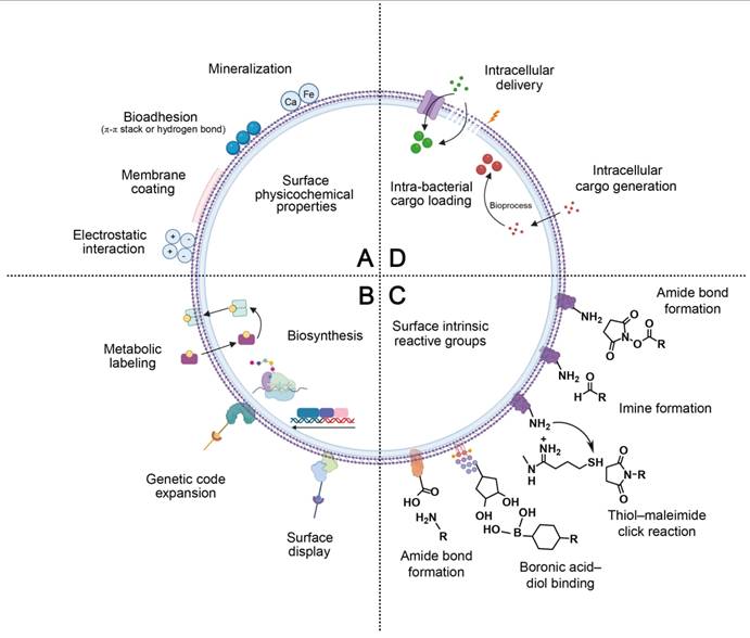
These externally exposed components provide the most accessible and chemically versatile interface for surface modification through biomaterial engineering, therefore, the bacterial surface has become the primary target for current bacterial engineering strategies (Figure 9). A variety of functional groups, such as carboxyl, amino, and cis-diol moieties, on surface-associated components [58, 119] provide tunable reaction sites for chemical modification of bacterial surfaces. Moreover, owing to the presence of abundant phosphate-containing components on bacterial surfaces, many bacteria exhibit an overall negative surface charge, which facilitates the adsorption of positively charged biomaterials through electrostatic interaction [45].
Schematic illustration of four principal approaches for bacterial engineering. (A) Surface modification through tuning physicochemical properties of the bacterial interface, including electrostatic interaction, membrane coating, bioadhesion, and biomineralization. (B) Biosynthesis-mediated modification through hijacking the endogenous bacterial biosynthetic machinery, such as metabolic labeling, genetic code expansion, and surface display. (C) Covalent conjugation to surface-intrinsic reactive groups, including amino, carboxyl, and cis-diol groups through amide bond formation, imine formation, thiol–maleimide click reaction, and boronic acid–diol binding, respectively. (D) Intra-bacterial cargo loading facilitated by cellular transporters, physical electroporation, and intracellular bioprocesses to strengthen their capacity to serve as carriers or in situ factories for therapeutic agents. Collectively, these strategies provide a versatile toolbox for engineering bacteria to enhance their stability, targeting capability, and therapeutic efficacy. Created in https://BioRender.com.
4.2 Negative surface charge-mediated electrostatic interaction
Owing to the presence of anionic interfacial components, including teichoic acids, lipoteichoic acids, lipopolysaccharides, and acidic polysaccharides, bacterial surfaces are generally negatively charged, enabling the adsorption of positively charged materials without prior chemical modification [120]. This feature provides a straightforward and cytocompatible foundation for bacterial surface engineering via electrostatic interaction. For example, Lactobacillus plantarum with a negatively charged bacterial surface was coated with cationic polymer poly-L-lysine through electrostatic interaction [121]. Building on the same principle, electrostatic deposition can be extended to layer-by-layer assembly through alternating deposition of oppositely charged materials. For instance, positively charged chitosan was first deposited onto Bacillus coagulans, followed by negatively charged alginate. The stepwise reversal of zeta potential after each deposition allowed the electrostatic formation of multilayers on the bacterial surface [122]. A notable advantage of charge-mediated interaction lies in its mild aqueous-reaction condition, which is generally benign for maintaining the bacterial viability [123]. The multilayer format offers a modular platform for additional surface functionalization. However, scale-up of this process remains challenging because of multiple repeated procedures of deposition, centrifugation and washing. Meanwhile, the formation of the resulting multilayer and their stability are sensitive to variations in pH and ionic strength, the deposition sequence, and the charge balance [124]. In addition, when the surface charge approaches neutralization, these coated bacteria may be aggregated, thus impairing the coating homogeneity. Future efforts should be devoted to refining the process and ensuring the coating integrity under formulation-associated stresses, such as drying and reconstitution [123].
4.3 Membrane-like surface-mediated assembly coating
Bacterial envelopes present a soft, hydrated, negatively charged, and membrane-like amphiphilic interface. Therefore, exogenous lipids or membrane structures can adsorb, reorganize, and self-assemble on the bacterial surface under mild conditions, forming an additional membrane-like coating without repeated deposition or prior chemical activation [93, 120, 125]. In this process, the assembled membrane serves as a physically stable outer cloak to improve bacterial viability and preserve their bioactivity. For example, EcN was coated with dioleoyl phosphatidic acid and cholesterol via simple vortex mixing for approximately 15 min, forming an additional self-assembled lipid membrane on the bacterial surface. This coating significantly improved bacterial survival against environmental stress and enhanced oral delivery efficiency and therapeutic efficacy in a murine colitis model [125]. This principle can be further extended to biologically derived membrane structures. For instance, EcN was wrapped with erythrocyte membranes by mechanical extrusion to generate cell-membrane-coated bacteria with a distinct outer lipid shell, reducing their inflammatory responses, mitigating phagocytic clearance, and improving their in vivo persistence and target-site accumulation [93]. The quality of membrane-based coatings is contingent upon the membrane source and the coating integrity. In addition, the coated bacteria remain sensitive to the lipid formulation and environmental conditions, such as the ionic milieu and the exposure to bile salts.
4.4 Chemically heterogeneous surface-mediated adhesive deposition
Adhesive deposition is a distinct strategy for bacterial surface engineering. Through interfacial adhesion, coating materials can be anchored onto bacterial surfaces. Bacterial envelopes exhibit a chemically heterogeneous interface characterized by multiple exposed functional groups, thus they serve as favorable substrates for adhesive deposition through hydrogen bonding, chelation, π–π stacking, and nonspecific interaction. In addition, covalent coupling can be applied to oxidized catechol-containing systems [120, 126, 127]. Catecholamine-derived coatings, inspired by mussel adhesive chemistry, can undergo oxidative self-polymerization under a mild condition to form conformal surface layers with strong interfacial adhesiveness, thereby achieving adhesive deposition on bacterial surfaces [126, 127]. For example, polydopamine nanoparticles have been deposited onto EcN to construct a living hybrid coating system for dopaminergic immunoregulation, thereby alleviating colitis [128]. Interaction of polyphenols with metal ions to form a metal-phenolic system can be an alternative method for adhesive deposition onto bacterial surfaces. Phenolic components on the bacterial surfaces mediate interfacial anchoring. The resulting metal-phenolic coordination can stabilize the coating and endow the bacteria with additional protective or therapeutic functions [129]. For example, the tannic acid/Fe(III) coordination was established on a bacterial surface to generate a single-cell nanoarmor. The transient protective shell enhanced bacterial resistance to antibiotics and improved therapeutic efficacy in antibiotic-associated diarrhea [82]. The strong adhesiveness of these materials enables broad surface attachment and facilitates their application across diverse engineering contexts. However, the adhesive interaction may promote non-specific binding and interparticle crosslinking, which may lead to bacterial aggregation and heterogeneous surface coating. In addition, the performance of bioinspired adhesive deposition is highly dependent on pH, the precursor concentration, oxidation or coordination conditions, and assembly kinetics. These parameters should be systematically optimized to preserve bacterial viability and their native function, meanwhile, a broader and safer reaction window can be defined for controllable coating formation.
4.5 Nucleation-prone surface-mediated mineralization
Bacterial envelopes bear negatively charged and ion-interacting groups that can locally enrich mineral precursors, providing heterogeneous nucleation sites for inorganic particle growth. This feature has been used for surface mineralization and protective mineral shells can be generated on the surface of living bacteria under mild conditions. Beyond robust physical protection, mineral layers introduce novel physicochemical and biological activities of inorganic components to bacteria, thereby empowering new functionalities for bacteria, including pH responsiveness, catalytic reactivity, magnetic resonance responsiveness, bioactive ion release, and integration with other therapeutic modalities [48, 57, 130]. For example, a pH-responsive biomineralized bacterial vaccine was developed by engineering recombinant Bacillus subtilis producing the HPV16 E7 antigen. In this vaccine, a calcium phosphate shell was formed on the bacterial surface to protect the bacteria [131]. The calcium phosphate shell acted as a physical shield and displayed an acid-labile property in a gastric environment, thereby improving the survival of the bacteria during gastrointestinal transit, prolonging their intestinal retention, and enhancing their oral bioavailability. In addition, Ca2+ released from the mineral coating induced bile acid aggregation and mitigated gastrointestinal stress, ultimately improving the bacterial survival and augmenting therapeutic efficacy in a colitis model [130]. Furthermore, an MnO2/Fe2O3 nanocoating was developed on a bacterial surface. The nanocoating served as a T1/T2 dual-mode MRI contrast layer for visualizing bacterial distribution in the gastrointestinal tract, and provided nanozyme-like antioxidant activity to scavenge ROS and alleviate inflammation [48].
From a translational perspective, surface mineralization offers the protection of bacteria against manufacture- and delivery-associated stresses, including oxygen exposure, ultraviolet irradiation, and ethanol treatment. Meanwhile, this strategy can be applicable to both obligate and facultative anaerobes. Despite these advantages, the effect of degradation products from the mineral layer on the local ionic environment and microbiota remains to be systematically evaluated. Meanwhile, a highly dense mineral shell may suppress bacterial proliferation and induce their transient dormancy, which should be robustly verified in in vivo models.
4.6 Surface intrinsic reactive groups for covalent modification
4.6.1 Surface amino groups
Surface amino groups are among the most accessible native reactive sites on bacterial cells, and they are associated with surface proteins and molecular components on the cell wall. Owing to their nucleophilicity, these primary amines can be employed for covalent surface engineering under a mild aqueous condition, while preserving the bacterial viability. Amide bond formation is a fundamental amino-directed strategy. In this route, carboxyl-containing molecules are introduced either as preactivated N-hydroxysuccinimide (NHS) esters or activated carbodiimide, and these molecules are coupled to primary amines on the bacterial surface to form stable amide linkages. The major advantage of this strategy lies in the robustness of the resulting bond, which is favorable for persistent surface display during gastrointestinal transit and subsequent host interaction. For example, after the croconium dye was converted into an NHS ester, the ester group was directly conjugated to the amine group on the surface of engineered EcN, endowing the bacteria with photothermal activity while preserving the bacterial viability and maintaining the immunostimulatory function [132]. A similar strategy was used to attenuate Salmonella Typhimurium VNP20009. The surface amine group was coupled with a carboxyl-containing JQ-1 derivative through 1-ethyl-3-(3-dimethylaminopropyl)carbodiimide (EDC)/Sulfo-NHS chemistry, generating a dual-drug-decorated bacterial biohybrid with enhanced tumor accumulation, photothermal ablation, and antitumor immune activation [133].
Primary amine groups on bacterial surfaces can also undergo amino-carbonyl coupling with aldehyde- or quinone-containing materials or molecules, forming imine-type covalent linkages [134-136]. This strategy was applied to Shewanella oneidensis MR-1. Aldehyde-bearing ZIF-90/MB particles were covalently hybridized onto the bacterial surface via acid-labile imine linkages formed between the aldehyde groups and the amine groups. The resulting stable bacteria–material biohybrid preserved the bacterial activity and facilitated acidic environment-triggered dissociation [137].
Beyond direct covalent conjugation, native surface amino groups can be converted into secondary reactive moieties for the subsequent functionalization. A representative example is 2-iminothiolane-mediated surface thiolation. In contrast to direct cargo conjugation, this strategy enabled covalent anchoring of bacteria to biological substrates by creating a dynamic mucoadhesive interface. Primary amines on surface-exposed proteins in EcN were transformed into free thiols under cyto-compatible conditions. The introduced thiols underwent catalyst-free thiol-disulfide exchange with disulfide-rich mucin, resulting in tunable mucin attachment. This approach resulted in a 170-fold increase in mucosal adhesion of bacteria in mucin-enriched jejunum, ultimately improving therapeutic efficacy in jejunal mucositis [67]. This design was subsequently extended to delivery of bacteria to the inflamed colon. Thiolated EcN showed enhanced mucus binding, prolonged intestinal retention, and improved restoration of microbiota homeostasis in inflammatory bowel disease [138].
4.6.2 Surface carboxyl groups
Surface carboxyl groups are another type of native reactive sites present on bacterial cells. These groups are predominantly associated with peptidoglycan components and acidic residues in proteins exposed at the bacterial interface. Carboxyl groups are first activated by carbodiimides through EDC/NHS chemistry to generate amine-reactive intermediates for subsequent amide bond formation [139]. This strategy has been used to attach amino-functionalized aptamers to EcN, including AS1411 for tumor targeting and an IL-6 aptamer for inflammation targeting. The modifications with AS1411 and the IL-6 aptamer improved bacterial localization at diseased sites and enhanced therapeutic efficacy in tumor-associated and colitis-associated disease models, respectively [105, 106].
4.6.3 Surface cis-diol groups
Surface cis-diol moieties are also an important type of native reactive sites on bacterial cells. These groups are predominantly associated with polysaccharide-containing structures exposed at the bacterial interface. These motifs can reversibly interact with boronic acid-containing materials to form boronate ester linkages, thereby establishing a dynamic covalent bond, which is distinct from amino- or carboxyl-directed coupling bonds [140]. The primary advantage of this strategy lies in its rapid assembly and environmental responsiveness, which enhances interfacial stabilization while retaining the capacity for stimulus-triggered dissociation or release. This strategy has been used to assemble artificial-enzyme-armed Bifidobacterium longum bacteria. The engineered bacteria exhibited enhanced intestinal targeting and retention, persistent ROS scavenging, and improved therapeutic efficacy in inflammatory bowel disease [58].
In general, the engineering strategies by harnessing the surface group of natural bacteria provide a flexible approach to constructing stable conjugation between bacteria and materials. However, these strategies have a few limitations. Because amino, carboxyl and cis-diol groups are widely distributed in bacterial surface proteins, cell wall components and polysaccharides, the modifications obtained are usually non-specific and may produce heterogeneous products. In addition, excessive coupling may undermine bacterial viability and mask natural adhesion molecules to impair bacterial movement or weaken the host-bacterial interaction.
4.7 Biosynthesis-enabled bacterial surface engineering
Bacteria undergo continuous remodeling of their surface structures through a biosynthesis process, such as glycans, proteins and polymers [141, 142]. This process has been harnessed to reshape surface structures [143, 144]. Both intrinsic components and novel functional molecules could be introduced into the bacterial outer surface for protection or functionalization [144, 145].
Metabolic labeling is a widely used strategy to functionalize bacterial surfaces. Exogenous, metabolically compatible precursor analogs can be processed by endogenous biosynthetic or remodeling machinery and their metabolic products are then incorporated into target surface structures during their formation or turnover [143, 146]. In bacteria, this approach has been used for metabolically labelling diverse surface components, including peptidoglycan, capsular polysaccharides, surface glycans, and teichoic acids. This approach can be further extended to proteins when the translational machinery accepts noncanonical amino acid surrogates [147-151]. For instance, D-alanine-derived precursors carrying oxaliplatin and a photosensitizer were simultaneously incorporated into peptidoglycan of tumor-targeting nonpathogenic E. coli MG1655, yielding a living platform for synergistic chemo-photodynamic therapy and immunogenic cell death induction [152]. In addition, bioorthogonal reactive groups can be introduced for subsequent conjugation by this strategy. Ji et al. used this metabolic method to incorporate 3-azido-D-alanine into the cell wall of Lactococcus lactis. The introduction of the azide group facilitated a subsequent copper-free click reaction with dopamine- and β-glucan-based components, which enhanced gastrointestinal tolerance, intestinal retention, colonization, and antidiabetic efficacy [153].
Orthogonal reactions can also be performed through genetic code expansion (GCE). In this approach, an orthogonal aminoacyl-tRNA synthetase/tRNA pair decodes a reassigned codon and a noncanonical amino acid is loaded into a growing peptide. Eventually, a reactive group is introduced at a predetermined residue of the target protein [145, 154, 155]. Praveschotinunt et al. placed an in-frame UAG codon in csgA, a gene for the curli monomer CsgA of E. coli, and co-expressed an orthogonal aaRS/tRNA pair that reads UAG as p-azido-L-phenylalanine (pAzF) [156] (Figure 10). Once the curli fibers were assembled on the cell surface, the azide group on these fibers reacted with DBCO-Cy5 via copper-free click chemistry. Since the Cy5 label was stable in vivo, the engineered strain could be real-time tracked within the mouse gut by monitoring fluorescence signals.
Representative examples of biosynthesis-enabled bacterial surface engineering. (A) The process of incorporating nonstandard amino acids into bacteria through an orthogonal translation system (OTS). These amino acids are targeted by an imaging probe. (B) Fluorescence imaging of live mice administered with either PBS (-) or engineered bacteria (+). Adapted with permission from [156], copyright 2018 American Chemical Society.
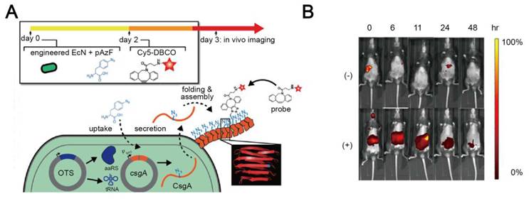
Surface display strategies have been developed to expose specific proteins, peptide ligands or antigenic epitopes by reshaping bacterial surfaces. By genetically programming the expression, secretion, trafficking, and anchoring of recombinant proteins or peptide modules, these artificial proteins or peptides can be transported and presented on cell membranes, cell walls, spore coats or biofilm matrices [157-159]. For example, Nguyen et al. genetically fused functional peptide domains into the C-terminus of CsgA, the major amyloid subunit of the E. coli curli system [160]. After translation, the final fusion protein was secreted and then self-assembled into extracellular nanofiber networks. By fusing different peptide tags to CsgA, distinguished functionalities were achieved: a metal-binding domain for stainless steel adhesion, and an A3 peptide to template silver nanoparticles. Furthermore, Praveschotinunt et al. adapted this platform to EcN and produced curli fibers with a pro-repair cytokine trefoil factor 3 (TFF3) [161]. Interestingly, the surface display strategy could also allow the incorporation of SpyTags for covalent capture of SpyCatcher-fused proteins, which resolves the issue of a large size for direct genetic fusion. For example, bacteria empowered through the surface display method by Zhang et al. remarkably alleviated colitis in a mouse model. Lactococcus lactis was engineered to display a SpyCatcher-AcmA fusion on the bacterial surface. In this system, AcmA acted as a cell-wall anchor. SpyTag-terminated polymers, such as polydopamine, were conjugated onto the surface through SpyTag/SpyCatcher covalent ligation. A dopamine shield was generated from this SpyTag/SpyCatcher complex to improve gastrointestinal stability, mucoadhesion, and local colonization, thus alleviating colitis in mice [162].
Biosynthesis-assisted surface engineering can introduce functional structural units in the process of surface remodeling, providing highly spatial control over the surface structure and stably displaying proteins, peptides or bio-orthogonal handles. However, their efficiency is convergent upon the metabolic activity, genetic operability and growth status of the bacterial chassis. In addition, genetic manipulation may raise concerns over the strain stability, biosafety and regulatory translation, which should be carefully addressed for their clinical application.
4.8 Intrabacterial cargo loading
In intrabacterial cargo loading, the intracellular space of bacterial cells serves as a protective compartment that shields functional cargoes from the surrounding milieu while preserving native surface structures involved in host interactions [163-165].
4.8.1 Intracellular delivery
The physicochemical properties of a cargo can facilitate its delivery into a cell via either native transport pathways or membrane permeabilization. For instance, silicon nanoparticles carrying indocyanine green (ICG) were modified with a glucose polymer, poly[4-O-(α-D-glucopyranosyl)-D-glucopyranose]. The resulting hybrid was internalized into attenuated Salmonella typhimurium or E. coli through an ATP-binding cassette (ABC) transporter. Since the envelope of both bacteria was undisturbed, the loaded bacteria successfully crossed the blood–brain barrier (BBB) and delivered ICG into the glioblastoma tissue [165] (Figure 11A-C). For cargos that cannot enter the bacterial cytoplasm through native transporters, permeabilization strategies, such as electroporation, offer an alternative loading route for a diverse range of cargoes. Zoaby et al. discovered that electroporation can improve the efficiency in loading doxorubicin-containing liposomes into bacteria [166, 167]. It is noted that the permeabilization process must be meticulously tuned to preserve the viability and the motility of the bacteria since the pores created through the process for the cargo entry may be detrimental to the bacterial cells.
Representative examples of intrabacterial cargo loading. (A) Schematic illustration of the construction of a Trojan bacteria system. (B) Crossing a blood–brain barrier (BBB) and targeting and penetrating glioblastoma (GBM) tissues by the Trojan bacteria system. (C) High-angle annular dark field-scanning transmission electron microscope (HAADF-STEM) images confirm intracellular localization of silicon nanoparticles in a bacteria cell, rather than nonspecific adsorption on the bacterial surface. Adapted with permission from [165], copyright 2022, Springer Nature. (D-E) The preparation and characterization of Se@EcN. Adapted with permission from [169], copyright 2025, American Chemical Society.
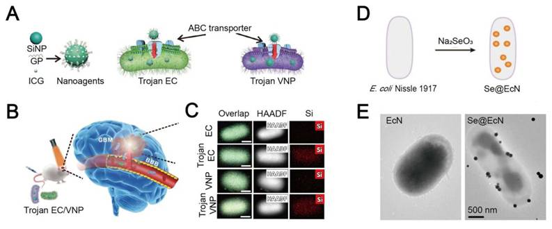
4.8.2 Intracellular cargo generation
Inspired by the biosynthetic and redox machinery of bacteria, an in situ cargo generation strategy has been developed. After an appropriate soluble precursor is introduced into the culture medium and enter a bacterial cell, the bacterial intracellular compartment can serve as a reactive microenvironment to generate the cargo with therapeutic function as well as a storage compartment for the generated cargo [168]. For example, bacterial cells internalize soluble selenium precursors and reduce them to selenium nanoparticles, which in turn scavenge ROS and amplify the therapeutic effect [49, 169] (Figure 11D-E). Combined metabolomic and proteomic analyses revealed that nanoSe biosynthesis was associated with metabolic remodeling, with enhanced redox adaptation and TCA cycle activity, but reduced carbohydrate catabolism and oxidative phosphorylation [49]. It is noteworthy that this process relies on a continuous precursor supply, and the supply may not be feasible in vivo. The biosynthesized product may be exported across the envelope, leading to an unforeseen biological consequence [170].
Intracellular cargo loading is a very useful strategy when bacterial cells are expected to act as live carriers to carry agents while preserving their outer structures and biological functions. The transporter-mediated loading method is relatively mild, and it can maintain the integrity of bacterial cell membranes. Therefore, it is suitable for applications that requires bacterial movement, tissue penetration or crossing barriers, such as tumor-targeting treatment. In contrast, permeability-based loading can accommodate a broad loading content, but this method should be meticulously optimized to prevent compromising the viability and hindering the motility of bacteria. In situ cargo generation of functional materials in cells is achieved by harnessing the metabolic process of bacteria, thus providing self-protection or therapeutic advantages under pressure conditions. However, this strategy is heavily contingent upon the availability of precedents, the metabolic activity of bacteria and the retention of products, which may impede its applicability in the human body. Meanwhile, metal precursors or uncontrolled particle formation may promote ROS generation and elevate the metabolic burden, which may compromise the genome integrity and increase the cost of selecting biomineralization traits. In contrast, controlled synthesis may detoxify soluble metal species and produce ROS scavenging nanomaterials, such as SeNPs. These context-dependent outcomes warrant further evaluation of bacterial growth performance and their long-term functional stability [170].
4.9 Bacterial engineering methods based on bulk encapsulation
Beyond engineering bacteria by harnessing their interfacial components and introducing new chemical functionalities, bulk encapsulation strategies have been developed to incorporate multiple bacteria cells and therapeutic agents within a biocompatible matrix. Bulk encapsulation methods not only provide collective protection against adverse environmental conditions prior to reaching target sites but also facilitate bio-responsive release of bacteria and therapeutic agents [49, 64, 171-175]. Encouragingly, the bulk encapsulation process is significantly more simplified compared to the modification of single bacteria cells. This process can be readily scaled up for large-scale production, exhibiting greater potential for clinical translation [176]. An array of techniques has been reported to encapsulate multiple bacterial cells as a population within one matrix to support their growth and proliferation. For instance, Luo et al. encapsulated EcN in a chitosan–alginate gel matrix via CaCl2 cross-linking [177]; IIomuanya et al. developed mucoadhesive PVA/PCL electrospun scaffolds for intravaginal delivery of Lactobacillus fermentum, metronidazole, and sodium cocoamphoacetate to manage bacterial vaginosis [178]; and Liu et al. encapsulated lactic acid bacteria in a 3D-printed biocompatible living hydrogel [179]. Furthermore, Yang et al. co-encapsulated EcN and indole-3-propionic acid in dextran (Dex)-TA hydrogel microspheres via droplet microfluidics, confirming this multifunctional delivery system was more effective in disease treatment than bacteria-only microspheres [180].
Compared with single-cell surface engineering, encapsulating bacteria in hydrogels, fibers, microcapsules, or other protective matrices is much simpler and more scalable. However, the control of the behavior of individual bacterium during the bulk encapsulation process is often very challenging. In addition, matrix design should strike a balance between protection of bacteria and their access to the surrounding microenvironment, because the encapsulation of bacteria within these matrices may impede the diffusion of nutrients and toxic metabolites, hinder the migration of bacteria, and ultimately diminish the treatment efficiency. Therefore, bulk encapsulation can be considered for scalable and protective formulation, but careful balance should be tailored between matrix protection, encapsulation efficiency, and bacterial viability.
5. Engineered bacteria in therapeutic applications
Despite multiple physiological and pathological barriers that hinder the survival and impair biological function of administered bacteria, recent engineering strategies have substantially improved their viability and functional performance during transport to diseased tissues. In this section, we summarize recent progress in the therapeutic application of engineered bacteria via different administration routes (Figure 12).
An overview of four administrative routes for engineered bacteria in therapeutic application. Oral administration of engineered bacteria not only allows the treatment of gastrointestinal (GI) diseases but also the extension of systemic therapeutic effects through multiple gut–organ axes via the production of beneficial metabolites. Topical administration is predominantly used for the treatment of vaginitis and wound healing. After intravenous administration, engineered bacteria home to tumor sites and exert their antitumor activity. Inhalational administration has shown promise for the treatment of pneumonia and lung cancer. Created in https://BioRender.com.
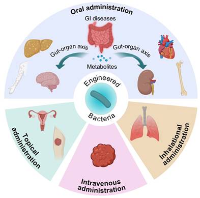
5.1 Oral engineered bacteria
The bioengineering-assisted approach for oral delivery of bacteria overcomes the physicochemical, biological, and ecological barriers encountered during gastrointestinal transit, improving their survival and colonization. By enhancing the stability and prolonging the functional persistence of bacteria in the gut, this approach not only augments bacterial efficacy in gastrointestinal disorders but also extends their therapeutic potential to diseases in distant organs through the gut-organ axis [181-186]. For example, Yang et al. coated EcN with tannic acid and mucin to obtain EcN@TA-Ca2+@Mucin [65]. These engineered bacteria displayed enhanced ROS-scavenging activity owing to the abundant phenolic hydroxyl groups in tannic acid, and the mucin coating improved bacterial colonization through multiple interactions with the intestinal mucus layer. Together, these acquired properties from tannic acid and mucin extended the colonic retention time of the bacteria to 96 h and improved their therapeutic efficacy in a murine colitis model. To target the gut microbiota–trimethylamine–trimethylamine N-oxide axis in atherosclerosis, Chen et al. developed nanoparticle-functionalized bacteria by loading fluoromethylcholine into nanoparticles and conjugating these nanoparticles onto polydopamine-coated LGG, yielding PDMF@LGG [182]. The polydopamine coating improved intestinal retention, while the methyl thioethanol ester in nanoparticles scavenged ROS and triggered responsive release of fluoromethylcholine in the gut. These engineered bacteria were administered to elevate the abundance of the Lachnospiraceae and Oscillospiraceae family to ameliorate dysbiosis, ultimately lowering the circulating trimethylamine N-oxide level and attenuating atherosclerotic progression in mice. Besides, oral bacteria can function as a vaccine delivery vehicle. Xia et al. developed a biomineralized Bacillus subtilis system to sustainably produce HPV16 E7 antigen in situ. Notably, the calcium phosphate mineral coating facilitated a 4-fold increase in intestinal retention of the bacteria compared with uncoated ones, while sustaining the antigen persistence over 4 days. This engineered bacterial vaccine systematically activated CD8+ T cells, resulting in the suppression of 64% of TC-1 tumors [131].
5.2 Topical engineered bacteria
Topical administration of bacteria is increasingly explored in the treatment of cutaneous and mucosal inflammatory disorders, particularly, wound healing and vaginitis. Under these chronically infected microenvironments, physicochemical alterations, pathogen colonization and sustained inflammation compromise the bacterial fitness and diminish their therapeutic efficacy. To overcome these barriers, bacteria can be encapsulated within protective biomaterials to enhance their viability and facilitate their targeted colonization at disease sites. For instance, Liu et al. engineered Lactobacillus rhamnosus CLK101 (LRh) to biosynthesize antioxidant selenium nanoparticles and then encapsulated the engineered bacteria within a phospholipid polymer hydrogel to prepare gel@F-LRh [49]. The hydrogel network mimicked a three-dimensional ECM structure, promoting bacterial metabolic activity and stability. The resulting gel@F-LRh effectively inhibited the proliferation of pathogenic Staphylococcus aureus and scavenged excess ROS in diabetic wounds, accelerating the healing of infected wounds in a mouse model. Zhang et al. developed a Bacillus subtilis-containing hydrogel platform, B. sub@HMHA, for the treatment of fungal vaginitis [187]. This living hydrogel locally produced high-molecular-weight hyaluronic acid and antimicrobial peptides, thereby reducing inflammation and inhibiting fungal growth. For bacterial vaginosis, bacterial therapy must overcome two major barriers: poor mucosal retention and intense competition with pathogenic microbiota. To address these issues, Ilomuanya et al. developed mucoadhesive electrospun scaffolds for intravaginal delivery of Lactobacillus fermentum together with metronidazole [178]. The engineered bacteria showed improved vaginal retention with a mucoadhesion value of 57.86% in a mouse model. Meanwhile, the co-delivered metronidazole was expected to suppress pathogenic bacteria, thereby creating a more favorable niche for Lactobacilli colonization and restoring the vaginal microbial balance. However, this combined strategy faces an important challenge: locally administered antibiotics may impair the efficacy of therapeutic bacteria. Since topical antibiotics are commonly used for biofilm-associated diseases such as infected wounds and vaginitis, future efforts should be devoted to protecting live therapeutic bacteria while maintaining antimicrobial efficacy against pathogens.
5.3 Intravenous engineered bacteria
Upon systemic administration, various live bacteria, including Salmonella typhimurium, EcN and Bifidobacterium, can intrinsically migrate to solid tumors due to their hypoxia tropism. However, the majority of systemically administered bacteria are rapidly cleared by the mononuclear phagocyte system, resulting in a low tumor colonization level and limited therapeutic efficacy [88]. Furthermore, compromised anti-tumor immune responses are often seen in an immunosuppressive TME, characterized by nutrient deprivation and accumulation of toxic metabolites [13]. Therefore, to improve bacteria-based tumor treatment outcomes, their tumor accumulation can be enhanced through evasion of immune clearance, and immune activation in the TME can be realized through their combination with complementary therapies. To improve tumor accumulation of intravenously delivered bacteria, efforts have been made to reduce systemic clearance and enhance tumor targeting. To reduce immune clearance, Cao et al. coated EcN with erythrocyte membranes to generate cell membrane-coated bacteria (CMCBs) [93]. The membrane coating facilitated a reduction in the immunogenicity of bacteria and the phagocytic uptake, while maintaining the bacterial viability. Although intravenously administered CMCBs majorly accumulated in liver and spleen at 1 h post-injection, the number of bacteria within healthy organs gradually decreased over time. Notably, this approach resulted in an approximately 42-fold increase in tumor accumulation compared to uncoated EcN post murine tail vein injection. To enhance tumor targeting, Xiao et al. covalently coupled a monomethyl auristatin E (MMAE)-based aptamer-drug conjugate to Salmonella typhimurium VNP20009, yielding VNP@Sgc8c-MMAE [104]. The intrinsic tumor tropism of the bacteria in combination with aptamer-mediated targeting resulted in less biodistribution in the liver, spleen and kidney, but a 3-fold increase in tumor accumulation and remarkable promotion of cytotoxic T cell infiltration in a Miapaca-2 tumor xenograft model.
5.4 Inhalational engineered bacteria
Living bacteria via pulmonary administration can modulate local microbial population and induce inflammatory disturbance in lung diseases, but its effectiveness is convergent upon preserving bacterial activity during the preparation and delivery process and engineering the formulation to overcome pulmonary barriers. In bacterial pneumonia, Fu et al. coated living Lactobacillus rhamnosus with chitosan, hyaluronic acid and ononin to generate OASCLR particles at approximately 1.32 μm in length and 0.51 μm in width [38]. The coating enhanced bacterial stability, facilitated ROS scavenging and promoted hyaluronic acid-mediated interaction with CD44-expressing inflammatory macrophages. Within OASCLR, living Lactobacillus rhamnosus retained its ability to produce lactic acid and inhibit Staphylococcus aureus. In murine models of Staphylococcus aureus pneumonia, nebulized OASCLR reshaped the lung microbiota and attenuated pulmonary inflammation. Nicola et al. developed a pulmonary formulation for chronic obstructive pulmonary disease using three live Lactobacillus strains, L. plantarum BAA 793, L. acidophilus 4356 and L. rhamnosus 53103 in combination with trehalose, leucine, sodium citrate and polysorbate. They prepared the powder formulation via spray drying [112]. In the resulting powder, approximately 99% of the bacterial viability was retained. The aerodynamic profile of the powder was applicable for distal lung delivery, with a mass median aerodynamic diameter of 2.4 μm and approximately 74% below 3.3 μm. In mouse models of chronic obstructive pulmonary disease, such as emphysema induced by porcine pancreatic elastase, with or without additional lipopolysaccharide-associated inflammation, administration of the spray dried formulation through an Insufflator resulted in a reduction in the alveolar enlargement and airway resistance, a decrease in the levels of MMP-9, neutrophil elastase, C reactive protein and IL-8, and an increase in bronchoalveolar lavage IgA. Interestingly, Nicola et al. also showed that prolonged pulmonary persistence of inhaled live Lactobacillus may not be essential for the therapeutic activity. Although whole lung colony counts of the administered three-strain Lactobacillus blend steadily decreased over 72 h, the local L(+) lactic acid level remained relatively stable. These findings suggest that transient bacterial residence may be sufficient to reshape the local microbial population and generate sustained exposure to bioactive metabolites. Therefore, an effective metabolic exposure window and a clearance profile of inhaled living bacterial therapeutics should be carefully defined.
6. Conclusion and perspectives
After the intricate relationship between microbial homeostasis and human health has been gradually unveiled over the past decades, bacteria have emerged as a promising candidate for living therapeutics for a wide spectrum of diseases. Bacterial therapeutics have been investigated across multiple administration routes, including oral, topical, intravenous, and inhalational delivery. However, these bacterial administration routes have encountered in vivo barriers, such as exposure to low pH and ROS, nutrient limitation, poor adhesion, competition from microorganisms within the body, immune clearance, and unsatisfied targetability. By addressing these challenges individually or in combination, engineering strategies can improve the survival, retention, and biodistribution of therapeutic bacteria, enhance immunomodulatory effects, and promote microbial homeostasis across various disease contexts, ultimately improving therapeutic efficacy. Nevertheless, although several in vivo barriers are shared among different administration routes, the feasibility and relevance of corresponding engineering strategies vary substantially depending on tissue-specific microenvironments and disease contexts. For instance, nutrient-supplying strategies have been validated in gastrointestinal bacterial delivery, but their potential value in other administration routes remains underexplored, despite nutrient availability representing a fundamental requirement. Moreover, strategies that improve bacterial adhesion after oral administration may not be applicable to other delivery routes, suggesting that the transferability of an effective engineering design across disease models requires further evaluation. Of note, these findings have largely emerged from preclinical studies across diverse disease models, and their clinical relevance remains to be further validated.
To achieve optimized bacterial therapy, future development should begin with rational selection and engineering of bacterial chassis. Strain-specific differences in the metabolic preference, the surface structure, colonization, stress resistance, and the immunomodulatory capacity, can have a substantial impact on biomaterial interactions and therapeutic outcomes [188]. A deep comprehension of the innate characteristics of individual bacterial cells is essential for biological engineering of these beneficial bacteria. In addition, efforts should be directed towards the design of genetically modified bacteria as living treatment factories, which offer additional advantages compared to non-modified ones in the field of disease treatment. Recent advances in synthetic biology, including programmable gene circuits and clustered regularly interspaced short palindromic repeats (CRISPR)-based editing, may further enable engineered probiotics to sense disease-associated signals, regulate therapeutic output, and improve biosafety through controllable functional modules. Beyond therapeutic potency, strain selection should also account for growth stability, surface characteristics, stress tolerance, and production reproducibility, as these properties directly influence subsequent material modification and clinical translation.
Beyond strain selection, biomaterial design should consider bacterial chassis, material interfaces, and pathological environments as an interconnected design space, and the design space can be optimized to improve therapeutic outcomes. Within this framework, integration of different materials onto bacteria may provide complementary properties and synergistic effects, while the incorporation of these materials may introduce the analytical and manufacturing complexity, and they may have an impact on biosafety and translational feasibility. Looking ahead, artificial intelligence (AI) guided material design may provide a new paradigm for developing biomaterials compatible with living bacteria. Moving beyond the empirical adaptation of existing materials, AI could help navigate the multidimensional relationships among interfacial properties, bacterial fitness, host compatibility, therapeutic efficacy, and manufacturability. This capability may facilitate the identification of material architectures that reconcile competing requirements and guide the rational integration of complementary components into synergistic systems tailored to specific disease contexts.
Although biomaterial engineering can substantially enhance bacterial delivery performance and therapeutic efficacy across multiple dimensions, clinical translation of bacterial therapeutics is governed by developability, manufacturability, and regulatory compliance in addition to functional performance in preclinical models. Bacterial therapeutic products with clinical potential must be produced from a reproducible, scalable manufacturing process, maintain stability throughout storage and administration, and meet predefined quality attributes consistent with Good Manufacturing Practice (GMP) and regulatory evaluation. From a translational perspective, bulk encapsulation does not require strain-specific modification of individual bacterial cells, therefore, it is applicable to a broad range of bacterial strains [189]. Its intrinsic limitation, however, arises from mass-transfer constraints. As the matrix thickness increases, the nutrient diffusion efficiency declines while toxic metabolites accumulate in the matrix core, resulting in reduced cell viability in the central region of large constructs and a small viable population concentrating at the periphery [114]. Therefore, the size and geometry of the matrix should be designed to balance between bacterial loading and the survival of cells in the interior of the matrix. Electrostatic adsorption and layer-by-layer assembly offer considerable design flexibility, but the multilayer assembly is sensitive to the ionic strength, deposition sequence and charge balance. This process also requires repeated deposition–centrifugation–washing cycles, complicating the reproducible scale-up process. The aggregation of coated bacteria may compromise the coating homogeneity as the surface charge approaches the neutrality. Membrane-based coatings can be applied to the bacterial surface through a comparatively simple process, such as coextrusion or sonication. The operational complexity is significantly reduced, while their translation raises distinct quality-control concerns. For synthetic lipid coatings, the batch-to-batch consistency may be compromised due to the lipid composition, the oxidation state and the membrane fluidity, whereas for cell-derived membranes, the donor source, the purification procedure, compositional variations, and residual biological impurities become the principal risks for manufacturing reproducibility and storage stability [190]. Covalent modification yields stable, site-defined functionalization of the bacterial surface, yet the coupling reagents and over-derivatization can impair the cell viability and function in a dose- and time-dependent manner. The covalent reaction conditions should be mild and they should be validated via functional-activity assays [176]. Adhesive deposition and controlled mineralization build a protective shell in few steps under mild conditions, but uncontrolled polymerization or coordination may lead to uneven coating and aggregation. Therefore, release testing should be conducted to determine the coating content, the percentage of aggregates, residual ions, and the mineral loading content, together with the dissolution kinetics. Biosynthesis-assisted surface engineering relies on the bacterial own biosynthetic machinery, the degree of functionalization depends on metabolic and expression activity and is highly sensitive to culture conditions and growth state, leading to batch-to-batch variability in effective ligand density. Intrabacterial cargo loading also faces challenges in increasing the loading efficiency and preventing payload leakage during storage and prior to use.
In addition to manufacturing concerns, long-term preservation is critical for maintaining bacterial viability, functionality, and practical applicability. For example, the freeze-drying preservation method is one of promising methods for stabilizing bacteria. However, drying, freeze-thaw, and prolonged storage may damage bacterial cell membranes and inactivate bacterial proteins, thereby diminishing bacterial viability. Although cryoprotectants and lyoprotectants help preserve bacterial viability [191], preservation-associated stresses may compromise the integrity of the engineered material interface as these stresses may disrupt electrostatic coatings, detach adsorbed lipid membranes, alter covalent conjugates, dissolve mineralized layers, or impede the release from encapsulating matrices. In the freeze-drying product, the bacteria should remain viable, meanwhile the coating structure, load stability, and the therapeutic function should be retained.
Equally important, the stability of the bacterial progeny should also be systematically examined [192]. The effects of prolonged culture and storage on bacterial cell division, genetic stability, phenotypic consistency, and functional fidelity should be systematically evaluated in working cell banks, as these attributes are critical for the safety and efficacy of bacterial therapeutics [193]. A stable therapeutic function after prolonged bacterial proliferation is essential to maintain the batch-to-batch reproducibility and long-term efficacy. Therefore, functional consistency after serial passages should be considered as an important quality control indicator.
Beyond manufacturing and preservation, another critical aspect of clinical translation is the characterization of pharmacokinetics and pharmacodynamics of engineered bacterial therapeutics, as these properties have a direct impact on the exposure response relationships underlying their efficacy and safety. Unlike a static molecular entity, a bacterial therapeutic is a dynamic population that may proliferate, die, mutate, exchange genetic materials, or alter its function in response to the host environment. Therefore, while an administered dose may be informative, it is insufficient to determine the resulting exposure. A meaningful assessment should distinguish the administered nominal dose, the viable dose at the target site, and the functionally active fraction during the treatment period. Strain identity testing, combined with culture-based enumeration and strain-specific qPCR or ddPCR can be applied to route-appropriate samples, such as feces, blood, local swabs, respiratory specimens, or tissue biopsies, to confirm the administered strain and quantify the viable and total bacterial abundance at the sampled sites. Meanwhile, serial sampling can be employed to characterize the persistence of colonization at the target site and the kinetics of its eventual clearance. A key limitation, however, is that the sampled material may not adequately represent the bacterial state in the region of interest. Feces, for example, reflect the distal lumen rather than the mucosal community. Quantification is complicated by the viable-but-non-culturable (VBNC) state, in which bacteria remain alive yet fail to grow under standard conditions [194], whereas DNA-based qPCR/ddPCR methods cannot distinguish live from dead bacteria [195]. Moreover, measurement of bacterial metabolites offers a functionally informative readout, but this measurement cannot be used to determine the abundance of bacteria. For example, butyrate, a short-chain fatty acid produced by commensal bacteria, induces colonic regulatory T-cell differentiation and ameliorates colitis [196]. Fecal butyrate levels have been proposed as a biomarker of treatment efficacy in fecal microbiota transplantation [197]. However, host absorption of the metabolite may deviate the measured level from the actual production level by bacteria. In addition, time-dependent drift from ongoing microbial metabolism after sampling and non-standardized measurement methods pose a compounded challenge to interpreting the metabolite data.
Pharmacokinetics and functional monitoring are also essential for evaluating effective elimination or functional silencing of engineered bacteria after completing their therapeutic action. This is particularly important because prolonged in vivo persistence after therapeutic treatment may lead to excessive colonization, ectopic migration, unintended dissemination, or opportunistic pathogenicity [70, 198]. Therefore, beyond post-treatment monitoring, the fate of engineered bacteria should be considered from early stages of product development. Programmable clearance mechanisms and functional silencing circuits should be incorporated during system design to enable controlled elimination or deactivation when treatment is completed [199].
To address the aforementioned challenges, comprehensive and sustained collaborative efforts are essential among researchers in the fields of microbiology, synthetic biology, biomaterials science, process development, manufacturing-associated science and technology, pharmaceutics, and clinical research. Through interdisciplinary efforts, engineered bacteria could enter clinical translation to offer broad therapeutic benefits for patients across a wide range of disease conditions (Figure 13).
Challenges for clinical translation of engineered bacteria. Created in https://BioRender.com.
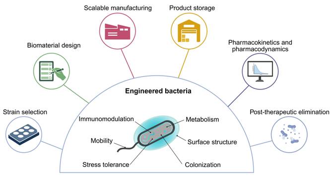
Abbreviations
FAO/WHO: Food and Agriculture Organization of the United Nations/World Health Organization; LGG: Lacticaseibacillus rhamnosus GG; IBD: inflammatory bowel disease; AMPs: antimicrobial peptides; SCFAs: short-chain fatty acids; ROS: reactive oxygen species; TLR2: Toll-like receptor 2; TME: tumor microenvironment; NK cells: natural killer cells; DCs: dendritic cells; PD-L1: programmed death-ligand 1; EcN: Escherichia coli Nissle 1917; E. coli: Escherichia coli; NHS: N-hydroxysuccinimide; EDC: 1-ethyl-3-(3-dimethylaminopropyl)carbodiimide; GCE: genetic code expansion; aaRS/tRNA: aminoacyl-transfer RNA synthetase/transfer RNA; pAzF: p-azido-L-phenylalanine; DBCO: dibenzocyclooctyne; Cy5: cyanine 5; DBCO-Cy5: dibenzocyclooctyne-cyanine 5; TFF3: trefoil factor 3; BBB: blood–brain barrier; ICG: indocyanine green; ATP: adenosine triphosphate; ABC transporter: ATP-binding cassette transporter; PELA: poly(ethylene glycol)-polylactide; Dex: dextran; TA: tannic acid; mGN: carboxymethylated β-glucan; Fh: ferrihydrite; GSH: glutathione; PRRs: pattern-recognition receptors; IL-6: interleukin-6; CD44: cluster of differentiation 44; CD47: cluster of differentiation 47; HPV16: human papillomavirus type 16; LRh: Lactobacillus rhamnosus CLK101; ECM: extracellular matrix; HMHA: high-molecular-weight hyaluronic acid; CMCBs: cell membrane-coated bacteria; MMAE: monomethyl auristatin E; PTT: photothermal therapy; CDT: chemodynamic therapy; PDT: photodynamic therapy; SDT: sonodynamic therapy; HMME: hematoporphyrin monomethyl ether; BiL: Bifidobacterium longum; ICD: immunogenic cell death; STING: stimulator of interferon genes; MRI: magnetic resonance imaging.
Acknowledgements
This work received funding from the Sichuan Provincial Science and Technology Plan Project (2024YFFK0048). Sichuan Provincial Foundation of Science and Technology (2023NSFSC1867, 2024NSFJQ0050). Department of Science and Technology of Sichuan Province (2025ZNSFSC1417). National Natural Science Foundation of China (32271445, 52203182), National Key Research and Development Program of China (2023YFB3810004, 2022YFC2009900), Central Government Guided Local Science and Technology Development Project for Regional Innovation System Construction (Project Name: Multifunctional Intelligent Photoelectric Energy, Platform Project Code: 2024ZYD0275). Diagrams, including the graphical abstract image and figures, were created with BioRender.com (https://www.biorender.com/). During the preparation of this work, Generative AI tool Claude was used in order to improve language and readability. After using this service, the authors reviewed and edited the content critically. The authors take full responsibility for the scientific accuracy, comprehensiveness, and impartiality of the work.
Contributions
Haoda Yu, Jieyu Liu, Shuxin Zhang and Bing Wang contributed to the manuscript drafting; Kui Luo, Xian Jiang and Bing Wang conducted critical review and revision of the manuscript; Kui Luo, Xian Jiang and Haoda Yu were responsible for the study conception and review of the entire manuscript. All authors have read the final manuscript and approved its publication.
Competing Interests
The authors have declared that no competing interest exists.
References
1. Uberoi A, McCready-Vangi A, Grice EA. The wound microbiota: Microbial mechanisms of impaired wound healing and infection. Nat Rev Microbiol. 2024;22:507-21
2. Gibson GR, Hutkins R, Sanders ME, Prescott SL, Reimer RA, Salminen SJ. et al. Expert consensus document: The international scientific association for probiotics and prebiotics (ISAPP) consensus statement on the definition and scope of prebiotics. Nat Rev Gastroenterol Hepatol. 2017;14:491-502
3. Jiang X, Zhang C, Cao S, Peng Y, Zhao M, Zhang X. et al. Analysis of intratumoural bacteria: Opportunities and challenges for cancer therapy. Acta Pharm Sin B. 2026;16:2877-902
4. Dong W, Li C, Lu J, Weng L, Xu Y, Xu M. et al. Engineered bacteria reprogram tumor microenvironment via cell senescence and neutrophil extracellular traps degradation. Acta Pharm Sin B. 2026;16:2332-56
5. Wang Y, Hu L, Wang L, Zhang C, Shen W, Yang H. et al. Engineered Escherichia coli Nissle 1917 targeted delivery of extracellular PD-L1-mFc fragment for treating inflammatory bowel disease. Acta Pharm Sin B. 2025;15:6019-33
6. Scharschmidt TC, Segre JA. Skin microbiome and dermatologic disorders. J Clin Invest. 2025;135:e184315
7. Monnerat N, Lambert AC, Genné D. What happens after an accidental intravenous probiotic injection? Clin Microbiol Infect. 2020;26:517-8
8. Zhang Y, Wu Y, Zhao X, Ye Q, Cao L, Liu M. et al. Progress and challenges of functionalized bacterial encapsulation: A novel biotechnology for next-generation biotherapeutics. Acta Pharm Sin B. 2025;15:5167-91
9. Zhang Y, Liu Y, Liang X, Wen Y, Zhao J, He Y. et al. Intestinal barrier in chronic gut and liver diseases: Pathogenesis and therapeutic targets. Acta Pharm Sin B. 2025;15:5515-36
10. Mazloom K, Siddiqi I, Covasa M. Probiotics: How effective are they in the fight against obesity? Nutrients. 2019;11:258
11. Naik S, Bouladoux N, Linehan JL, Han SJ, Harrison OJ, Wilhelm C. et al. Commensal-dendritic-cell interaction specifies a unique protective skin immune signature. Nature. 2015;520:104-8
12. Lai Y, Di Nardo A, Nakatsuji T, Leichtle A, Yang Y, Cogen AL. et al. Commensal bacteria regulate toll-like receptor 3-dependent inflammation after skin injury. Nat Med. 2009;15:1377-82
13. Liu J, Bai Y, Li Y, Li X, Luo K. Reprogramming the immunosuppressive tumor microenvironment through nanomedicine: An immunometabolism perspective. EBioMedicine. 2024;107:105301
14. Li Z, Zhang Q, Li Z, Ren L, Pan D, Gong Q. et al. Branched glycopolymer prodrug-derived nanoassembly combined with a STING agonist activates an immuno-supportive status to boost anti-PD-L1 antibody therapy. Acta Pharm Sin B. 2024;14:2194-209
15. Li Y, Shen X, Ding H, Zhang Y, Pan D, Su L. et al. Dendritic nanomedicine enhances chemo-immunotherapy by disturbing metabolism of cancer-associated fibroblasts for deep penetration and activating function of immune cells. Acta Pharm Sin B. 2024;14:3680-96
16. Lin L, Fang Z, Liu G, Liu Y, Li Z, Pan D. et al. Prodrug-based combinational nanomedicine remodels lipid metabolism for reinforced ferroptosis and immune activation. Acta Pharm Sin B. 2025;15:2746-63
17. Cheng X, Cai H, Li X, Zhang Y, Li S, Li Y. et al. Reversing adenosine-mediated immunosuppression in triple-negative breast cancer by synergistic chemo-immunotherapy via stimuli-responsive nanomedicines. EBioMedicine. 2026;123:106059
18. Yang X, Guo Y, Chen C, Shao B, Zhao L, Zhou Q. et al. Interaction between intestinal microbiota and tumour immunity in the tumour microenvironment. Immunology. 2021;164:476-93
19. Belkaid Y, Hand TW. Role of the microbiota in immunity and inflammation. Cell. 2014;157:121-41
20. Gopalakrishnan V, Spencer CN, Nezi L, Reuben A, Andrews MC, Karpinets TV. et al. Gut microbiome modulates response to anti-PD-1 immunotherapy in melanoma patients. Science. 2018;359:97-103
21. Liu W, Lan Z, Lin Z, Zhao Y, Lian J, Yu G. The microbiome-immune cell interaction network: Advancing tumor immunotherapy. Semin Cancer Biol. 2025;114:128-37
22. Xin Y, Yu Y, Wu M, Su M, Elsabahy M, Qu X. et al. Tumor and intratumoral pathogen cascade-targeting photothermal nanotherapeutics for boosted immunotherapy of colorectal cancer. J Control Release. 2025;379:574-91
23. Park SY, Hwang BO, Lim M, Ok SH, Lee SK, Chun KS. et al. Oral-gut microbiome axis in gastrointestinal disease and cancer. Cancers (Basel). 2021;13:2124
24. Vonaesch P, Anderson M, Sansonetti PJ. Pathogens, microbiome and the host: Emergence of the ecological Koch's postulates. FEMS Microbiol Rev. 2018;42:273-92
25. Jiang J, Ma Y, Zhou L, Han W, Liang Y, Dong J. et al. Synergistic genetic and chemical engineering of probiotics for enhanced intestinal microbiota regulation and ulcerative colitis treatment. Adv Mater. 2025;37:2417050
26. Lee Y, Sugihara K, Gillilland MG 3rd, Jon S, Kamada N, Moon JJ. Hyaluronic acid-bilirubin nanomedicine for targeted modulation of dysregulated intestinal barrier, microbiome and immune responses in colitis. Nat Mater. 2020;19:118-26
27. Oh J, Voigt AY. The human skin microbiome: From metagenomes to therapeutics. Nat Rev Microbiol. 2025;23:771-87
28. Bloom SM, Symul L, Elsherbini J, Xu J, Hussain S, Shih J. et al. Vaginal microbiota impacts of a Lactobacillus crispatus live biotherapeutic and predictors of colonization in randomized controlled trial. Cell Host Microbe. 2026;34:734-50.e10
29. Hunt M, Torres M, Bachar-Wikstrom E, Wikstrom JD. Cellular and molecular roles of reactive oxygen species in wound healing. Commun Biol. 2024;7:1534
30. Amabebe E, Tatiparthy M, Kammala AK, Richardson LS, Taylor BD, Sharma S. et al. Vaginal pharmacomicrobiomics modulates risk of persistent and recurrent bacterial vaginosis. NPJ Biofilms Microbiomes. 2025;11:115
31. Hahn J, Ding S, Im J, Harimoto T, Leong KW, Danino T. Bacterial therapies at the interface of synthetic biology and nanomedicine. Nat Rev Bioeng. 2024;2:120-35
32. Laarman A, Milder F, van Strijp J, Rooijakkers S. Complement inhibition by gram-positive pathogens: Molecular mechanisms and therapeutic implications. J Mol Med (Berl). 2010;88:115-20
33. Chakravarty A, Panchagnula MV, Mohan A, Patankar NA. Pulmonary drug delivery and retention: A computational study to identify plausible parameters based on a coupled airway-mucus flow model. PLoS Comput Biol. 2022;18:e1010143
34. Byun AS, Vitetta L, Chan H-K, Kwok PCL. Respiratory delivery of Lacticaseibacillus rhamnosus GG by vibrating-mesh and jet nebulisation. Pharmaceutics. 2024;16:1326
35. Tran T-T, Cheow WS, Pu S, Park J-W, Hadinoto K. Dry powder inhaler formulation of Lactobacillus rhamnosus GG targeting Pseudomonas aeruginosa infection in bronchiectasis maintenance therapy. Pharmaceutics. 2024;16:980
36. Pangeni R, Meng T, Poudel S, Sharma D, Hutsell H, Ma J. et al. Airway mucus in pulmonary diseases: Muco-adhesive and muco-penetrating particles to overcome the airway mucus barriers. Int J Pharm. 2023;634:122661
37. Paudel KR, Dharwal V, Patel VK, Galvao I, Wadhwa R, Malyla V. et al. Role of lung microbiome in innate immune response associated with chronic lung diseases. Front Med (Lausanne). 2020;7:554
38. Fu J, Liu X, Cui Z, Zheng Y, Jiang H, Zhang Y. et al. Probiotic-based nanoparticles for targeted microbiota modulation and immune restoration in bacterial pneumonia. Natl Sci Rev. 2023;10:nwac221
39. Charbonneau MR, Isabella VM, Li N, Kurtz CB. Developing a new class of engineered live bacterial therapeutics to treat human diseases. Nat Commun. 2020;11:1738
40. Li M, Liu N, Zhu J, Wu Y, Niu L, Liu Y. et al. Engineered probiotics with sustained release of interleukin-2 for the treatment of inflammatory bowel disease after oral delivery. Biomaterials. 2024;309:122584
41. Peng P, Feng T, Yang X, Ding R, Wang J, Chen P. et al. Bioorthogonal conjugation and responsive nanocoating of probiotics for inflammatory bowel disease. J Control Release. 2024;374:538-49
42. Zhou J, Li M, Chen Q, Li X, Chen L, Dong Z. et al. Programmable probiotics modulate inflammation and gut microbiota for inflammatory bowel disease treatment after effective oral delivery. Nat Commun. 2022;13:3432
43. Zhu YX, You Y, Chen Z, Xu D, Yue W, Ma X. et al. Inorganic nanosheet-shielded probiotics: A self-adaptable oral delivery system for intestinal disease treatment. Nano Lett. 2023;23:4683-92
44. Huang Y, Niu W, He Y, Zhuang X, Qu Z, Lan X. et al. Flash nano-encapsulation of probiotics: Bolstering environmental resistance and intestinal colonization to prevent necrotizing enterocolitis. J Control Release. 2026;389:114481
45. Zeng Z, Wang T, Yang Y, He Y, Deng S, Dai F. et al. Novel silk fibroin/chitosan microgel for enhanced probiotic delivery: Improved stability, viability, and targeted release in gastrointestinal conditions. Carbohydr Polym. 2025;368:124191
46. Zhang C, Gao X, Ren X, Xu T, Peng Q, Zhang Y. et al. Bacteria-induced colloidal encapsulation for probiotic oral delivery. ACS Nano. 2023;17:6886-98
47. Xie A, Ji H, Liu Z, Wan Y, Zhang X, Xiong H. et al. Modified prebiotic-based “shield” armed probiotics with enhanced resistance of gastrointestinal stresses and prolonged intestinal retention for synergistic alleviation of colitis. ACS Nano. 2023;17:14775-91
48. Chen S, Sun B, Lin L, Zhao X, Zhou Y, Wu F. et al. Generating theranostic bacteria by depositing a protective visual therapeutic nanocoating via in situ sequential mineralization. Sci Adv. 2025;11:eady9183
49. Liu Y, Yang M, Li Y, Liu Y, Su H, Zhang W. et al. A multifunctional living hydrogel for the synergistic management of infected diabetic wounds. Mater Today Bio. 2025;32:101787
50. Campbell EL, Colgan SP. Control and dysregulation of redox signalling in the gastrointestinal tract. Nat Rev Gastroenterol Hepatol. 2019;16:106-20
51. Zhou S, Hou S, Lu Q. A microparticle sunscreen with highly effective ROS scavenging and nonpenetration. Polym Sci Technol. 2025;1:716-26
52. Liu J, Wang Y, Heelan WJ, Chen Y, Li Z, Hu Q. Mucoadhesive probiotic backpacks with ROS nanoscavengers enhance the bacteriotherapy for inflammatory bowel diseases. Sci Adv. 2022;8:eabp8798
53. Yin L, Han S, Xiaowen J, Liu J, Chen Z, Yang X. et al. Probiotic-drug conjugates achieve synchronized site-specific probiotic colonization and on-demand drug release against ulcerative colitis and its complication. J Am Chem Soc. 2025;147:30924-42
54. Su Z, Li W, Li S, Wang R, Sheng D, Zhao G-p. et al. Harnessing Akkermansia muciniphila membrane coating for probiotic therapy in inflammatory bowel disease. ACS Appl Mater Interfaces. 2025;17:11653-66
55. Dai C, Liu Y, Li W, Gu Q, Han J, Ma C. et al. Design of probiotic-nanozyme synergistic systems: A new bio-hybrid strategy for disease therapy. Coord Chem Rev. 2026;549:217228
56. Chen X, Guo H, Li Y, Lin Q, Liu H, Hong L. et al. Carbon-coated probiotics restore intestinal homeostasis to promote damage repair in inflammatory bowel disease. Adv Sci (Weinh). 2025;12:e09812
57. Chen T, Meng W, Li Y, Li X, Yu X, Qi J. et al. Probiotics armed with in situ mineralized nanocatalysts and targeted biocoatings for multipronged treatment of inflammatory bowel disease. Nano Lett. 2024;24:7321-31
58. Cao F, Jin L, Gao Y, Ding Y, Wen H, Qian Z. et al. Artificial-enzymes-armed Bifidobacterium longum probiotics for alleviating intestinal inflammation and microbiota dysbiosis. Nat Nanotechnol. 2023;18:617-27
59. Qiao G, Li S, Liu J, Pan X, Gong Z, Jiang J-H. et al. Engineered probiotics secreting IL-18BP and armed with dual single-atom catalysts for synergistic therapy in ulcerative colitis. ACS Appl Mater Interfaces. 2025;17:50337-48
60. Cui T, Li B, Lou Z, Yang K, Yan K, Ding D. et al. Oral administration of nanozyme-armed probiotic Escherichia coli Nissle 1917 with ROS scavenging for inflammatory bowel disease therapy. Chem Eng J. 2025;519:164949
61. Zhang R, Chen L, Liang Q, Xi J, Zhao H, Jin Y. et al. Unveiling the active sites on ferrihydrite with apparent catalase-like activity for potentiating radiotherapy. Nano Today. 2021;41:101317
62. Liang W, Chen G, Zheng B, Zhou H, Ouyang Y, Wang X. et al. Biocompatible nanozyme shell-armed probiotic with inflammation targeting and scavenging properties enables effective treatment of colitis. Adv Mater. 2026;38:e08532
63. Baldim V, Bedioui F, Mignet N, Margaill I, Berret JF. The enzyme-like catalytic activity of cerium oxide nanoparticles and its dependency on Ce3+ surface area concentration. Nanoscale. 2018;10:6971-80
64. Minooei F, Gilbert NM, Zhang L, Sarah NeCamp M, Mahmoud MY, Kyser AJ. et al. Rapid-dissolving electrospun nanofibers for intra-vaginal antibiotic or probiotic delivery. Eur J Pharm Biopharm. 2023;190:81-93
65. Yang X, Yang J, Ye Z, Zhang G, Nie W, Cheng H. et al. Physiologically inspired mucin coated Escherichia coli Nissle 1917 enhances biotherapy by regulating the pathological microenvironment to improve intestinal colonization. ACS Nano. 2022;16:4041-58
66. Yang J, Zhang G, Yang X, Peng M, Ge S, Tan S. et al. An oral “super probiotics” with versatile self-assembly adventitia for enhanced intestinal colonization by autonomous regulating the pathological microenvironment. Chem Eng J. 2022;446:137204
67. Luo H, Chen Y, Kuang X, Wang X, Yang F, Cao Z. et al. Chemical reaction-mediated covalent localization of bacteria. Nat Commun. 2022;13:7808
68. Song W-F, Yao W-Q, Chen Q-W, Zheng D, Han Z-Y, Zhang X-Z. In situ bioorthogonal conjugation of delivered bacteria with gut inhabitants for enhancing probiotics colonization. ACS Cent Sci. 2022;8:1306-17
69. Wang Y, Su X, Liu Y, Hu L, Kang L, Xu C. et al. Improved prebiotic-based “shield” equipped probiotics for enhanced colon cancer therapy by polarizing M1 macrophages and regulating intestinal microbiota. Acta Pharm Sin B. 2025;15:4225-47
70. Liu H, Chen Z, Lin Q, Chen Y, Hong L, Zhong J. et al. A multicellular self-organized probiotic platform for oral delivery enhances intestinal colonization. Nat Commun. 2025;16:7060
71. Pan C, Jiang X, Wei J, Liu C, Zhang M, Gao C. et al. Ameba-inspired strategy enhances probiotic efficacy via prebound nutrient supply. Nat Commun. 2025;16:1827
72. Woelfel S, Silva MS, Stecher B. Intestinal colonization resistance in the context of environmental, host, and microbial determinants. Cell Host Microbe. 2024;32:820-36
73. Zmora N, Zilberman-Schapira G, Suez J, Mor U, Dori-Bachash M, Bashiardes S. et al. Personalized gut mucosal colonization resistance to empiric probiotics is associated with unique host and microbiome features. Cell. 2018;174:1388-405.e21
74. García-Bayona L, Comstock LE. Bacterial antagonism in host-associated microbial communities. Science. 2018;361:eaat2456
75. Zeng MY, Inohara N, Nuñez G. Mechanisms of inflammation-driven bacterial dysbiosis in the gut. Mucosal Immunol. 2017;10:18-26
76. Do H, Li Z-R, Tripathi PK, Mitra S, Guerra S, Dash A. et al. Engineered probiotic overcomes pathogen defences using signal interference and antibiotic production to treat infection in mice. Nat Microbiol. 2024;9:502-13
77. Drolia R, Amalaradjou MAR, Ryan V, Tenguria S, Liu D, Bai X. et al. Receptor-targeted engineered probiotics mitigate lethal listeria infection. Nat Commun. 2020;11:6344
78. Xu H, Li Y, Song J, Zhou L, Wu K, Lu X. et al. Highly active probiotic hydrogels matrixed on bacterial EPS accelerate wound healing via maintaining stable skin microbiota and reducing inflammation. Bioact Mater. 2024;35:31-44
79. Chen G, Wang F, Zhang X, Shang Y, Zhao Y. Living microecological hydrogels for wound healing. Sci Adv. 2023;9:eadg3478
80. Wei G, Liu Q, Wang X, Zhou Z, Zhao X, Zhou W. et al. A probiotic nanozyme hydrogel regulates vaginal microenvironment for Candida vaginitis therapy. Sci Adv. 2023;9:eadg0949
81. Aghamohammad S, Rohani M. Antibiotic resistance and the alternatives to conventional antibiotics: The role of probiotics and microbiota in combating antimicrobial resistance. Microbiol Res. 2023;267:127275
82. Pan J, Gong G, Wang Q, Shang J, He Y, Catania C. et al. A single-cell nanocoating of probiotics for enhanced amelioration of antibiotic-associated diarrhea. Nat Commun. 2022;13:2117
83. Zhou C, Zou Y, Xu R, Han X, Xiang Z, Guo H. et al. Metal-phenolic self-assembly shielded probiotics in hydrogel reinforced wound healing with antibiotic treatment. Mater Horiz. 2023;10:3114-23
84. Wu Q, Lu Z, Wang L, Peng S, Wang Z, Qiu Y. et al. Konjac glucomannan/xanthan gum hydrogels loaded with metal-phenolic networks encapsulated probiotic to promote infected wound healing. Carbohydr Polym. 2025;353:123243
85. Li Z, Behrens AM, Ginat N, Tzeng SY, Lu X, Sivan S. et al. Biofilm-inspired encapsulation of probiotics for the treatment of complex infections. Adv Mater. 2018;30:e1803925
86. Li D, Wu M. Pattern recognition receptors in health and diseases. Signal Transduct Target Ther. 2021;6:291
87. Chen R, Zou J, Chen J, Zhong X, Kang R, Tang D. Pattern recognition receptors: Function, regulation and therapeutic potential. Signal Transduct Target Ther. 2025;10:216
88. Toso JF, Gill VJ, Hwu P, Marincola FM, Restifo NP, Schwartzentruber DJ. et al. Phase Ⅰ study of the intravenous administration of attenuated Salmonella Typhimurium to patients with metastatic melanoma. J Clin Oncol. 2002;20:142-52
89. Harimoto T, Hahn J, Chen Y-Y, Im J, Zhang J, Hou N. et al. A programmable encapsulation system improves delivery of therapeutic bacteria in mice. Nat Biotechnol. 2022;40:1259-69
90. Windemuth S, Hahn J, You J, Wang Z, Ding S, Tarrab S. et al. Iron-tannin coating reduces clearance and increases tumor colonization of systemically delivered bacteria. ACS Synth Biol. 2024;13:3948-60
91. Guan Q, Chen J, Liu Z, Xin J, Chen Y, Wang P. et al. Biocompatible cloaking of bacteria for effective tumor imaging and therapy. Mater Today Bio. 2025;32:101788
92. Mao L, Xarpidin B, Shi R, Lin Y, Hu H, Wu C. et al. Natural enzyme-loaded polymeric stealth coating-armed engineered probiotics by disrupting tumor lactate homeostasis to synergistic metabolism-immuno-enzyme dynamic therapy. Adv Sci (Weinh). 2025;12:2417172
93. Cao Z, Cheng S, Wang X, Pang Y, Liu J. Camouflaging bacteria by wrapping with cell membranes. Nat Commun. 2019;10:3452
94. Akanny E, Bourgeois S, Bonhommé A, Commun C, Doleans-Jordheim A, Bessueille F. et al. Development of enteric polymer-based microspheres by spray-drying for colonic delivery of lactobacillus rhamnosus gg. Int J Pharm. 2020;584:119414
95. Chen W, Guo Q, Li H, Chi X, Ma X, Tang Y. et al. Engineered probiotics mitigate gut barrier dysfunction induced by nanoplastics. Adv Sci (Weinh). 2025;12:e2417283
96. Fan H, Li C, Zhang Z, Hu M, Liu S, Wu A. et al. pH-responsive probiotic-liposome hybrid system synergistically treats radiation-induced injury via dual mechanisms of anti-inflammatory and microbiome modulation. Adv Healthc Mater. 2025;14:2501642
97. Huang L, Wang J, Kong L, Wang X, Li Q, Zhang L. et al. ROS-responsive hyaluronic acid hydrogel for targeted delivery of probiotics to relieve colitis. Int J Biol Macromol. 2022;222:1476-86
98. Shen J, Zhu Y, Li T, Wu X, Zheng JH, Shi J. Glutathione-responsive self-assembling peptide-coated Salmonella for antitumor therapy. J Control Release. 2026;390:114547
99. Hua S, Li K, Shang P, Liu L, Pan J, Zhu B. et al. An inflammation-targeting engineered probiotic Escherichia coli Nissle 1917 with high anti-TNF-α nanobody secretion efficacy alleviates ulcerative colitis. Adv Sci (Weinh). 2025;12:e12360
100. Ding W, Qiao F, Xuan S, Liu M, Yang F, De Souza C. et al. Hyaluronic acid-functionalized alginate microspheres via electrospraying for CD44-mediated targeted delivery of probiotics. J Agric Food Chem. 2025;73:31984-97
101. Qiao F, Luo Y, Lei P, Wang S, Duan Z, Cui H. et al. Microfluidic fabrication of hyaluronic acid-cysteine-modified chitosan microspheres for CD44-targeted probiotic delivery in colitis treatment. ACS Appl Mater Interfaces. 2025;17:41852-67
102. Li W, Zhang Z, Liu J, Wang B, Pu G, Li J. et al. Nanodrug-loaded Bifidobacterium bifidum conjugated with anti-death receptor antibody for tumor-targeted photodynamic and sonodynamic synergistic therapy. Acta Biomater. 2022;146:341-56
103. Xu X, Ding Y, Dong Y, Yuan H, Xia P, Qu C. et al. Nanobody-engineered biohybrid bacteria targeting gastrointestinal cancers induce robust STING-mediated anti-tumor immunity. Adv Sci (Weinh). 2024;11:e2401905
104. Xiao Y, Pan T, Da W, Liu Y, Chen S, Chen D. et al. Aptamer-drug conjugates-loaded bacteria for pancreatic cancer synergistic therapy. Signal Transduct Target Ther. 2024;9:272
105. Geng Z, Cao Z, Liu R, Liu K, Liu J, Tan W. Aptamer-assisted tumor localization of bacteria for enhanced biotherapy. Nat Commun. 2021;12:6584
106. Chen Y, Shui M, Li H, Guo M, Yuan Q, Hao W. et al. Inflammation-targeted delivery of probiotics for alleviation of colitis and associated cognitive disorders through improved vitality and colonization. Biomaterials. 2025;318:123163
107. Felfoul O, Mohammadi M, Taherkhani S, de Lanauze D, Zhong Xu Y, Loghin D. et al. Magneto-aerotactic bacteria deliver drug-containing nanoliposomes to tumour hypoxic regions. Nat Nanotechnol. 2016;11:941-7
108. Ma X, Liang X, Li Y, Feng Q, Cheng K, Ma N. et al. Modular-designed engineered bacteria for precision tumor immunotherapy via spatiotemporal manipulation by magnetic field. Nat Commun. 2023;14:1606
109. Yang Y, Wang Y, Zeng F, Chen Y, Chen Z, Yan F. Ultrasound-visible engineered bacteria for tumor chemo-immunotherapy. Cell Rep Med. 2024;5:101512
110. Abedi MH, Yao MS, Mittelstein DR, Bar-Zion A, Swift MB, Lee-Gosselin A. et al. Ultrasound-controllable engineered bacteria for cancer immunotherapy. Nat Commun. 2022;13:1585
111. Pan H, Li L, Pang G, Han C, Liu B, Zhang Y. et al. Engineered NIR light-responsive bacteria as anti-tumor agent for targeted and precise cancer therapy. Chem Eng J. 2021;426:130842
112. Nicola T, Wenger N, Xu X, Evans M, Qiao L, Rezonzew G. et al. A lactobacilli-based inhaled live biotherapeutic product attenuates pulmonary neutrophilic inflammation. Nat Commun. 2024;15:7113
113. Centurion F, Merhebi S, Baharfar M, Abbasi R, Zhang C, Mousavi M. et al. Cell-mediated biointerfacial phenolic assembly for probiotic nano encapsulation. Adv Funct Mater. 2022;32:2200775
114. Rathore S, Desai PM, Liew CV, Chan LW, Heng PWS. Microencapsulation of microbial cells. J Food Eng. 2013;116:369-81
115. Zhou P, Sun Q, Huang L, Xia Y, Wang J, Mo D. et al. Dual-targeting Mn@CeO2 nanozyme-modified probiotic hydrogel microspheres reshape gut homeostasis in inflammatory bowel disease. ACS Nano. 2025;19:31619-42
116. Zhang X, Yang L, He X, Zhao M, Fang Z, Xu G. et al. Advanced drug delivery platforms targeting cellular senescence: A promising strategy for cancer therapy. Acta Pharm Sin B. 2026;16:3473-505
117. Li Y, Zhang X, Li Y, Sun L, Hu N, Lui S. et al. Neutrophil-based delivery platforms: From natural mechanisms to engineered therapeutics. Theranostics. 2026;16:123-55
118. Wang L, Jin C, Zhou J, Yin J, Xu G, Duan Z. et al. Nanomedicine in organ transplantation: From graft preservation and repair to immunomodulation and monitoring. Theranostics. 2026;16:3286-320
119. Jiang W, Saxena A, Song B, Ward BB, Beveridge TJ, Myneni SCB. Elucidation of functional groups on gram-positive and gram-negative bacterial surfaces using infrared spectroscopy. Langmuir. 2004;20:11433-42
120. Peng L, Wang X, Wang Y, Guo J, Yang J. Surface modification of probiotics at single-cell resolution: Strategies, applications, and future directions in inflammatory bowel disease. Mater Today Bio. 2025;35:102485
121. Hlaing SP, Kim J, Lee J, Kwak D, Kim H, Yoo JW. Enhanced viability of probiotics against gastric acid by one-step coating process with poly-l-lysine: In vitro and in vivo evaluation. Pharmaceutics. 2020;12:662
122. Anselmo AC, McHugh KJ, Webster J, Langer R, Jaklenec A. Layer-by-layer encapsulation of probiotics for delivery to the microbiome. Adv Mater. 2016;28:9486-90
123. Sun Q, Yin S, He Y, Cao Y, Jiang C. Biomaterials and encapsulation techniques for probiotics: Current status and future prospects in biomedical applications. Nanomaterials (Basel). 2023;13:2185
124. Guzmán E, Rubio RG, Ortega F. A closer physico-chemical look to the layer-by-layer electrostatic self-assembly of polyelectrolyte multilayers. Adv Colloid Interface Sci. 2020;282:102197
125. Cao Z, Wang X, Pang Y, Cheng S, Liu J. Biointerfacial self-assembly generates lipid membrane coated bacteria for enhanced oral delivery and treatment. Nat Commun. 2019;10:5783
126. Ding YH, Floren M, Tan W. Mussel-inspired polydopamine for bio-surface functionalization. Biosurf Biotribol. 2016;2:121-36
127. Ryu JH, Messersmith PB, Lee H. Polydopamine surface chemistry: A decade of discovery. ACS Appl Mater Interfaces. 2018;10:7523-40
128. Li J, Hou W, Lin S, Wang L, Pan C, Wu F. et al. Polydopamine nanoparticle-mediated dopaminergic immunoregulation in colitis. Adv Sci (Weinh). 2022;9:e2104006
129. Ejima H, Richardson JJ, Caruso F. Metal-phenolic networks as a versatile platform to engineer nanomaterials and biointerfaces. Nano Today. 2017;12:136-48
130. Geng Z, Wang X, Wu F, Cao Z, Liu J. Biointerface mineralization generates ultraresistant gut microbes as oral biotherapeutics. Sci Adv. 2023;9:eade0997
131. Xia D, Gao Z, Li J, Chen C, Xi Y, Li S. et al. pH-responsive biomineralized probiotic for self-amplifying mucosal vaccination: Gut-engineered antigen factories drive targeted cervical tumor regression. Adv Mater. 2026;38:e10404
132. Wang H, Zheng L, Yang C, Jia L, Zhou R, Liu H. et al. Probiotic-mediated tumor microenvironment reprogramming with protease-sensitive interleukin-15 and photothermal therapy. Cell Rep Med. 2025;6:102191
133. Chen W, He C, Qiao N, Guo Z, Hu S, Song Y. et al. Dual drugs decorated bacteria irradiate deep hypoxic tumor and arouse strong immune responses. Biomaterials. 2022;286:121582
134. Zhao S, Chen Q, Saiding Q, An S, Zhou Z, Kong N. et al. Live bacterial chemistry in biomedicine. Chem. 2025;11:102436
135. Dai X, Liu Z, Zhao X, Guo K, Ding X, Xu F-J. et al. NIR-II-responsive hybrid system achieves cascade-augmented antitumor immunity via genetic engineering of both bacteria and tumor cells. Adv Mater. 2024;36:2407927
136. Omer R, Mohsin MZ, Mohsin A, Mushtaq BS, Huang X, Guo M. et al. Engineered bacteria-based living materials for biotherapeutic applications. Front Bioeng Biotechnol. 2022;10:870675
137. Chen Q-W, Liu X-H, Fan J-X, Peng S-Y, Wang J-W, Wang X-N. et al. Self-mineralized photothermal bacteria hybridizing with mitochondria-targeted metal-organic frameworks for augmenting photothermal tumor therapy. Adv Funct Mater. 2020;30:1909806
138. Wang H, Li Q, Zhang X, Wang J, Li B, Pang Y. et al. Mucosal colonized engineered Nissle 1917 for targeted gut delivery and restored microbiota homeostasis in inflammatory bowel disease. Small. 2026;22:e07578
139. Nakajima N, Ikada Y. Mechanism of amide formation by carbodiimide for bioconjugation in aqueous media. Bioconjug Chem. 1995;6:123-30
140. Gennari A, Gujral C, Hohn E, Lallana E, Cellesi F, Tirelli N. Revisiting boronate/diol complexation as a double stimulus-responsive bioconjugation. Bioconjug Chem. 2017;28:1391-402
141. Calles-Garcia D, Dube DH. Chemical biology tools to probe bacterial glycans. Curr Opin Chem Biol. 2024;80:102453
142. Schneewind O, Missiakas DM. Protein secretion and surface display in gram-positive bacteria. Philos Trans R Soc Lond B Biol Sci. 2012;367:1123-39
143. Siegrist MS, Swarts BM, Fox DM, Lim SA, Bertozzi CR. Illumination of growth, division and secretion by metabolic labeling of the bacterial cell surface. FEMS Microbiol Rev. 2015;39:184-202
144. Yang S, Yang M, Jennings M, Timek H, Haley AE, Romee R. et al. Repurposing the bacterial surface display technology for drug delivery. Adv Drug Deliv Rev. 2025;227:115701
145. Dunkelmann DL, Chin JW. Engineering pyrrolysine systems for genetic code expansion and reprogramming. Chem Rev. 2024;124:11008-62
146. Banahene N, Kavunja HW, Swarts BM. Chemical reporters for bacterial glycans: Development and applications. Chem Rev. 2022;122:3336-413
147. Zheng Y, Zhu X, Jiang M, Cao F, You Q, Chen X. Development and applications of D-amino acid derivatives-based metabolic labeling of bacterial peptidoglycan. Angew Chem Int Ed Engl. 2024;63:e202319400
148. Wang YJ, Li L, Yu J, Hu HY, Liu ZX, Jiang WJ. et al. Imaging of Escherichia coli K5 and glycosaminoglycan precursors via targeted metabolic labeling of capsular polysaccharides in bacteria. Sci Adv. 2023;9:eade4770
149. Andolina G, Wei R, Liu H, Zhang Q, Yang X, Cao H. et al. Metabolic labeling of pseudaminic acid-containing glycans on bacterial surfaces. ACS Chem Biol. 2018;13:3030-7
150. Bonnet J, Wong YS, Vernet T, Di Guilmi AM, Zapun A, Durmort C. One-pot two-step metabolic labeling of teichoic acids and direct labeling of peptidoglycan reveals tight coordination of both polymers inserted into pneumococcus cell wall. ACS Chem Biol. 2018;13:2010-5
151. Ignacio BJ, Bakkum T, Bonger KM, Martin NI, van Kasteren SI. Metabolic labeling probes for interrogation of the host-pathogen interaction. Org Biomol Chem. 2021;19:2856-70
152. Nie C, Feng T, Wang P, Jiang X, Peng P, Yang X. et al. Dual metabolic modification of tumor-targeted bacteria for synergistic chemo-photodynamic therapy and immunotherapy. Chem Eng J. 2024;497:154567
153. Ji H, Wan Y, Li S, Zhou D, Gu F, Sun J. et al. Remolding probiotics for effective treatment of type 2 diabetes via oral administration. Biomaterials. 2025;315:122970
154. Yi HB, Lee S, Seo K, Kim H, Kim M, Lee HS. Cellular and biophysical applications of genetic code expansion. Chem Rev. 2024;124:7465-530
155. Ozer E, Yaniv K, Chetrit E, Boyarski A, Meijler MM, Berkovich R. et al. An inside look at a biofilm: Pseudomonas aeruginosa flagella biotracking. Sci Adv. 2021;7:eabg8581
156. Praveschotinunt P, Dorval Courchesne N-M, den Hartog I, Lu C, Kim JJ, Nguyen PQ. et al. Tracking of engineered bacteria in vivo using nonstandard amino acid incorporation. ACS Synth Biol. 2018;7:1640-50
157. Xie R, Fan D, Cheng X, Yin Y, Li H, Wegner SV. et al. Living therapeutics: Precision diagnosis and therapy with engineered bacteria. Biomaterials. 2025;321:123342
158. Michon C, Langella P, Eijsink V, Mathiesen G, Chatel J-M. Display of recombinant proteins at the surface of lactic acid bacteria: Strategies and applications. Microb Cell Fact. 2016;15:70
159. Isticato R, Cangiano G, Tran HT, Ciabattini A, Medaglini D, Oggioni MR. et al. Surface display of recombinant proteins on Bacillus subtilis spores. J Bacteriol. 2001;183:6294-301
160. Nguyen PQ, Botyanszki Z, Tay PKR, Joshi NS. Programmable biofilm-based materials from engineered curli nanofibres. Nat Commun. 2014;5:4945
161. Praveschotinunt P, Duraj-Thatte AM, Gelfat I, Bahl F, Chou DB, Joshi NS. Engineered E. coli Nissle 1917 for the delivery of matrix-tethered therapeutic domains to the gut. Nat Commun. 2019;10:5580
162. Zhang D, Wang T, Zhang S, Song Q, Xie L, Liu J. et al. Promoting probiotic delivery and colonization through controllable polymer surface display. J Control Release. 2026;390:114507
163. Kim KS. Mechanisms of microbial traversal of the blood-brain barrier. Nat Rev Microbiol. 2008;6:625-34
164. Kim KJ, Elliott SJ, Di Cello F, Stins MF, Kim KS. The k1 capsule modulates trafficking of E. coli-containing vacuoles and enhances intracellular bacterial survival in human brain microvascular endothelial cells. Cell Microbiol. 2003;5:245-52
165. Sun R, Liu M, Lu J, Chu B, Yang Y, Song B. et al. Bacteria loaded with glucose polymer and photosensitive ICG silicon-nanoparticles for glioblastoma photothermal immunotherapy. Nat Commun. 2022;13:5127
166. Zoaby N, Shainsky-Roitman J, Badarneh S, Abumanhal H, Leshansky A, Yaron S. et al. Autonomous bacterial nanoswimmers target cancer. J Control Release. 2017;257:68-75
167. Xie S, Zhang P, Zhang Z, Liu Y, Chen M, Li S. et al. Bacterial navigation for tumor targeting and photothermally-triggered bacterial ghost transformation for spatiotemporal drug release. Acta Biomater. 2021;131:172-84
168. Rahn-Lee L, Komeili A. The magnetosome model: Insights into the mechanisms of bacterial biomineralization. Front Microbiol. 2013;4:352
169. Li P, Zhu L, Song C, Wu M, Zhu X, He S. et al. Triple-functional probiotics with intracellularly synthesized selenium nanoparticles for colitis therapy by regulating the macrophage phenotype and modulating gut microbiota. ACS Nano. 2025;19:14213-32
170. Campaña AL, Saragliadis A, Mikheenko P, Linke D. Insights into the bacterial synthesis of metal nanoparticles. Front Nanotechnol. 2023;5:1216921
171. Li W, Liu Y, Zheng X, Han J, Shi A, Wong CC. et al. Rewiring tryptophan metabolism via programmable probiotic integrated by dual-layered microcapsule protects against inflammatory bowel disease in mice. ACS Nano. 2024;18:35443-64
172. Huang Y, Xu J, Sun G, Cheng X, An Y, Yao X. et al. Enteric-coated cerium dioxide nanoparticles for effective inflammatory bowel disease treatment by regulating the redox balance and gut microbiome. Biomaterials. 2025;314:122822
173. Fu Y, Wang T, Ge X, Wen H, Fei Y, Li M. et al. Orally-deliverable liposome-microgel complexes dynamically remodel intestinal environment to enhance probiotic ulcerative colitis therapy via TLR4 inhibition and tryptophan metabolic crosstalk. Biomaterials. 2025;321:123339
174. Tuğcu-Demiröz F, Saar S, Tort S, Acartürk F. Electrospun metronidazole-loaded nanofibers for vaginal drug delivery. Drug Dev Ind Pharm. 2020;46:1015-25
175. Gao P, Duan Z, Xu G, Gong Q, Wang J, Luo K. et al. Harnessing and mimicking bacterial features to combat cancer: From living entities to artificial mimicking systems. Adv Mater. 2024;36:2405075
176. Zhao L, Niu M, Ma Z, He F, Liu X, Gong X. et al. Modified probiotics and the related combinatorial therapeutics. Acta Pharm Sin B. 2025;15:2431-53
177. Luo X, Song H, Yang J, Han B, Feng Y, Leng Y. et al. Encapsulation of Escherichia coli strain Nissle 1917 in a chitosan-alginate matrix by combining layer-by-layer assembly with CaCl2 cross-linking for an effective treatment of inflammatory bowel diseases. Colloids Surf B Biointerfaces. 2020;189:110818
178. Ilomuanya MO, Bassey PO, Ogundemuren DA, Ubani-Ukoma UN, Tsamis A, Fan Y. et al. Development of mucoadhesive electrospun scaffolds for intravaginal delivery of lactobacilli spp, a tenside, and metronidazole for the management of bacterial vaginosis. Pharmaceutics. 2023;15:1263
179. Liu Y, Yin X, Xia X, Liu Z, Chen L, Dong M. 3D printed lactic acid bacteria hydrogel: Cell release kinetics and stability. Food Sci Hum Wellness. 2023;12:477-87
180. Yang X, Nie W, Wang C, Fang Z, Shang L. Microfluidic-based multifunctional microspheres for enhanced oral co-delivery of probiotics and postbiotics. Biomaterials. 2024;308:122564
181. Wu R, Liu M, Gao C, Zhao C, Zhao F, Dong S. et al. Polydopamine-encapsulated probiotics restore gut homeostasis and reinstate macrophage efferocytosis in systemic lupus erythematosus. Adv Sci (Weinh). 2026;13:e22637
182. Chen Z, Zhu Q, Xu H, Hu Y, Wang Y, Chen Z. et al. Nano-functionalized probiotic treats atherosclerosis via inhibiting intestinal microbiota-TMA-TMAO axis. Nat Commun. 2025;16:11294
183. Deng J, Hu Y, Zhu P, Yu Y, Chen Q, Wu H. et al. Probiotic delivery for editing of the gut microbiota to mitigate colitis and maintain hepatic homeostasis via gut-liver axis. ACS Nano. 2025;19:10500-14
184. Gao X, Sun Y, Yang Y, Yang X, Liu Q, Guo X. et al. Directed evolution of hydroxylase XCP4H for enhanced 5-HTP production in engineered probiotics to treat depression. Int J Biol Macromol. 2025;307:142250
185. Qian S, Zhang X, Zheng X, Li R, Hao X, Tang Z. et al. Development of interleukin-27 recombinant Lactococcus lactis and its efficacy in treating psoriasis and colitis in mice. Int J Biol Macromol. 2024;282:137113
186. Jin ES, Kim JY, Yang JM, Kim JS, Min J, Jeon SR. et al. The effect of genetically modified Lactobacillus plantarum carrying bone morphogenetic protein 2 gene on an ovariectomized rat. J Korean Neurosurg Soc. 2022;65:204-14
187. Zhang X, Pu R, Zhang C, Zhan M, Chen L, Ma L. et al. Engineered probiotic hydrogel alleviates vaginitis via antimicrobial and anti-inflammatory pathways. ACS Appl Bio Mater. 2025;8:7037-48
188. Byun D, Lee LT, Zhang C, Aggarwal N, Chang MW. Microbial chassis selection for niche-specific engineered live biotherapeutics. Curr Opin Microbiol. 2026;91:102748
189. Lyu Y, Huang H, Su Y, Ying B, Liu W-C, Dong K. et al. Macroencapsulated bacteria for in vivo sensing and therapeutics. Matter. 2024;7:1440-65
190. Kim Y-A, Lee MH, Sohn HS, Kim HY. From circulation to regeneration: Blood cell membrane-coated nanoparticles as drug delivery platform for immune-regenerative therapy. Pharmaceutics. 2026;18:66
191. Chávez BE, Ledeboer AM. Drying of probiotics: Optimization of formulation and process to enhance storage survival. Dry Technol. 2007;25:1193-201
192. Brittain NY, Finbloom JA. Bioinspired approaches to encapsulate and deliver bacterial live biotherapeutic products. Adv Drug Deliv Rev. 2025;225:115663
193. Brunelli L, Perotti S, Gargari G, De Vitis V, Mantegazza G, Ferrari R. et al. Genetic and phenotypic stability of Lacticaseibacillus paracasei DG (DSM 34154) over 10 years of industrial production. Appl Environ Microbiol. 2025;91:e0239424
194. Oliver JD. Recent findings on the viable but nonculturable state in pathogenic bacteria. FEMS Microbiol Rev. 2010;34:415-25
195. Emerson JB, Adams RI, Román CMB, Brooks B, Coil DA, Dahlhausen K. et al. Schrödinger’s microbes: Tools for distinguishing the living from the dead in microbial ecosystems. Microbiome. 2017;5:86
196. Smith PM, Howitt MR, Panikov N, Michaud M, Gallini CA, Bohlooly-Y M. et al. The microbial metabolites, short-chain fatty acids, regulate colonic Treg cell homeostasis. Science. 2013;341:569-73
197. Xu H-M, Huang H-L, Xu J, He J, Zhao C, Peng Y. et al. Cross-talk between butyric acid and gut microbiota in ulcerative colitis following fecal microbiota transplantation. Front Microbiol. 2021;12:658292
198. Xu R, Yu Y, Chen T. Exploring the dark side of probiotics to pursue light: Intrinsic and extrinsic risks to be opportunistic pathogens. Curr Res Food Sci. 2025;10:101044
199. Xiang X, Zhou Z, Rao X, Jiang X-R. Recent advances of engineered bacteria for therapeutic applications. Mol Ther. 2026;34:714-33
Author contact
Corresponding author: wangbedu.cn (Prof. Wang), luokuiedu.cn (Prof. Luo), jiangxianedu.cn (Prof. Jiang).