Impact Factor




Theranostics 2026; 16(14):8284-8301. doi:10.7150/thno.133735 This issue Cite
Research Paper
Clinically accessible drug-based nano-assemblies with self-targeting ability for NIR-II fluorescence imaging-guided surgery in triple-negative breast cancer
Ruiqin Yang1,#, Kangliang Lou3,#, Shuangyan Bao4,#, Chen Yang4, Yating Gai5, Shuanglong Chen1, Qingmo Yang1, Siqi Qiu6, Yilong Lin1, Hairong Zhao4,  , Yang Li2,7,
1. Department of Breast Surgery, The First Affiliated Hospital of Xiamen University, School of Medicine, Xiamen University, Xiamen 361003, China.
2. State Key Laboratory of Structural Chemistry & CAS Key Laboratory of Design and Assembly of Functional Nanostructures, Fujian Institute of Research on the Structure of Matter, Chinese Academy of Sciences, Fuzhou 350108, China.
3. Department of Breast Surgery, The First Affiliated Hospital, Zhejiang University School of Medicine, Hangzhou 310003, China.
4. Yunnan Provincial Key Laboratory of Entomological Biopharmaceutical R&D, Dali University, Dali 671003, China.
5. Xiamen Institute for Food and Drug Control, Xiamen 361012, China.
6. Diagnosis and Treatment Center of Breast Diseases, Shantou Central Hospital, Shantou 515031, China.
7. Key Laboratory of Biological Nanotechnology NHC. No. 87 Xiangya Road, Changsha, Hunan 410008, China.
# Ruiqin Yang, Kangliang Lou, and Shuangyan Bao contributed equally to this work.
Received 2026-3-1; Accepted 2026-7-4; Published 2026-7-22
Abstract
Rationale: Accurate intraoperative visualization is critical for reducing margin positivity during breast-conserving surgery for triple-negative breast cancer (TNBC). Second near-infrared (NIR-II) fluorescence imaging represents a promising approach for precision surgery by combining lesion detection with real-time guidance. Nevertheless, the clinical translation of most fluorescence agents remains hampered by carrier-related toxicity and complex synthesis. Therefore, a “green” drug-repurposing strategy was adopted here to construct carrier-free pure-drug nano-assemblies (PDNAs), aiming to provide a biocompatible and precise intraoperative navigation tool for TNBC resection.
Methods: We developed a novel PDNA system (CF-ICG) formed by the simple self-assembly of two clinically employed drugs: calcium folinate and indocyanine green. The targeting specificity of CF-ICG and the feasibility of NIR-II fluorescence-guided surgery were validated using MDA-MB-231-Luc xenograft and MMTV-PyVT transgenic models. A rapid ex vivo incubation protocol was developed to differentiate breast cancer from para-cancer tissues.
Results: Driven by intrinsic Ca2+ from CF, CF-ICG was assembled through π-π stacking and electrostatic interactions, demonstrating stable physicochemical properties and FRα self-targeting ability. In vivo imaging provided high-contrast NIR-II signals for real-time surgical navigation and enabled precise identification of residual submillimeter tumor lesions (diameter ~0.9 mm) in MDA-MB-231-Luc xenograft models. It also clearly differentiated malignant from normal breast tissues in MMTV-PyVT transgenic mice (AUC = 0.941). Furthermore, the diagnostic performance of the rapid ex vivo incubation protocol was preliminarily validated using surgical specimens from TNBC patients (n = 11). Notably, this approach effectively differentiated tumors from para-cancer tissues within 12 min (AUC = 0.926).
Conclusions: By combining a “green” fabrication process with a drug-repurposing strategy, we developed CF-ICG as a carrier-free PDNA with tumor self-targeting capability, enabling precise intraoperative navigation in preclinical models and ex vivo tissues. These findings support the further development of this approach for more accurate tumor visualization and surgical decision-making in TNBC.
Keywords: NIR-II fluorescence imaging-guided surgery, pure-drug nano-assemblies, surgical margin, FRα-targeted imaging, breast cancer
Background
Breast-conserving surgery (BCS) is currently a standard surgical option for early-stage triple-negative breast cancer (TNBC) patients [1], where securing a negative surgical margin is paramount to reducing local recurrence [2]. Nevertheless, solely relying on subjective intraoperative assessments often compromises surgical precision, resulting in high positive margin rates of 15%-50% [3, 4]. Although frozen section analysis and other intraoperative diagnostic methods have been applied in clinical practice, they are limited by complicated workflows and long turnaround times [5, 6]. Therefore, it is critical to develop rapid and high-accuracy technologies for intraoperative margin assessment.
With high sensitivity, superior spatiotemporal resolution and instant visualization, fluorescence imaging has been widely explored for surgical navigation. Unlike conventional anatomical imaging, it can provide microstructural information at the molecular level [7-9]. Indocyanine green (ICG), a clinically approved fluorophore, has been used in fluorescence-guided surgical procedures for several types of cancer [10-13]. In addition, ICG shows tail emission in the second near-infrared region (NIR-II, 1000-1700 nm) [14, 15] and has demonstrated potential for intraoperative evaluation of tumor margins [16, 17]. However, when used alone, ICG lacks the ability to actively target tumors, which limits its imaging specificity [18]. Therefore, introducing targeting moieties that recognize tumor-associated surface biomarkers may help improve its tumor selectivity.
Folate receptor alpha (FRα) is rarely expressed in most normal tissues but is frequently upregulated in multiple solid tumors, including TNBC [19]. This expression pattern makes FRα an attractive molecular target for tumor-specific imaging and promotes the development of FRα-targeted probes [20, 21]. Among these agents, the folate-based fluorescent probe EC17 has been assessed in clinical trials for margin evaluation during BCS [22]. However, the clinical translation of many molecular probes is still limited by strict regulatory requirements and high manufacturing costs. Conventional nanoprobes usually involve complex, multi-step synthetic routes, which increase the difficulty and cost of production. In addition, carrier-based preparations often require comprehensive long-term toxicity assessment, which further prolongs clinical development and raises the risk of translational failure.
Carrier-free pure drug nano-assemblies (PDNAs) have recently been explored as a simple strategy for building nanoscale therapeutic or imaging systems [23, 24]. The formation of these nano-assemblies is mainly driven by non-covalent intermolecular forces, including hydrophobic effects, π-π stacking, hydrogen bonds and electrostatic interactions. Compared to traditional nanoprobes, PDNAs offer simplified fabrication, lower production costs, and favorable biocompatibility [25-27]. ICG is particularly suitable for this type of assembly because of its amphiphilic structure, which contains hydrophilic sulfonate groups and hydrophobic indole rings. This structure enables ICG to co-assemble with compatible molecules, such as methotrexate [28]. In addition, coordination interactions mediated by metal ions (e.g., Cu2+, Fe3+, Ca2+) have been used to facilitate the controlled formation of PDNAs [29].
Herein, we developed a spontaneous Ca2+-driven PDNA (CF-ICG) by simply co-assembling two clinically utilized intravenous injections: ICG (for NIR-II imaging) and calcium folinate (CF, serving as the Ca2+ source and FRα-targeting ligand). The entire preparation process is rapid and “green”, requiring only simple physical stimulation in a sterile water environment without organic solvents or additional non-FDA-approved components. Based on inherent FRα-targeting of CF, CF-ICG enabled accurate intraoperative identification of residual tumor lesions under NIR-II fluorescence imaging, thereby facilitating a more complete surgical resection. By bypassing the synthetic complexities and carrier-related toxicities of conventional probes, this carrier-free self-assembly strategy provides a promising optical guidance tool, paving a potential route for precise breast cancer surgery.
Materials and Methods
Preparation of the CF-ICG probe
Clinically employed CF injection and ICG injection were mixed under continuous shaking to facilitate Ca2+-driven self-assembly between the two components. Specifically, 400 μL of CF injection (10 mg/mL) and 400 μL of ICG injection (5 mg/mL) were introduced into 2 mL of sterile water for injection (SWFI). The mixture was continuously vortexed for 12 min to obtain the PDNAs (CF-ICG). To remove unassembled free CF and free ICG molecules, the CF-ICG suspension was purified via dialysis against SWFI using a membrane with a 2000 Da MWCO. Dialysis was performed at 4 ℃ in the dark for 24 h, with the dialysis medium replaced with SWFI every 6 h. Following purification, the CF-ICG suspension was collected for subsequent experiments.
Characterization of CF-ICG
The absorption and emission spectra of CF-ICG in SWFI (with the ICG concentration equivalent to 50 μg/mL) were measured using a Varioskan Flash multimodal microplate reader and a FLS980 spectrometer (Edinburgh Instruments), respectively. Elemental mapping and compositional analyses were performed via high-resolution transmission electron microscope coupled with energy-dispersive X-ray spectroscopy (FEI Tecnai F20). JEM-1400 transmission electron microscopy (TEM) was employed to visualize the detailed morphology of CF-ICG. The mean hydrodynamic diameter (Dh) and zeta potential were characterized utilizing a Horiba nanoPartica SZ-100 nanoparticle analyzer.
Moreover, to evaluate the stability of CF-ICG within different physiological environments, the samples were incubated in various media, including PBS, 10% FBS, SWFI, and human plasma (purchased from Sigma-Aldrich), and stored in the dark at 25 ℃. The Dh and zeta potential of the CF-ICG in the corresponding solutions were measured at 0, 24, 48, and 72 h. Concurrently, TEM images of CF-ICG were acquired after storage in SWFI at identical intervals. The stability of absorption spectra of the CF-ICG in SWFI was compared with free ICG over the same duration. Furthermore, the fluorescence stability of CF-ICG and ICG was compared in SWFI upon continuous 808 nm laser irradiation (0.1 W/cm2).
In order to elucidate the underlying mechanisms by which intermolecular interactions dictate the self-assembly process, we performed competitive binding assays using different chemical agents to induce disassembly: NaCl (a competitive inhibitor for electrostatic interactions), SDS (a competitive inhibitor for hydrophobic interactions), and urea (a competitive inhibitor for hydrogen bonds). The Dh variations of CF-ICG after incubation with SDS, urea, and NaCl were monitored. Quantum chemical calculations and dissipative particle dynamics (DPD) simulations were used to further elucidate the supramolecular interactions during the self-assembly of CF-ICG.
Cellular uptake study
The targeting capability of CF-ICG toward tumor cells was assessed under the Beckman Coulter CytoFlex S flow cytometer and an upright fluorescence microscope (DM2700 P, Leica). For flow cytometry (FCM), the excitation wavelength was 638 nm and the emission was collected at 780 nm. For upright fluorescence microscopy, fluorescence excitation was provided by a Leica EL6000 external light source, and the emission signal of ICG was collected over the range of 771-829 nm.
The MDA-MB-231 (human-derived) and 4T1 (murine-derived) TNBC cell lines [30, 31] with high FRα expression [32] were used here. Specifically, cells were randomized into Control, CF-ICG, CF-ICG + folic acid (FA). For the CF-ICG group, cells were treated with CF-ICG (with the ICG concentration equivalent to 2 μg/mL) for 6 h. The CF-ICG + FA group were pre-treated with 200 µg/mL FA for 2 h before the addition of CF-ICG. The Control group were treated with an equivalent volume of PBS. Following incubation, cells were rinsed, fixed and subsequently stained with DAPI (to label nuclei, blue pseudocolor) and DiI (to label membranes, red pseudocolor). Cells were then imaged using an upright fluorescence microscope (with green pseudocolor for the ICG channel). To further quantify the targeted cellular uptake, cells from each group were collected and analyzed via FCM.
To comprehensively evaluate the cellular uptake characteristics of CF-ICG, cells were incubated with CF-ICG for different durations (2, 4 and 6 h) or at different equivalent ICG concentrations (0, 0.5, 1, 2 and 4 μg/mL), and the cellular fluorescence intensity was then compared. In addition, CF-ICG uptake in FRα-high MDA-MB-231 cells was compared with ICG uptake in MDA-MB-231 cells and CF-ICG uptake in MCF-10A cells with low FRα expression [32].
In vivo NIR-II fluorescence imaging of subcutaneous tumor-bearing mice
NIR-II imaging were performed utilizing a Series III 900/1700 NIR-II fluorescence imaging system (Suzhou Yingrui Optical Technology Co., Ltd.) under 808 nm excitation, with images collected through a 1000 nm long-pass (LP) filter. Subcutaneous tumor-bearing mice were randomized into three groups (n = 4 per group) and intravenously injected with CF-ICG (2 mg/kg ICG equivalent), free ICG (at the same ICG dose), or CF-ICG following 1 h of FA pre-treatment (50 mg/kg). Fluorescence images were acquired at 3, 6, 12, 24, 36, 48, and 72 h post-injection.
In vivo NIR-II fluorescence-guided surgery in various mouse models
Following the intravenous injection of CF-ICG, MMTV-PyVT and wild-type FVB/N mice (n = 3) were euthanized at 36 h. Guided by the real-time NIR-II images, the mammary glands were then surgically excised. Following ex vivo NIR-II imaging, the tissue slices were prepared for hematoxylin and eosin (H&E), immunohistochemistry (IHC) staining, and fluorescence scanning under the Odyssey® CLX imaging system (LI-COR, excitation: 785 nm, collection: 812-832 nm). The mean fluorescence intensities (MFIs) of histologically confirmed tumor tissues and normal tissues were utilized to generate the receiver operating characteristic (ROC) curves. The optimal MFI threshold (Youden’s index) was subsequently identified to maximize both sensitivity and specificity by the formula: Youden’s index = sensitivity + specificity - 1 [33].
To evaluate the feasibility of identifying micro-lesions, mice (n = 6) bearing multifocal microtumors (20-40 mm3) were subjected to bioluminescence imaging using the IVIS Spectrum imaging system (Caliper Life Sciences) and intravenously administered CF-ICG. At 36 h post-injection, these microtumors were resected under NIR-II imaging guidance and collected for subsequent ex vivo NIR-II imaging as well as pathological staining.
Moreover, mice bearing a single tumor were intravenously injected with CF-ICG at 36 h before surgery and randomized into two groups: NIR-II fluorescence- or white light-guided resection (n = 5 for each). Tumor resection was carried out under continuous isoflurane anesthesia. Tumors were initially resected under conventional white light visualization. Subsequently, those mice in the fluorescence-guided group received real-time NIR-II fluorescence imaging to identify any residual fluorescence in the tumor bed. Any tissue with detectable NIR-II fluorescence was removed until no further signal was observed. At 15 days post-operation, mice were monitored for recurrence via bioluminescence imaging. Mice were sacrificed once the recurrent tumor volumes reached 1500 mm3 or if they displayed moribund behavior accompanied by significant body weight reduction.
Ex vivo CF-ICG incubation within freshly excised human breast cancer specimens
This clinical study enrolled 11 TNBC patients from The First Affiliated Hospital of Xiamen University, and written informed consent was obtained from all participants. The study was strictly approved by the Institutional Review Board of The First Affiliated Hospital of Xiamen University (No. 2025KY311-01). Ex vivo incubation and imaging protocols were performed utilizing paired cancer and adjacent para-cancer samples resected from these patients during surgery. Para-cancer tissues were defined as morphologically normal breast tissues harvested at least 1 cm away from the macroscopic margins of the primary tumor mass. Histologically, these para-cancer tissues contained normal mammary ducts and adipose components without obvious tumor infiltration or atypical hyperplasia. To ensure reproducibility, we strictly followed a Standard Operating Procedure (SOP) during the ex vivo incubation and imaging process.
All freshly resected clinical specimens were processed within 30 min post-excision to strictly maintain their biological activity. Tissues were trimmed into 2-mm-thick blocks to minimize potential diffusion-related bias caused by excessive tissue thickness. The rapid incubation process (utilizing CF-ICG or free ICG in SWFI, with the ICG concentration equivalent to 100 μg/mL) was performed in a shaker (25 ℃, 60 rpm) for exactly 7 min. Following incubation, a 5-min rinse in PBST (PBS containing 0.1% Tween-20) was performed in a shaker (25 ℃, 120 rpm) to remove tissue debris and the non-specifically bound probes. NIR-II images were then obtained (excitation wavelength: 808 nm, emission collection wavelength range: above 1000 nm using a 1000 nm LP filter). To ensure consistency across highly variable breast tissue specimens, the sampling regions were strictly defined based on the macroscopic outline of the tissue under white light guidance. The imaging parameters (exposure time: 100 ms, excitation power: 0.1 W/cm2) were kept constant across all samples.
Following imaging, the tissue slices were prepared for H&E, IHC staining, and fluorescence scanning under the Odyssey® CLX imaging system (excitation: 785 nm, collection: 812-832 nm). The MFIs of histologically confirmed cancer tissues and para-cancer tissues were used to generate the ROC curves. The optimal MFI threshold (Youden’s index) was then determined.
Use of AI-assisted tools
ChatGPT (OpenAI) was used during manuscript preparation solely for English language polishing and wording refinement to improve grammar and readability in portions of the Introduction and Discussion sections. The AI tool was not used to generate original scientific content, design the study, collect or analyze data, generate images or figures, interpret results, or draw conclusions.
Statistical analysis
The NIR-II fluorescence intensities were quantified by Image Studio (LI-COR) and PSLViewer. To assess inter-group differences, independent t-tests and one-way or two-way ANOVA were applied. Mouse survival was analyzed using the Kaplan-Meier curves. All data were derived from a minimum of three independent replicates. Significance was defined at p < 0.05. Graph generation and curve fitting were conducted using GraphPad Prism.
Results
Synthesis and characterization of CF-ICG
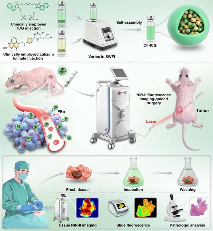
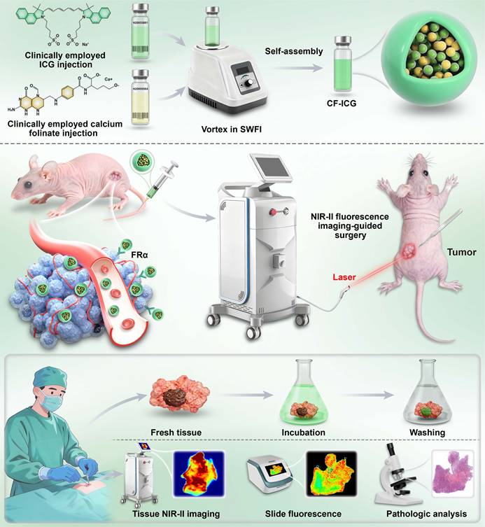
CF-ICG was prepared through a simple “green” synthesis method, as illustrated in Figure 1. To optimize the formulation ratio of CF to ICG, we monitored the Dh, polydispersity index (PDI) and ICG encapsulation efficiency of the assemblies at various CF-to-ICG mass ratios. Our results demonstrated that both the Dh and PDI reached a minimum at a CF-to-ICG mass ratio of 2:1, indicating that the nano-assemblies formed a highly uniform and monodisperse population with PDI < 0.2 (Figure S1A and S1B). Meanwhile, at this 2:1 ratio, the nanoparticles exhibited high stability with minimal variation in Dh during 72 h of storage in SWFI (Figure S1C). Additionally, the encapsulation efficiency of ICG reached ~88.75 ± 2.00% under this ratio (Figure S1D). Therefore, the 2:1 CF-to-ICG mass ratio was selected for all subsequent experiments. Routine physicochemical characterization revealed that CF-ICG formed a clear and transparent green solution, and TEM images demonstrated that the nano-assemblies were monodisperse and spherical (Figure 2A). As shown in Figure 2B, CF-ICG displayed prominent elemental signals of Ca, S, C, N, and O, which are the major constituents of CF and ICG. According to the quantitative elemental analysis based on EDS atomic fractions (Ca: 2.67% and S: 1.93%), a molar ratio of approximately 2.77:1 between CF and ICG was determined within the nano-assemblies (Table S1). Meanwhile, dynamic light scattering analysis indicated that CF-ICG possessed an average Dh of 203.5 ± 22.5 nm (Figure 2C). In contrast to the physical mixture of CF and ICG, CF-ICG exhibited an additional peak at 1081 cm-1 in Fourier transform infrared (FTIR) spectroscopy, suggesting the newly formed electrostatic interactions (Figure S2). Moreover, absorbance spectroscopy revealed that the absorption peak of CF-ICG (790 nm) exhibited a 10-nm red shift compared to both free ICG and the CF/ICG physical mixture (both at 780 nm) (Figure 2D). Furthermore, CF-ICG also demonstrated the first near-infrared (NIR-I) and NIR-II emission in SWFI (Figure 2E and 2F). Correspondingly, the NIR-I emission peak of CF-ICG (823 nm) was red-shifted by 4 nm relative to that of free ICG (819 nm). With free ICG serving as the standard reference (quantum yield ~2.9%), the relative NIR-I fluorescence quantum yield of CF-ICG was evaluated to be approximately 3.49 ± 0.23% (Figure S3). This slight enhancement suggested that the ICG molecules within the CF-ICG assembly may be appropriately spaced, thereby effectively suppressing aggregation-caused quenching.
Schematic illustration of CF-ICG fabrication and its application in NIR-II fluorescence imaging-guided breast cancer surgery. The top panel illustrates the preparation of CF-ICG via the self-assembly of clinically approved ICG injection and calcium folinate (CF) injection. The middle panel demonstrates the in vivo NIR-II imaging-guided evaluation of surgical margins in preclinical models. The bottom panel displays the clinical translation of CF-ICG-based ex vivo tissue incubation imaging for the intraoperative evaluation of surgical margins.
Characterization of CF-ICG and schematic illustration of its self-assembly mechanism. (A) Representative transmission electron microscopy (TEM) micrograph of CF-ICG and a digital photograph of the corresponding aqueous solution (Top-left inset). The scale bar represents 400 nm. (B-C) EDS element mapping (B) and hydrodynamic diameter (Dh) distribution (C) of CF-ICG. (D-F) Absorption (D), NIR-I (E), and NIR-II emission (F) spectra of ICG, CF, the physical mixture of CF and ICG (CF/ICG), and CF-ICG in sterile water for injection (SWFI). Scale bars represent 100 nm in (B). (G) Variation of Dh of CF-ICG during storage (0, 24, 48, and 72 h) in different media (PBS, 10% FBS, SWFI, and human plasma). (H) Representative TEM micrograph of CF-ICG during storage in SWFI. Scale bars represent 400 nm and 100 nm, respectively. (I) Variation of zeta potential of CF-ICG during storage in different media (PBS, 10% FBS, SWFI, and human plasma). (J-K) Variations of absorption spectra of ICG (J) and CF-ICG (K) in SWFI during storage. (L) Dh variations of CF-ICG after incubation with SDS, urea, and NaCl at different concentrations. (M) Self-assembly mechanism of CF-ICG. Quantum chemical calculations revealed salt bridges, cation-π interactions, and π-π stacking between CF and ICG, with a binding energy of ΔE = -168.14 kcal/mol. DPD simulations showed the CF/ICG self-assembly process in water over 0-200 ns (yellow represents ICG, and red represents CF). (N-Q) Quantitative analysis of potential energy (N), π-π stacking number (O), hydrogen bond number (P), and solvent-accessible surface area (SASA, Q) during the self-assembly process of CF-ICG.
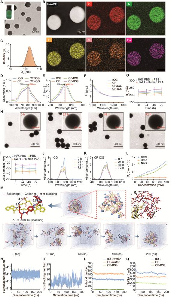
To assess the stability of the probe, the Dh, surface charge and morphological evolution of CF-ICG were monitored. The Dh and surface charge remained relatively stable in different media (PBS, 10% FBS, SWFI and human plasma) across the observation period (Figure 2G and 2I). TEM results also showed that the nanostructure maintained an intact and uniform spherical morphology throughout the storage period without noticeable aggregation or disassembly (Figure 2H). In addition, absorption spectral monitoring confirmed that the absorbance of CF-ICG decayed more slowly over time than that of ICG (Figure 2J and 2K). Furthermore, under continuous 808 nm laser irradiation, the fluorescence intensity of free ICG decreased significantly over time, while CF-ICG retained a higher proportion of its initial fluorescence intensity (Figure S4). These results confirmed the favorable physical and optical stability of CF-ICG, supporting its reliable performance in real-time surgical navigation.
To study the role of intermolecular forces in CF-ICG self-assembly, CF-ICG was treated with SDS, urea, or NaCl to disrupt hydrophobic interactions, hydrogen bonding, or electrostatic interactions, respectively. As the concentrations of NaCl and SDS increased, the Dh of CF-ICG gradually increased (Figure 2L), indicating that the disruption of electrostatic or hydrophobic interactions destabilized the structural integrity of the nano-assembly. In contrast, the Dh remained stable in the presence of urea. These results revealed that hydrophobic and electrostatic interactions were the main forces driving CF-ICG self-assembly.
Through quantum chemical calculations and DPD simulations, the mechanism of supramolecular self-assembly was further clarified. As shown in Figure 2M, CF and ICG formed a stable complex mainly through salt bridges, cation-π interactions and π-π stacking, and the calculated binding energy (ΔE) of the complex was -168.14 kcal/mol. Snapshots from DPD simulations revealed that randomly dispersed CF and ICG molecules progressively assembled into compact nano-aggregates during the 200 ns simulation. Moreover, the total potential energy of the system stayed relatively stable, and the number of π-π stacking interactions between CF and ICG gradually increased. At the same time, hydrogen bonding within the CF-ICG complex remained negligible (Figure 2N-P). Furthermore, the solvent-accessible surface area (SASA) of CF-ICG was significantly lower than that of ICG or CF alone (Figure 2Q), indicating that CF-ICG formed a compact and tightly aggregated structure. These simulation results further confirmed that electrostatic interactions and π-π stacking are the primary driving forces for CF-ICG self-assembly.
FRα-targeted cellular uptake of CF-ICG in vitro
The FRα affinity of CF-ICG was first evaluated by incubating MDA-MB-231 cells (high FRα expression) with escalating concentrations of the nano-probe, followed by quantification of cell-associated fluorescence. CF-ICG exhibited a high binding avidity toward cellular FRα, with a calculated dissociation constant (Kd) of ~87.74 nM (Figure S5). FRα-mediated internalization of CF-ICG was subsequently evaluated. As illustrated in Figure 3A, MDA-MB-231 cells actively internalized CF-ICG into the cytoplasm, which was markedly suppressed upon pre-treatment with an excess of free FA to block FRα, suggesting that cellular internalization of CF-ICG is specifically mediated by FRα targeting. Consistent with this microscopic observation, MFI analysis showed a similar trend (Figure 3B). Quantitative FCM analysis further confirmed that the uptake of CF-ICG by tumor cells was significantly inhibited after FA blocking (Figure 3C). In addition, prolonging the incubation duration (Figure 3D-F) and increasing the probe concentration (Figure 3G) both led to an enhancement in the fluorescence signal within the MDA-MB-231 cell line. A similar time- and concentration- dependent internalization was also displayed in the 4T1 cell line (Figure 3H and 3I). Besides, the CF-ICG uptake was markedly higher in MDA-MB-231 cells than in normal MCF-10A mammary epithelial cells and was substantially higher than ICG uptake in MDA-MB-231 cells (Figure S6). These findings validated that CF-ICG possesses highly efficient FRα-mediated targeting capabilities in breast cancer cells.
FRα-targeted cellular uptake of CF-ICG in vitro. (A-B) Representative upright fluorescence micrographs illustrating the CF-ICG uptake in MDA-MB-231 cells with/without FA blocking (A) and the corresponding mean fluorescence intensity (MFI) analysis (B, n = 4). (C) Flow cytometric (FCM) evaluation of the cellular uptake of CF-ICG with/without FA blocking in MDA-MB-231 cells. (D-E) Representative micrographs illustrating the CF-ICG uptake in MDA-MB-231 cells at different times (2, 4, and 6 h, D) and the corresponding quantitative fluorescence analysis (E, n = 4). (F-G) Cellular uptake evaluation of CF-ICG in MDA-MB-231 cells at different times (2, 4, and 6 h, F) and concentrations (0, 0.5, 1, 2, and 4 μg/mL equivalent ICG, G) via FCM analysis. (H-I) Cellular uptake evaluation of CF-ICG in 4T1 cells at various incubation times (H) and concentrations (I) via FCM analysis. (J) Cell viability of different cell lines after treatment with different concentrations of CF-ICG (0, 0.5, 1, 2, 4, 8, and 16 μg/mL equivalent ICG) as assessed by the CCK-8 assay (n = 5). All scale bars represent 50 μm. (For FCM, excitation: 638 nm, collection: 780 nm; for upright fluorescence microscopy, excitation was provided by a Leica EL6000 external light source, and the corresponding emission signals were collected at 771-829 nm for ICG, 430-490 nm for DAPI, and 570-590 nm for DiI; mean ± s.d.).
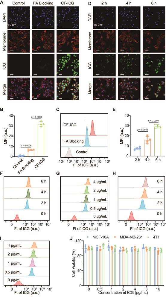
The biocompatibility of CF-ICG was evaluated across various cell lines using the CCK-8 assay. As illustrated in Figure 3J, CF-ICG showed negligible cytotoxicity in normal mammary epithelial cells and breast cancer cells, even at a high ICG-equivalent concentration of 16 μg/mL. The cell viability remained above 90% across all tested concentrations, demonstrating the favorable in vitro biocompatibility of the nano-assemblies.
In vivo NIR-II fluorescence imaging and biodistribution
To validate the in vivo tumor-targeting capability of CF-ICG, CF-ICG or free ICG was intravenously administered to MDA-MB-231-Luc tumor-bearing mice. For the blocking study, excess FA was injected before CF-ICG administration to occupy FRα binding sites. NIR-II imaging revealed that in both the free ICG and FA-blocking groups, prominent fluorescence signals were detected primarily in the liver, with only negligible accumulation observed in the tumor. In contrast, the CF-ICG group exhibited the most robust and persistent fluorescence signal within the tumor, which remained detectable even at 72 h post-injection (Figure 4A and 4B). Furthermore, the signal-to-noise ratio (SNR) in the CF-ICG group at 36 h post-injection demonstrated a significant enhancement over that of the ICG and FA-blocking groups, highlighting the enhanced tumor-specific accumulation of CF-ICG (Figure 4C).
In vivo NIR-II fluorescence imaging and biodistribution of CF-ICG. (A) Representative in vivo NIR-II images of mice at 3, 6, 12, 24, 36, 48, and 72 h after intravenous administration of ICG, CF-ICG or CF-ICG with excess FA pre-blocking. (B) Tumor MFI quantification during NIR-II imaging. (C) SNR analysis at 36 h post-injection. (D-E) Representative ex vivo NIR-II images (D) and corresponding MFI quantification (E) of major organs and tumors at 72 h post-injection. NIR-II images were acquired using a 1000 nm LP filter under 808 nm excitation. (mean ± s.d., n = 4).
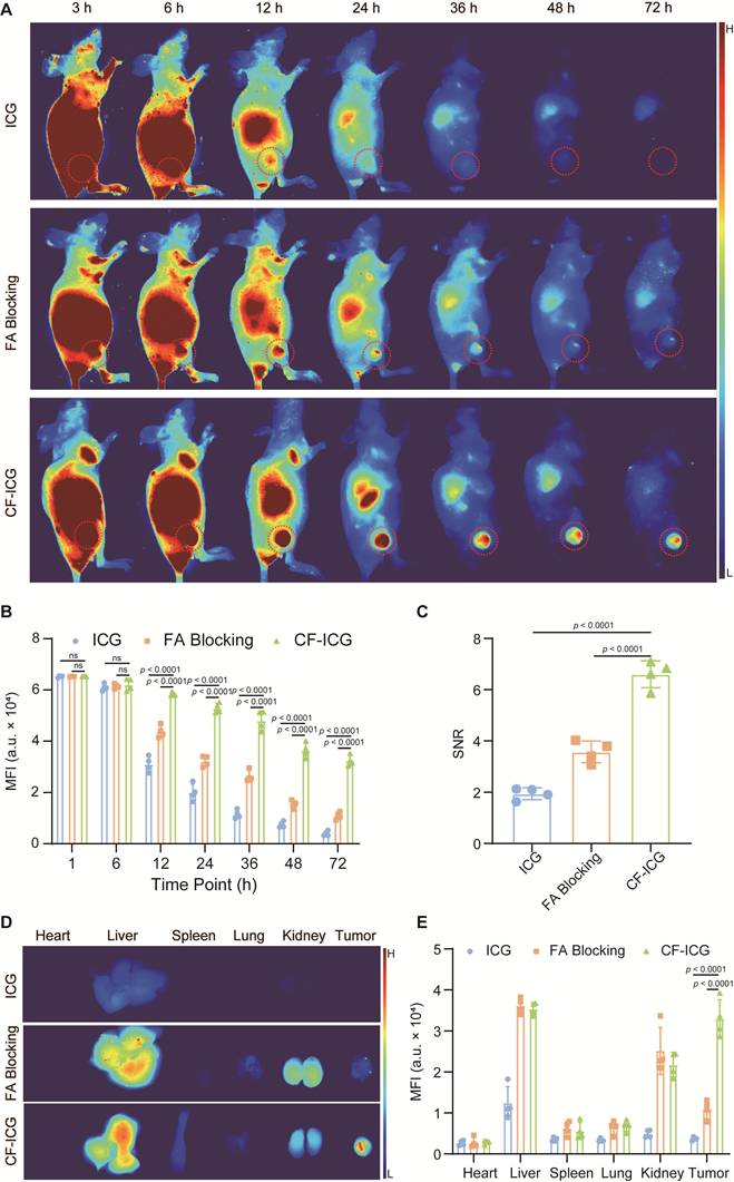
To further evaluate the systemic biodistribution profile of CF-ICG, major organs and tumors were harvested at 72 h post-administration. Ex vivo NIR-II imaging demonstrated that the tumors in the CF-ICG group possessed a significantly higher MFI compared to those in the other groups (Figure 4D). To precisely quantify the tissue distribution, inductively coupled plasma mass spectrometry (ICP-MS) was performed, revealing that the tumor accumulation of the nanoprobes reached approximately 9.66 ± 1.15 %ID/g at 36 h post-administration (Figure S7). Combined fluorescence imaging and ICP-MS results further verified predominant probe enrichment in tumors and livers, with only low-level accumulation detected in other major organs (Figure 4E and S7).
In vivo NIR-II fluorescence-guided surgery in the MMTV-PyVT transgenic mouse model
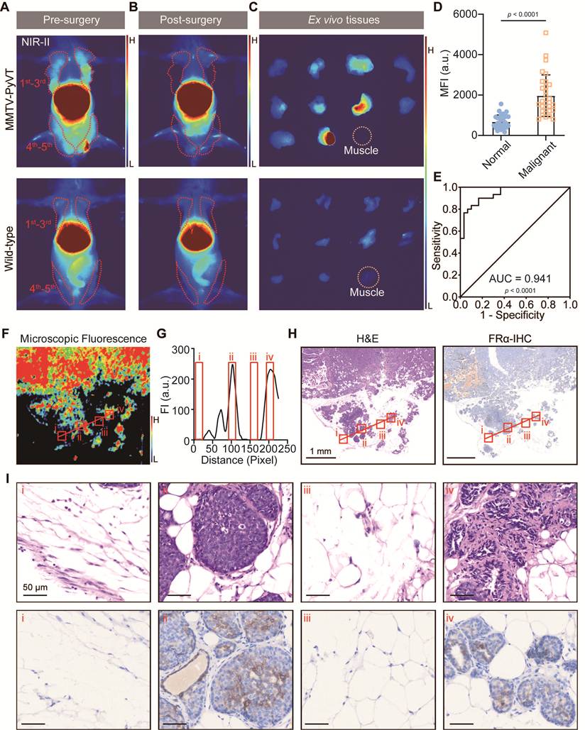
To assess the diagnostic efficacy of CF-ICG, NIR-II imaging was conducted on an MMTV-PyVT transgenic model with spontaneous breast cancer. In vivo NIR-II imaging revealed intense fluorescence signals in malignant mammary glands from transgenic mice. In contrast, normal mammary tissues from wild-type mice exhibited only minimal fluorescence (Figure 5A). After resection under NIR-II fluorescence imaging guidance (Figure 5B), the ex vivo malignant tissues also demonstrated an approximately 2.9-fold higher MFI compared to normal controls (Figure 5C and 5D). The diagnostic accuracy was further validated by an ROC curve, yielding an area under the curve (AUC) of 0.941 (95% CI: 0.887-0.995; p < 0.0001; Figure 5E). Furthermore, quantitative analysis along the line of interest (LOI) in Figure 5F and 5G revealed that regions ii and iv exhibited superior fluorescence intensity compared to regions i and iii. This fluorescence contrast was corroborated by histopathological assessment: H&E and FRα-IHC staining confirmed that the high-fluorescence regions (ii and iv) contained malignant lesions with high FRα expression, whereas the low-fluorescence regions (i and iii) consisted of normal tissues with low FRα expression (Figure 5H and 5I).
NIR-II fluorescence-guided surgery in the MMTV-PyVT transgenic mouse model. (A-C) Representative in vivo NIR-II images of an MMTV-PyVT mouse (top) and a wild-type mouse (bottom) before (A) and after (B) fluorescence-guided surgery, as well as the corresponding ex vivo NIR-II images of resected mammary glands (C). The red dotted regions in (A and B) represent the positions of mammary glands, and the yellow dotted regions in (C) represent the muscle tissues. (D-E) Quantification of the MFI (D) of ex vivo normal and malignant mammary glands and the ROC curve (E) for identifying malignancies. (F-G) Representative NIR fluorescence slide scanning result (excitation: 785 nm, collection: 812-832 nm) of the malignant tissue (F) and the corresponding fluorescence intensity (G) along the line of interest (LOI, the red line in F). (H) H&E and FRα immunohistochemistry (IHC) results of the tissue from (F). Scale bars represent 1 mm. (I) H&E and FRα-IHC results of the tissues marked with red squares (i-iv) along the LOI in (F and H). Scale bars represent 50 μm. NIR-II images were acquired using a 1000 nm LP filter under 808 nm excitation. (mean ± s.d., n = 30).
In vivo NIR-II fluorescence-guided surgery in MDA-MB-231-Luc tumor-bearing mice
In order to evaluate the feasibility of CF-ICG to visualize tiny malignant foci during surgery, a multifocal microtumor model was established. As illustrated in Figure 6A and 6B, NIR-II fluorescence signals from microtumors demonstrated a high degree of spatial consistency with bioluminescence signals. Among the 37 microtumors identified by bioluminescence imaging, 35 lesions (94.6%) were detected and removed under intraoperative NIR-II fluorescence guidance. The resected tissues with NIR-II signal were confirmed by pathological examination, revealing that the tumor margins could be clearly delineated under real-time surgical navigation (Figure 6C and 6D). Moreover, the smallest detected lesions were less than 2 mm in diameter, indicating that CF-ICG-based NIR-II imaging could effectively identify microtumors and guide precise resection during surgery.
NIR-II fluorescence-guided surgery in MDA-MB-231-Luc tumor-bearing mice. (A-B) Representative preoperative bioluminescent (BL, A), intraoperative and postoperative in vivo NIR-II images (B) of a multifocal microtumor mouse model. The red dotted regions represent the tumors. (C-D) Ex vivo NIR-II images of resected tumor and muscle tissues (C) and the corresponding H&E-stained histological results (D). The yellow dotted region in (C) represents the muscle tissue, while the red dotted regions in (D) outline the border between the tumor and healthy tissues. Scale bars represent 100 μm. (E) Representative preoperative, intraoperative, and postoperative bioluminescent (BL) and NIR-II images of MDA-MB-231-Luc tumor-bearing mice during NIR-II fluorescence-guided surgery, along with the corresponding ex vivo NIR-II images, H&E-stained images and NIR fluorescence slide scanning images of resected tissues including the primary tumor (P1), residual tumor (P2) and muscle tissue. The red dotted regions and the red arrow represent the tumors, and the yellow dotted region represents the muscle tissue. NIR fluorescence slide scanning was performed under 785 nm excitation with emission collection at 812-832 nm. (F) Preoperative and postoperative BL images of mice undergoing NIR-II fluorescence-guided surgery. (G) Kaplan-Meier survival analysis of the NIR-II fluorescence-guided and white light-guided groups. NIR-II images were acquired using a 1000 nm LP filter under 808 nm excitation. (mean ± s.d., n = 5).
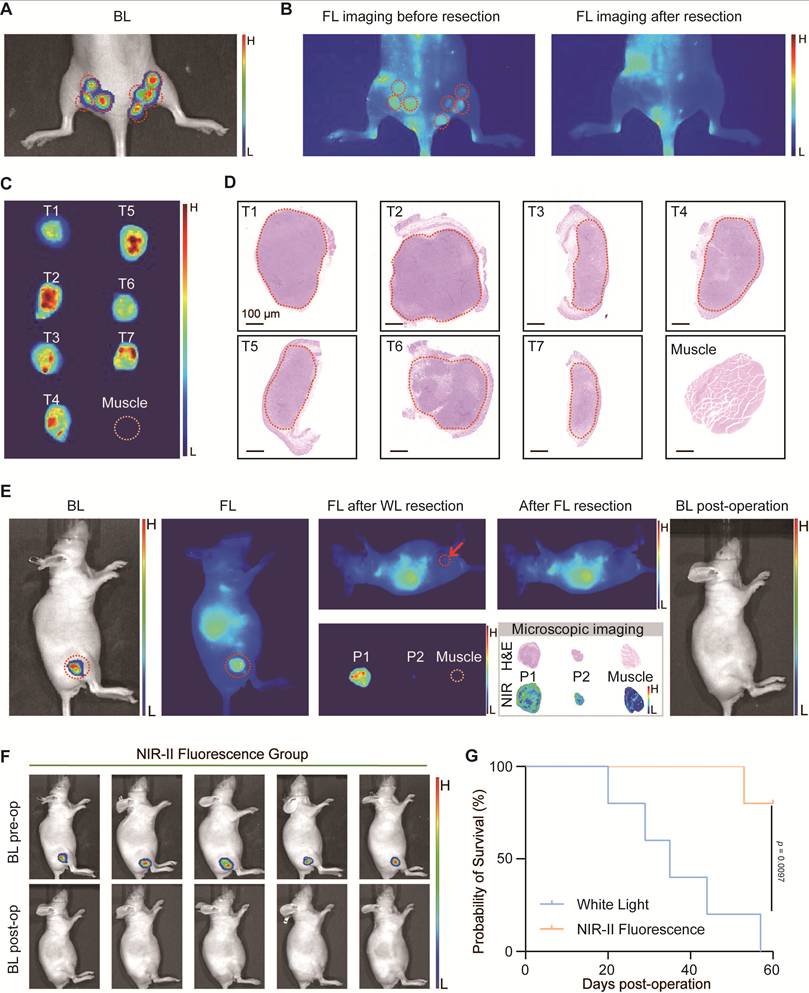
To further evaluate its surgical applicability, NIR-II fluorescence-guided surgery was performed in mice bearing a single tumor to better mimic clinical tumor resection (Movie S1). As shown in Figure 6E, a small residual tumor that was missed under white light, with a diameter of approximately 0.9 mm, was detected by CF-ICG-based NIR-II imaging. This finding was further confirmed by H&E staining. At 15 days post-surgery, local recurrence was evaluated using bioluminescence imaging. In the NIR-II fluorescence-guided group, no tumor recurrence with bioluminescence signals was observed postoperatively (Figure 6F). However, all five mice (5/5, 100%) in the white light-guided group exhibited recurrent tumor bioluminescence signals (Figure S8). Kaplan-Meier survival analysis further revealed that CF-ICG-guided surgery facilitated a more thorough tumor resection and significantly prolonged overall survival relative to the white-light controls (Figure 6G), supporting the potential value of CF-ICG in real-time surgical navigation.
Ex vivo incubation and NIR-II fluorescence imaging of fresh patient-derived breast cancer tissues
To facilitate intraoperative pathological diagnosis and margin assessment, an ex vivo incubation protocol using CF-ICG was evaluated on fresh specimens to differentiate tissue properties and delineate malignancy boundaries. Paired breast cancer and adjacent para-cancer tissues harvested from 11 TNBC patients were divided into CF-ICG and free ICG incubation groups.
As demonstrated in Figure 7A and 7B, CF-ICG exhibited a superior ability to discriminate breast cancer tissues from para-cancer tissues, with a significantly elevated cancer-to-para-cancer fluorescence ratio compared to the free ICG group. Meanwhile, cancer tissues in the CF-ICG group exhibited markedly stronger NIR-II fluorescence relative to matched para-cancer counterparts (Figure 7C), yielding an AUC of approximately 0.926 (95% CI: 0.814-1.000; p = 0.0007; Figure 7D). In addition, as shown in Figure 7E, the cancer tissue with high FRα expression demonstrated higher fluorescence than the low-FRα para-cancer and the low-FRα cancer tissue. Owing to the intrinsic heterogeneity of breast cancer, rare FRα-low cancer tissues with diminished fluorescence occasionally led to false-negative outcomes.
Ex vivo incubation and NIR-II fluorescence imaging of fresh patient-derived breast cancer tissues. (A-B) Ex vivo NIR-II fluorescence and white-light images of freshly resected cancer and para-cancer tissues after incubation with ICG or CF-ICG (A) and corresponding cancer-to-para-cancer fluorescence ratios (B). (C-D) Quantification of the MFI (C) of cancer and para-cancer tissues after incubation with CF-ICG, and the corresponding ROC curve (D) for identifying malignancies. (E) Representative white-light and NIR-II fluorescence images of para-cancer, FRα-high and FRα-low cancer tissues after incubation with CF-ICG, along with the H&E staining, FRα-IHC staining and NIR fluorescence slide scanning results. Scale bars represent 1 mm. (F-G) Representative NIR fluorescence slide scanning result (excitation: 785 nm, collection: 812-832 nm) of cancer tissue (F) and corresponding fluorescence intensity (G) along the LOI (the red line in F). (H) H&E (left) and FRα-IHC (right) results of the tissue from (F). Scale bars represent 1mm. (I) H&E (top) and FRα-IHC (bottom) results of the tissues marked with red squares (i-iv) along the LOI in (F and H). Scale bars represent 50 μm. NIR-II images were acquired using a 1000 nm LP filter under 808 nm excitation. (mean ± s.d., n = 11).
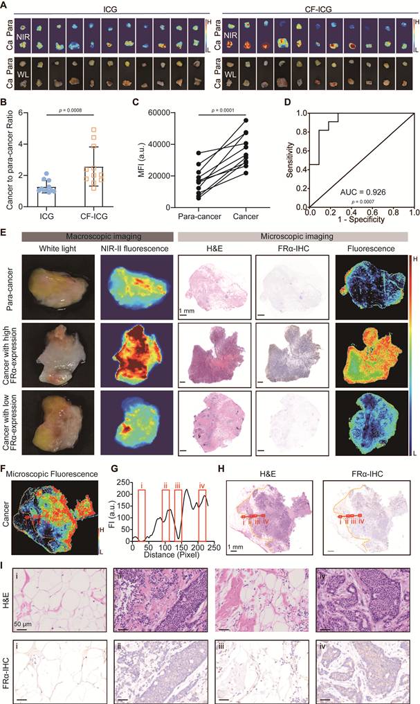
Furthermore, we analyzed the fluorescence distribution along the LOI within the cancer tissue (Figure 7F). According to Figure 7F-I, the fluorescence signal clearly delineated the boundaries of the cancer, with regions containing malignant cells (ii and iv) exhibiting significantly stronger fluorescence and higher FRα expression compared to normal tissue areas (i and iii), further supporting the feasibility of CF-ICG for targeted imaging of breast cancer.
Biocompatibility and systemic toxicity assessment of CF-ICG
To comprehensively evaluate the systemic biocompatibility of CF-ICG, safety assessments were performed. The hemolysis assay verified that CF-ICG did not induce erythrocyte rupture, with the hemolysis rate well below the 5% standard threshold, even at high concentrations (Figure 8A and 8B).
Biocompatibility and systemic toxicity evaluation of CF-ICG. (A-B) Digital photographs (A) and the corresponding quantification of the hemolysis ratios (B) of red blood cells after incubation with CF-ICG at different concentrations (2, 4, 8, and 16 μg/mL equivalent ICG). (C) Changes in body weight of BALB/c mice administered PBS or CF-ICG over a 28-day period. (D) Representative H&E staining results of major organs collected from BALB/c mice at 1, 7, and 28 days after CF-ICG or PBS administration. Scale bars represent 200 μm. (E) Hematological and serum biochemical parameters of mice at 1, 7, and 28 days after CF-ICG or PBS administration. (n = 4, mean ± s.d.).
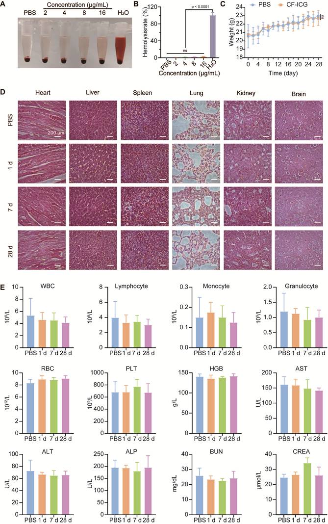
For in vivo safety evaluation, body weight monitoring showed no obvious difference between the CF-ICG and PBS groups (Figure 8C). Following CF-ICG administration, blood samples and major organs were collected at 1, 7, and 28 days for subsequent analysis. Histopathological examination of major organs revealed no apparent pathological abnormalities, including necrosis, inflammation, or tissue damage, in either the CF-ICG or PBS group (Figure 8D). Hematological and serum biochemical parameters remained within normal physiological ranges (Figure 8E). These results suggested that CF-ICG possesses favorable biocompatibility and a satisfactory safety profile for in vivo applications in surgical navigation.
Discussion
Fluorescence-guided surgery has been increasingly adopted to enhance intraoperative tumor visualization, facilitating tumor detection and reducing the risk of positive margins [34]. Herein, we developed a carrier-free PDNA through the spontaneous Ca2+-driven self-assembly between ICG, a clinically approved NIR fluorophore, and CF, a clinically available folate derivative. This design integrates the superior NIR-II fluorescence characteristics of ICG with the inherent FRα-targeting capability of CF, providing high-contrast and real-time tumor visualization in TNBC.
Although PDNAs and ICG-based self-assembled probes have advanced rapidly in recent years [38-40], most current formulations still rely on raw bulk drug powders, toxic organic solvents, surfactants, or non-clinical excipients, which may significantly impede their clinical translation [35-37]. In addition, some systems depend on multi-step fabrication procedures or synthetic targeting ligands to achieve tumor specificity, increasing structural complexity and raising additional biosafety concerns, which compromises the original design intention of PDNAs [41-43]. In contrast, our approach overcomes these limitations by utilizing two commercially available clinical intravenous injections as the sole starting materials. CF-ICG is prepared in SWFI through a simple and “green” physical process, which can avoid carrier-related toxicity and organic solvent residues. In addition, the assembly is mainly driven by π-π stacking and electrostatic interactions, which support the structural stability of CF-ICG and thereby fulfill an essential prerequisite for reliable surgical guidance. Importantly, a key advantage of this system lies in its multifunctional collaborative assembly strategy: CF serves as both a Ca2+ assembly driver (self-driven) and an inherent FRα-targeted ligand (self-targeted), while ICG provides stable NIR-II imaging characteristics. Therefore, this assembly strategy avoids additional chemical modification and achieves multifunctional integration, which is rarely realized in conventional PDNAs.
Because FRα is selectively upregulated in a variety of malignant tissues [44], it has become an important molecular target for tumor-specific imaging and treatment, providing a feasible strategy for precision oncology applications [45]. The NIR fluorescence probe pafolacianine (OTL38) has been approved by the FDA and can be used to identify ovarian cancer lesions during surgery [46]. However, pafolacianine mainly emits in the NIR-I window (700-900 nm), where high autofluorescence, limited penetration depth, and relatively low SNR may affect imaging performance [47]. In contrast, CF-ICG enables NIR-II (1000-1700 nm) imaging, which can provide higher imaging resolution, enhanced penetration depth, and reduced background signals [48-50]. Additionally, by combining specific FRα targeting with the optical advantages of NIR-II, CF-ICG can visualize tumor lesions with higher accuracy and is particularly suitable for detecting submillimeter lesions, which is crucial for surgical navigation. Therefore, this method achieved accurate identification of malignant foci in the preclinical model (AUC = 0.941) and enabled more complete removal of malignant tissues than traditional white light-guided surgery. In summary, the NIR-II fluorescence characteristics and FRα-targeting ability of CF-ICG support its potential application in precise margin evaluation during FRα-positive breast cancer surgery.
Moreover, the development of efficient and reliable verification methods is crucial for evaluating the clinical translation potential of new imaging agents. Therefore, we established an ex vivo CF-ICG incubation method to rapidly assess fresh tissues obtained during breast cancer surgery. According to our results, this method effectively distinguished between breast cancer and adjacent para-cancerous tissues, with an AUC value of 0.926. The whole process takes only 12 min (7 min for incubation and 5 min for rinsing), which is expected to simplify intraoperative margin evaluation and reduce the time burden of traditional techniques such as frozen section pathology. Future research will focus on pharmacokinetics, biodistribution, and the compatibility of CF-ICG with clinical imaging systems to further evaluate its clinical translational potential.
Several limitations of this study should be noted. First, although FRα is overexpressed in many TNBC cases, its heterogeneous expression may lead to false-negative results in tumors with low FRα levels. Future studies may consider multi-target imaging strategies to address this limitation. Second, CF-ICG showed nonspecific retention in the liver and other reticuloendothelial system (RES) organs, indicating that further refinement of surface properties may be required to minimize off-target accumulation. Third, the clinical validation was based on a relatively limited number of human specimens. Larger cohorts and multicenter studies will be required to establish reliable diagnostic thresholds for standardized surgical decision-making. In addition, although biocompatibility was preliminarily evaluated in animal models, the long-term safety of CF-ICG remains to be further investigated. Furthermore, given the reported photothermal and photodynamic effects of ICG, the therapeutic potential of CF-ICG may be explored in future studies.
Conclusions
In summary, we developed CF-ICG as a carrier-free NIR-II fluorescent probe through a drug-repurposing strategy and a “green” self-assembly process using clinically available components. CF-ICG shows excellent FRα-mediated tumor targeting ability and enables high-contrast tumor margin delineation as well as precise pathological evaluation in preclinical models and patient-derived tissues. This novel strategy provides a preliminary foundation for advancing precision BCS and may contribute to improved surgical outcomes in patients with TNBC.
Abbreviations
BCS: breast-conserving surgery; PDNAs: pure-drug nano-assemblies; CF: calcium folinate; NIR-II: second near-infrared; TNBC: triple-negative breast cancer; ICG: indocyanine green; FRα: folate receptor alpha; SNR: signal-to-noise ratio; SWFI: sterile water for injection; MWCO: molecular weight cutoff; ICP-MS: inductively coupled plasma mass spectrometry; DPD: dissipative particle dynamics; FCM: flow cytometry; FA: folic acid; TEM: transmission electron microscopy; Dh: hydrodynamic diameter; SASA: solvent-accessible surface area; MFI: mean fluorescence intensity; LP: long-pass; FTIR: Fourier transform infrared; RES: reticuloendothelial system; ROC: receiver operating characteristic; LOI: line of interest; H&E: hematoxylin and eosin; IHC: immunohistochemistry; AUC: area under the curve; NIR-I: first near-infrared; SD: standard deviation.
Supplementary Material
Supplementary methods, figures and table.

Supplementary video.
Acknowledgements
Funding
This work was supported by the Natural Science Foundation of Fujian Province of China (Grant No.2023J05273), Xiamen Natural Science Foundation of China (Grant No.3502Z202372071), Fujian Provincial Health Commission science and technology plan project youth research project (Grant No.2022QNB012), Yunnan Fundamental Research Project (Grant No.202401AS070029), Natural Science Foundation of China (No. 62475264, No. 82303078), Fujian Provincial Natural Science Foundation of China (No. 2025Y0056), XMIREM autonomously deployment project (No. 2026CX01), and the Open Research Fund of Key Laboratory of Biological Nanotechnology NHC (No. NBT2025N02).
Authorship contribution statement
Conceptualization: Ruiqin Yang, Kangliang Lou, Shuangyan Bao, Hairong Zhao, and Yang Li; Methodology: Ruiqin Yang, Kangliang Lou, Shuangyan Bao, Chen Yang, Yating Gai, Shuanglong Chen, and Qingmo Yang; Formal analysis and investigation: Ruiqin Yang, Kangliang Lou, Shuangyan Bao, Chen Yang, Yating Gai, Hairong Zhao, and Yang Li; Writing - original draft preparation: Ruiqin Yang, Kangliang Lou and Shuangyan Bao; Writing - review and editing: Hairong Zhao and Yang Li; Funding acquisition: Ruiqin Yang and Hairong Zhao; Resources: Ruiqin Yang, Shuanglong Chen, Qingmo Yang, Siqi Qiu, Yilong Lin, Hairong Zhao, and Yang Li; Supervision: Yang Li.
Data availability
All data that support the findings are included within the paper. Other source data related to this study are available from the corresponding authors upon reasonable request.
Artificial Intelligence (AI) tools
The authors acknowledge the use of ChatGPT (OpenAI) for language polishing and wording refinement of selected parts of the Introduction and Discussion sections. No AI tools were used for data collection and analysis, image generation, figure preparation, interpretation of results, or conclusion generation. The authors carefully reviewed, edited, and approved all AI-assisted text and take full responsibility for the accuracy, integrity, and originality of the manuscript.
Competing Interests
The authors have declared that no competing interest exists.
References
1. Fancellu A, Houssami N, Sanna V, Porcu A, Ninniri C, Marinovich ML. Outcomes after breast-conserving surgery or mastectomy in patients with triple-negative breast cancer: meta-analysis. Br J Surg. 2021;108:760-8
2. Buchholz TA, Somerfield MR, Griggs JJ, El-Eid S, Hammond ME, Lyman GH. et al. Margins for breast-conserving surgery with whole-breast irradiation in stage I and II invasive breast cancer: American Society of Clinical Oncology endorsement of the Society of Surgical Oncology/American Society for Radiation Oncology consensus guideline. J Clin Oncol. 2014;32:1502-6
3. Tamanuki T, Namura M, Aoyagi T, Shimizu S, Suwa T, Matsuzaki H. Effect of Intraoperative Imprint Cytology Followed by Frozen Section on Margin Assessment in Breast-Conserving Surgery. Ann Surg Oncol. 2021;28:1338-46
4. Laws A, Brar MS, Bouchard-Fortier A, Leong B, Quan ML. Intraoperative Margin Assessment in Wire-Localized Breast-Conserving Surgery for Invasive Cancer: A Population-Level Comparison of Techniques. Ann Surg Oncol. 2016;23:3290-6
5. St John ER, Al-Khudairi R, Ashrafian H, Athanasiou T, Takats Z, Hadjiminas DJ. et al. Diagnostic Accuracy of Intraoperative Techniques for Margin Assessment in Breast Cancer Surgery: A Meta-analysis. Ann Surg. 2017;265:300-10
6. Pradipta AR, Tanei T, Morimoto K, Shimazu K, Noguchi S, Tanaka K. Emerging technologies for real-time intraoperative margin assessment in future breast-conserving surgery. Adv Sci. 2020;7:1901519
7. Voskuil FJ, Vonk J, van der Vegt B, Kruijff S, Ntziachristos V, van der Zaag PJ. et al. Intraoperative imaging in pathology-assisted surgery. Nat Biomed Eng. 2022;6:503-14
8. Rowe SP, Pomper MG. Molecular imaging in oncology: Current impact and future directions. CA Cancer J Clin. 2022;72:333-52
9. Yang Y, Chen Y, Pei P, Fan Y, Wang S, Zhang H. et al. Fluorescence-amplified nanocrystals in the second near-infrared window for in vivo real-time dynamic multiplexed imaging. Nat Nanotechnol. 2023;18:1195-204
10. Chen QY, Xie JW, Zhong Q, Wang JB, Lin JX, Lu J. et al. Safety and Efficacy of Indocyanine Green Tracer-Guided Lymph Node Dissection During Laparoscopic Radical Gastrectomy in Patients with Gastric Cancer: A Randomized Clinical Trial. JAMA Surg. 2020;155:300-11
11. Doshi CP, Wozniak A, Quek ML. Near-infrared Fluorescence Imaging of Ureters with Intravenous Indocyanine Green During Radical Cystectomy to Prevent Ureteroenteric Anastomotic Strictures. Urology. 2020;144:220-4
12. Dip F, LoMenzo E, Sarotto L, Phillips E, Todeschini H, Nahmod M. et al. Randomized Trial of Near-infrared Incisionless Fluorescent Cholangiography. Ann Surg. 2019;270:992-9
13. Vidal Fortuny J, Belfontali V, Sadowski SM, Karenovics W, Guigard S, Triponez F. Parathyroid gland angiography with indocyanine green fluorescence to predict parathyroid function after thyroid surgery. Br J Surg. 2016;103:537-43
14. Antaris AL, Chen H, Diao S, Ma Z, Zhang Z, Zhu S. et al. A high quantum yield molecule-protein complex fluorophore for near-infrared II imaging. Nat Commun. 2017;8:15269
15. Carr JA, Franke D, Caram JR, Perkinson CF, Saif M, Askoxylakis V. et al. Shortwave infrared fluorescence imaging with the clinically approved near-infrared dye indocyanine green. Proc Natl Acad Sci U S A. 2018;115:4465-70
16. Hu Z, Fang C, Li B, Zhang Z, Cao C, Cai M. et al. First-in-human liver-tumour surgery guided by multispectral fluorescence imaging in the visible and near-infrared-I/II windows. Nat Biomed Eng. 2019;4:259-71
17. Cao C, Deng S, Wang B, Shi X, Ge L, Qiu M. et al. Intraoperative near-infrared II window fluorescence imaging-assisted nephron-sparing surgery for complete resection of cystic renal masses. Clin Transl Med. 2021;11:e604
18. Wang C, Wang Z, Zhao T, Li Y, Huang G, Sumer BD. et al. Optical molecular imaging for tumor detection and image-guided surgery. Biomaterials. 2018;157:62-75
19. Gonzalez T, Muminovic M, Nano O, Vulfovich M. Folate Receptor Alpha-A Novel Approach to Cancer Therapy. Int J Mol Sci. 2024;25:1046
20. Fernández M, Javaid F, Chudasama V. Advances in targeting the folate receptor in the treatment/imaging of cancers. Chem Sci. 2018;9:790-810
21. Rana A, Bhatnagar S. Advancements in folate receptor targeting for anti-cancer therapy: A small molecule-drug conjugate approach. Bioorg Chem. 2021;112:104946
22. Tummers QR, Hoogstins CE, Gaarenstroom KN, de Kroon CD, van Poelgeest MI, Vuyk J. et al. Intraoperative imaging of folate receptor alpha positive ovarian and breast cancer using the tumor specific agent EC17. Oncotarget. 2016;7:32144-55
23. Zhang X, Li N, Zhang S, Sun B, Chen Q, He Z. et al. Emerging carrier-free nanosystems based on molecular self-assembly of pure drugs for cancer therapy. Med Res Rev. 2020;40:1754-75
24. Fu S, Li G, Zang W, Zhou X, Shi K, Zhai Y. Pure drug nano-assemblies: A facile carrier-free nanoplatform for efficient cancer therapy. Acta Pharm Sin B. 2022;12:92-106
25. Liu HW, Chen L, Xu C, Li Z, Zhang H, Zhang XB. et al. Recent progresses in small-molecule enzymatic fluorescent probes for cancer imaging. Chem Soc Rev. 2018;47:7140-80
26. Li Z, Liang PZ, Ren TB, Yuan L, Zhang XB. Orderly Self-Assembly of Organic Fluorophores for Sensing and Imaging. Angew Chem Int Ed Engl. 2023;62:e202305742
27. Wang R, Xi T, Li K, Wang C, Gao X, Yang H. et al. Self-Assembled Fluorescent Peptide Nanoprobes for Disease Diagnosis. Small Methods. 2025;9:e2500518
28. Xue K, Wei F, Lin J, Tian H, Zhu F, Li Y. et al. Tumor acidity-responsive carrier-free nanodrugs based on targeting activation via ICG-templated assembly for NIR-II imaging-guided photothermal-chemotherapy. Biomater Sci. 2021;9:1008-19
29. Zou R, Wang Q, Wu J, Wu J, Schmuck C, Tian H. Peptide self-assembly triggered by metal ions. Chem Soc Rev. 2015;44:5200-19
30. Salembier R, De Haes C, Demeyere K, Van Laere S, Cervenkova L, Van Renne N. et al. Tractable mouse TNBC models capture the heterogeneous tumor immune microenvironment and adaptation to PD-L1 blockade. NPJ Breast Cancer. 2025;12:15
31. Lehmann BD, Colaprico A, Silva TC, Chen J, An H, Ban Y. et al. Multi-omics analysis identifies therapeutic vulnerabilities in triple-negative breast cancer subtypes. Nat Commun. 2021;12:6276
32. Wei M, Bai J, Shen X, Lou K, Gao Y, Lv R. et al. Glutathione-Exhausting Nanoprobes for NIR-II Fluorescence Imaging-Guided Surgery and Boosting Radiation Therapy Efficacy via Ferroptosis in Breast Cancer. ACS nano. 2023;17:11345-61
33. Youden WJ. Index for rating diagnostic tests. Cancer. 1950;3:32-5
34. Mieog JSD, Achterberg FB, Zlitni A, Hutteman M, Burggraaf J, Swijnenburg R-J. et al. Fundamentals and developments in fluorescence-guided cancer surgery. Nat Rev Clin Oncol. 2022;19:9-22
35. Chauhan N, Cabrera M, Chowdhury P. et al. Indocyanine Green-based Glow Nanoparticles Probe for Cancer Imaging. Nanotheranostics. 2023;7:353-67
36. Lu Z, Wang T, Xu X, Hu Q, Wang L, Jin Y. Carrier-free nano-integrated strategy of phototherapy and chemotherapy for synergetic AKT targeted therapy to inhibit cancer metastasis and recurrence. J Nanobiotechnology. 2025;23:520
37. Gao YY, Lou KL, Lin LL, Li CX, Lin SJ, Chen YX. et al. A NIR-Ⅱ fluorescent probe for real-time visualization and early assessment of responses to CDK4/6 inhibitors in breast cancer. Cancer Lett. 2026;646:218429
38. Fang F, Chen X. Carrier-Free Nanodrugs: From Bench to Bedside. ACS nano. 2024;18:23827-41
39. Karaosmanoglu S, Zhou M, Shi B, Zhang X, Williams GR, Chen X. Carrier-free nanodrugs for safe and effective cancer treatment. J Control Release. 2021;329:805-32
40. Petrovic S, Bita B, Barbinta-Patrascu ME. Nanoformulations in Pharmaceutical and Biomedical Applications: Green Perspectives. Int J Mol Sci. 2024;25:5842
41. Ji H, Wang W, Qiao O, Hao X. Review of Carrier-Free Self-Assembly of Anticancer Nanodrugs. ACS Appl Nano Mater. 2024;7:4564-87
42. An J, Zhang Z, Zhang J, Zhang L, Liang G. Research progress in tumor therapy of carrier-free nanodrug. Biomed Pharmacother. 2024;178:117258
43. Zhang X, Hu S, Huang L, Chen X, Wang X, Fu YN. et al. Advance Progress in Assembly Mechanisms of Carrier-Free Nanodrugs for Cancer Treatment. Molecules. 2023;28:7065
44. Norton N, Youssef B, Hillman DW, Nassar A, Geiger XJ, Necela BM. et al. Folate receptor alpha expression associates with improved disease-free survival in triple negative breast cancer patients. NPJ Breast Cancer. 2020;6:4
45. Scaranti M, Cojocaru E, Banerjee S, Banerji U. Exploiting the folate receptor α in oncology. Nat Rev Clin Oncol. 2020;17:349-59
46. Tanyi JL, Randall LM, Chambers SK, Butler KA, Winer IS, Langstraat CL. et al. A Phase III Study of Pafolacianine Injection (OTL38) for Intraoperative Imaging of Folate Receptor-Positive Ovarian Cancer (Study 006). J Clin Oncol. 2023;41:276-84
47. Yang R-Q, Lou K-L, Wang P-Y, Gao Y-Y, Zhang Y-Q, Chen M. et al. Surgical Navigation for Malignancies Guided by Near-Infrared-II Fluorescence Imaging. Small Methods. 2021;5:2001066
48. Zhang Z, Du Y, Shi X, Wang K, Qu Q, Liang Q. et al. NIR-II light in clinical oncology: opportunities and challenges. Nat Rev Clin Oncol. 2024;21:449-67
49. Azari F, Zhang K, Kennedy GT, Chang A, Nadeem B, Delikatny EJ. et al. Precision Surgery Guided by Intraoperative Molecular Imaging. J Nucl Med. 2022;63:1620-7
50. Hong G, Antaris AL, Dai H. Near-infrared fluorophores for biomedical imaging. Nat Biomed Eng. 2017;1:0010
Author contact
Corresponding authors: Yang Li, CAS Key Laboratory of Design and Assembly of Functional Nanostructures, Fujian Institute of Research on the Structure of Matter, Chinese Academy of Sciences, Fuzhou 350108, China, Email: li.yangac.cn; Hairong Zhao, Yunnan Provincial Key Laboratory of Entomological Biopharmaceutical R&D, Dali University, Dali 671003, China, Email: hr_zhaoxmucom.