Impact Factor




Theranostics 2026; 16(15):8461-8477. doi:10.7150/thno.134397 This issue Cite
Research Paper
Radiocopper in BCMA-targeted immunotheranostics of myeloma
Martin Ullrich1  , Kristof Zarschler2, Manja Kubeil1,3, Markus Laube1, Jens Pietzsch1,4*, Birgit Belter1*
1. Helmholtz-Zentrum Dresden-Rossendorf, Institute of Radiopharmaceutical Cancer Research, Department of Radiopharmaceutical and Chemical Biology, Bautzner Landstrasse 400, D-01328 Dresden, Germany.
2. Helmholtz-Zentrum Dresden-Rossendorf, Institute of Radiopharmaceutical Cancer Research, Department of Medicinal Radiochemistry, Bautzner Landstrasse 400, D-01328 Dresden, Germany.
3. Helmholtz-Zentrum Dresden-Rossendorf, Institute of Resource Ecology, Department of Radiation Research on Biological Systems, Bautzner Landstrasse 400, D-01328 Dresden, Germany.
4. Technische Universität Dresden, School of Science, Faculty of Chemistry and Food Chemistry, D-01062 Dresden, Germany.
*J.P. and B.B. share senior authorship
Received 2026-3-13; Accepted 2026-6-30; Published 2026-7-29
Abstract
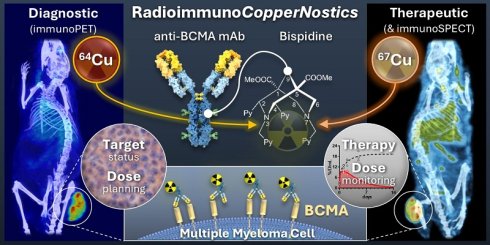
Background: The theranostic potential of copper-64 (PET imaging) and copper-67 (SPECT imaging and therapy) is increasingly recognized. This work investigates the performance of this ‘true’ matched pair in a preclinical radioimmunotheranostic setting that utilizes experimental monoclonal antibodies (mAbs) directed against B cell maturation antigen (BCMA), a transmembrane glycoprotein that has emerged as a critical target for the treatment of multiple myeloma (MM).
Methods: The commercially available BCMA-directed mAbs MAB193 and Vicky-1 were modified with the bispidine N2py4 enabling chelation of radiocopper, and their binding affinity was determined using surface plasmon resonance and flow cytometry. Pharmacokinetics and tumor uptake of the copper-64-labeled mAbs were determined in mice bearing subcutaneous myeloma xenografts, using PET. The best-performing mAb was included in a pilot study on therapeutic use with copper-67 in U266 myeloma-bearing mice, involving dose monitoring and dose predictions for humans based on quantitative SPECT.
Results: After bioconjugation, both [64Cu]Cu-N2py4-MAB193 and [64Cu]Cu-N2py4-Vicky-1 maintained low nanomolar BCMA binding affinity. In PET imaging, [64Cu]Cu-N2py4-MAB193 showed the highest uptake in U266 myeloma xenografts and the lowest normal tissue background, e.g., achieving a tumor-to-muscle contrast of 14.9 within 48 h. [67Cu]Cu-N2py4-MAB193 delivered absorbed doses up to 1.26 Gy/MBq in U266 myeloma xenografts and reduced the tumor mass at total doses above 52 Gy. Predicted human effective doses were below 35.3 µSv/MBq for immunoPET and 162 µSv/MBq for radioimmunotherapy/immunoSPECT.
Conclusion: The fundamental efficacy of copper-64 and copper-67 in BCMA-targeted immunotheranostics of MM promises both precise PET-based dose planning as well as radioimmunotherapy in combination with highly sensitive SPECT-based dose monitoring, as demonstrated by the theranostic capabilities of [64Cu/67Cu]Cu-N2py4-MAB193 in tumor-bearing mice. The results provide a strong incentive for incorporating the CopperNostics approach into the further development of BCMA-targeted radioimmunotheranostic agents, including precise tailoring of their pharmacokinetic properties to the physical half-life of copper-67.
Keywords: multiple myeloma, B cell maturation antigen, small animal PET, small animal SPECT, xenograft model, copper-64, copper-67, bispidine chelators
Introduction
As radionuclides of the same element, copper-64 (PET imaging, physical half-life 12.7 h) and copper-67 (β‾ therapy and SPECT imaging, physical half-life 61.8 h) are increasingly recognized as a ‘true’ matched pair with potential for medical use in cancer theranostics [1, 2]. The physical half-life of copper-64 is sufficiently consistent with the slow pharmacokinetics of monoclonal antibodies (mAbs) and thus offers advantages over shorter-lived radionuclides in immunoPET imaging [3]. The mean energies of β‾ particles emitted by copper-67 (150 keV) provide the same radiotherapeutic efficacy as those of lutetium-177 (147 keV) [4, 5]. Concomitantly, a high relative abundance of γ photons enables high-sensitivity SPECT imaging [6, 7]. Commercial production via accelerator-based photonuclear reaction yields copper-67 in sufficient quantities and purity, opening up realistic prospects for expanding the portfolio of therapeutic radiopharmaceuticals that can be produced independent of nuclear reactors [7].
To date, the theranostic value of copper-64-labeled agents addressing critical cancer targets has been demonstrated in vivo for numerous classes of compounds, including small molecules, peptides, antibody fragments, mAbs, and nanoparticles [1, 7]. In contrast, the theranostic potential of copper-67 has been demonstrated primarily for peptide receptor radionuclide therapies targeting critical cell surface receptors in cancer, most frequently somatostatin type 2 receptors (SSTR2) [1, 5, 7]. For a limited range of cancer-relevant antigens, several preclinical studies have demonstrated the therapeutic efficacy of copper-67-labeled radioimmunoconjugates, e.g., mAbs targeting human leucocyte antigen DR10, human epidermal growth factor receptor 2 (HER-2), and mucin 1 (MUC1) [6, 8-12]. The promising therapeutic effects reported therein suggest expanding the potential applications of copper-67-labeled mAbs to other cancer-relevant antigens and warrant a thorough analysis of their therapeutic dose delivery.
B cell maturation antigen (BCMA, also known as TNFRSF17 or CD269), a transmembrane glycoprotein belonging to the tumor necrosis factor receptor superfamily, has emerged as a critical target for innovative immunotherapies of multiple myeloma (MM), a malignancy characterized by the uncontrolled proliferation of plasma cells within the bone marrow [13-18]. A fraction of BCMA is cleaved from the surface of the myeloma cells by γ-secretase, resulting in the release of soluble BCMA (sBCMA) into the bloodstream [19, 20]. BCMA-directed therapeutics, encompassing chimeric antigen receptor T cells, bispecific T cell engagers, and antibody-drug conjugates, have undergone testing in MM patients [21, 22]. However, despite high objective response rates, relapses do occur, and tumor cells develop resistance to current BCMA-targeted immunotherapies [23], challenging further development and improvement of treatment strategies.
Myeloma cells display inherent sensitivity to radiation, with the most extensive clinical experience gained from external beam radiation therapy and from peptide receptor radionuclide therapy targeting the chemokine receptor CXCR4 [24-26]. Given the clinically proven efficacy of the existing non-radioactive BCMA-directed treatments, several radiolabeled agents for BCMA-specific targeting of myeloma cells have been successfully tested in preclinical settings so far. These developments were based on peptides, single-domain antibodies (sdAbs), mAbs, and one mAb-nanoparticle conjugate, and involved radiolabeling with gallium-68, copper-64, zirconium-89, fluorine-18, lutetium-177, and iodine-131 [27-32]. Recently, a gallium-68-labeled nanobody-based BCMA-tracer has been successfully tested in PET imaging of MM in a prospective first-in-human trial (NCT06717113) [33]. These promising results provide a strong incentive to expand BCMA-directed radioimmunotheranostic approaches.
For the immunotheranostic application of radiocopper, chelators that form stable complexes and enable fast radiolabeling of the biological vector molecules under mild reaction conditions are of particular interest. Due to their preorganized geometry, hexadentate bispidines (3,7-diazabicyclo[3.3.1]nonane derivatives) are particularly suitable for the selective complex formation with CuII radionuclides, resulting in metal complexes with high thermodynamic stability and kinetic inertness [34-38].
This work investigates the potential and practical applicability of copper-64 and copper-67 in radioimmunotheranostics within an experimental setting that utilizes mAbs targeting BCMA in models of human myeloma. The preclinical study reports on (i) the radiochemical and binding properties of two human BCMA-selective mAbs, MAB193 and Vicky-1, labeled with radiocopper trough complexation with a covalently linked bispidine (N2py4), (ii) the distribution and pharmacokinetic properties of [64Cu]Cu-N2py4-MAB193 and [64Cu]Cu-N2py4-Vicky-1 in mice measured by quantitative PET imaging, including their performance in visualizing subcutaneous U266 and L363 myeloma xenografts, and (iii) radiation doses from treatment with [67Cu]Cu-N2py4-MAB193, estimated based on quantitative SPECT imaging in U266 tumor-bearing mice. A critical discussion on therapeutic dose delivery by the copper-67-labeled anti-BCMA bispidine-mAb conjugate is provided.
Materials and Methods
Chemicals, solvents, and antibodies
Chemicals and solvents were purchased from common vendors (Merck, Darmstadt, Germany; Thermo Scientific, Waltham, MA, USA; VWR, Radnor, PA, USA) and used without further purification. Deuterated solvents were purchased from Deutero. Milli-Q® water (resistivity 18.2 MΩ × cm at 25°C) was used for high performance liquid chromatography (HPLC) purification and radiochemical experiments.
Anti-human BCMA mAbs were purchased from commercial suppliers: MAB193 (rat monoclonal IgG2A Clone 335004, Cat. No. MAB193, Bio-Techne, Minneapolis, MN, USA); Vicky-1 (rat monoclonal IgG1 Clone Vicky-1, Cat. No. ALX-804-151, ENZO Life Sciences, Lausen, Switzerland); E6D7B (rabbit monoclonal IgG Clone E6D7B, Cat. No. 88183, Cell Signaling Technology Europe, Leiden, Netherlands), and IgG2A-ITC (rat monoclonal IgG2A isotype control, Cat. No. MAB006, Bio-Techne).
The anti-human BCMA sdAb 269A37948 (patent WO-2018028647-A1 [39]) was produced by cloning the nucleotide sequence (GenBank: LQ 790725.1) into a pET-28b vector (Merck) followed by functional gene expression and purification of the recombinant protein as described recently [37, 40].
Synthesis of bispidine-mAb conjugates
The functionalized hexadentate bispidine N2py4-benzyl-isothiocyanate (N2py4-Bn-NCS) was synthesized, purified, and underwent quality control as described previously [35, 37, 41].
The anti-human BCMA mAbs MAB193 and Vicky-1 were reconstituted in freshly prepared sodium bicarbonate saline buffer (50 mM NaHCO3, 150 mM NaCl, pH 8.5) and functionalized with a 10-fold molar excess of N2py4-Bn-NCS, M = 741.9 g/mol, c = 10 nmol/µL DMSO) at 37 °C for 4 h with gentle shaking. The bispidine-mAb conjugates N2py4-MAB193 and N2py4-Vicky-1 were purified by size-exclusion chromatography using ZebaSpin Desalting Columns (7K MWCO, 5 mL, Thermo Scientific, Waltham, MA, USA) with elution in ammonium acetate buffer (0.2 M NH4OAc, pH 6) as well as spin filtration using Amicon Ultra centrifugal filters (30 K MWCO, 0.5 mL, Merck). Protein concentrations were determined using the DC Protein Assay (Cat. No. 5000116, Bio-Rad Laboratories, Hercules, CA, USA). Bispidine-mAb conjugates underwent quality control using matrix-assisted laser desorption ionization time-of-flight (MALDI-TOF) mass spectrometry as described in the Supporting information and were stored at 4 °C until further use.
To ensure batch-to-batch consistency, reaction conditions for bioconjugation (molar ratio of antibody-to-chelator, buffer composition, pH, reaction volume, time, and temperature) were kept constant, and freshly prepared chelator batches as well as freshly reconstituted antibody lyophilizates were used at all times.
Radiolabeling of bispidine-mAb conjugates
Copper-64 and copper-67 were produced with a TR-FLEX cyclotron (Advanced Cyclotron Systems Inc., Richmond, BC, Canada) by a 64Ni(p,n)64Cu and a 70Zn(p,α)67Cu nuclear reaction, respectively, as described previously.[42-44] For radiolabeling of bispidine-mAb conjugates, a substance-to-[64Cu]CuCl2 ratio of 1 nmol : 100 MBq and a substance-to-[67Cu]CuCl2 ratio of 1 nmol : 25 MBq was maintained. The reactions were set up in ammonium acetate buffer (0.2 M NH4OAc, pH 6) and incubated at 37 °C for 30 min with gentle shaking. The radiolabeled antibody-to-[64Cu/67Cu]CuCl2 ratio was assessed by radio-TLC using iTLC-SG and 0.05 M aq. EDTA (pH 5.5) and reported as radiochemical purity (%). Radiolabeled bispidine-mAb conjugates underwent quality control using radio-thin layer chromatography (radio-TLC) and denaturing sodium dodecyl sulfate-polyacrylamide gel electrophoresis (SDS-PAGE) as described in the Supporting information.
Cells
The human myeloma cell lines U266 (Cat.No. ACC 9) and L363 (Cat.No. ACC 49) were purchased from the DSMZ (Braunschweig, Germany) and cultured in Roswell Park Memorial Institute 1640 Medium (Thermo Scientific, Cat No. 61870036,) supplemented with 10% (v/v) fetal bovine serum (Merck) and 1 U/mL penicillin/streptomycin (Thermo Scientific, Cat No. 11548876,). The human melanoma cell line A375 (Cat.No. CRL-1619) was purchased from ATCC (LGC, Wesel, Germany) and cultured in Dulbecco’s modified Eagle medium (Thermo Scientific, Cat No. 12077549) supplemented with 10% (v/v) fetal bovine serum and 1 U/mL penicillin/streptomycin. The cells were cultivated in a CO2 incubator (37 °C, 5% (v/v) CO2, 95% (v/v) humidity).
Tumor xenograft models
Animal experiments were carried out according to the guidelines of the German Regulations for Animal Welfare. The protocols were approved by the local Ethical Committee for Animal Experiments (license DD24.1-5131/449/49 and 25-5131/562/52). 8 to 12 weeks old female Rj:NMRI-Foxn1nu/nu mice (Janvier Labs, Saint Berthevin Cedex, France) were subcutaneously injected with 5 × 10⁶ U266 or L363 cells in 100 µL isotonic NaCl (aq.) containing 50% (v/v) VitroGel® Hydrogel Matrix (TheWell Bioscience, Monmouth Junction, NJ, USA), or with 5 × 10⁶ A375 cells in 100 µL isotonic NaCl (aq.) into the right hind leg. Tumor size was monitored by caliper measurements and tumor volume was calculated using the formula VTumor = π/6 × (length × width2). Tumor-bearing mice were included in the experiments when tumors reached a volume of 150 mm3. After the final experiment, anesthetized animals were sacrificed under desflurane anesthetic (Baxter, Deerfield, IL; USA) using CO2 inhalation and cervical dislocation.
Gamma secretase inhibitor treatment
To reduce the release of sBCMA from myeloma xenografts into the blood stream, U266 and L363 tumor-bearing mice were treated with a γ secretase inhibitor (GSI, Crenigacestat LY3039478, MedChemExpress, Monmouth Junction, NJ, USA), dissolved in corn oil containing 10% (v/v) DMSO. Animals received four consecutive GSI treatments in 24 h-intervals, each with a dose of 2.5 mg/kg administered in 150 µL through oral gavage.
Immunohistochemistry
Formalin-fixed and paraffin-embedded sections of tumor xenografts (5 µm) were prepared according to standard procedures. Antigen-demasking was performed in sodium citrate buffer (10 mM, pH 6.0, 95°C, 20 min), followed by blocking of endogenous peroxidase and biotin using Biotin Blocking System (Agilent Technologies, Santa Clara, CA, USA, Cat. No. X0590) and blocking of non-specific binding sites with gelatin blocker (50 mM Tris, pH 8.0, 60 mM NaCl, 0.3% (v/v) gelatin, 2% (w/v) bovine serum albumin, 3% (w/v) milk powder, 0.5% (v/v) Tween20). Samples were incubated with primary antibody (polyclonal rabbit anti-human BCMA/TNFRSF17, Abcam, Cambridge, UK, Cat. No. ab5972) or mAb isotype control (normal Rabbit IgG, Abcam, Cat. No. ab27478) at 4°C overnight, followed by secondary antibody (biotinylated goat anti-rabbit antibody, Cat. No. 111-065-003, Jackson ImmunoResearch, Baltimore Pike, PA, USA) at room temperature for 1 h. Sections were incubated with ExtrAvidin® peroxidase for 30 min (Cat. No. E2886, Merck), stained with AEC substrate (Cat. No. 551015, BD Biosciences, Franklin Lakes, NJ, USA) and counterstained with Mayer’s Hematoxylin.
Enzyme-linked immunosorbent assay (ELISA)
For quantification of BCMA concentrations in tumor explants as well as in blood serum (sBCMA) tumor-bearing mice were anesthetized, blood was collected by cardiac puncture followed by immediate cervical dislocation. Blood was allowed to coagulate for 20 min at room temperature, followed by centrifugation at 3000 × g for 5 min at 4 °C. Clear serum was transferred into fresh microtubes and stored at -80°C until further use. Tumors were excised, cut into halves, and pieces of 2-4 mm from the central part of the tumor were quickly frozen in liquid nitrogen. Tissue pieces were transferred to a 2-mL-microtube containing 600 µL of lysis buffer (150 mM NaCl, 50 mM Tris pH 7.5, 1% NP-40, freshly added protease inhibitor cocktail tablets, Cat. No. 04693159001, Roche, Basel, Switzerland), and two steel balls, and disrupted in a TissueLyser device (Qiagen, Venlo, The Netherlands) at 25 Hz at room temperature for 3 min. Samples were treated with ultrasound for 15 s, stored on ice for 20 min, followed by centrifugation at 16.000 × g at 4 °C for 10 min. Protein content was determined using a BCA protein assay kit (Fisher Scientific) according to the manufacturer’s instructions. All samples were diluted to 1 mg/mL total protein in lysis buffer and stored at -80 °C.
The amount of BCMA in whole-cell lysates and sBCMA in mouse blood serum was determined using the human BCMA/TNFRSF17 DuoSet ELISA (Cat. No. DY193, Bio-Techne). Samples were thawed on ice and diluted 1:40 (tumors, finally 25 µg of protein/mL) or 1:40-1:200 (serum) in Reagent Diluent. ELISA was performed according to the manufacturer’s instructions.
Positron emission tomography
Small-animal positron emission tomography (PET) was performed using the nanoScan PET/CT (Mediso Medical Imaging Systems, Budapest, Hungary). Elevated serum concentrations of myeloma-related sBCMA were reduced by daily treatment with GSI (-1 d prior to 3 d after injection of the radiolabeled mAb). Each mouse received between 10 and 15 MBq of the radiolabeled mAb (eq. to 0.11-0.17 nmol) delivered in isotonic NaCl (aq.) via intravenous injection through a tail vein catheter within the initial 30 s after scan start. A series of PET scans were performed at defined time points (with scan durations) after injection of the radiolabeled mAb: 1 h (0-1 h), 5 h (4.5- 5.5 h), 24 h (23-25 h), and 48 h (47-49 h). With each scan, a corresponding CT image was captured and used for anatomical referencing and attenuation correction. Binning and time framing were performed as reported previously [45]. Images were reconstructed using the Tera-Tomo™ three-dimensional (3D) algorithm with a voxel size of 0.4 mm applying corrections for attenuation, scattering, and decay.
Saturation binding on tumor cryosections
Cryosections (10 µm) were prepared from tumor explants and mounted onto SuperFrost® plus glass slides (Thermo Scientific). Tissue sections were warmed to room temperature, incubated in Dulbecco’s phosphate-buffered saline for 15 min, air-dried, and incubated with [64Cu]Cu-N2py4-MAB193 serially diluted in binding buffer (Dulbecco’s phosphate-buffered saline containing 2.5% (w/v) bovine serum albumin) to final concentrations increasing from 0.025 to 50 nM for 1.5 h at room temperature. Non-specific binding was measured in presence of a BCMA-specific competitor (sdAb 269A37948) at final concentrations increasing from 0.25 to 500 nM (10-fold molar excess for each concentration). Tissue sections were washed twice in binding buffer for 5 min, dipped in distilled water, and air-dried. BAS-SR phosphoimaging plates (Fujifilm, Tokyo, Japan) were exposed to tissue sections and standards (increasing molar amounts of the radiolabeled mAb) for 45 min and imaged using an Amersham TyphoonTM FLA 9500 (Cytiva). Photostimulated luminescence (PSL) intensities were extracted from radioluminographic images using AIDA 5.1 (Elysia-raytest, Straubenhardt, Germany). Intensities per tissue section were converted into molar amounts of bound radiolabeled mAb using the standard series. Binding constants were calculated from non-linear regression analysis using the ‘one-site specific binding’ model implemented in Prism 11 (GraphPad Software, Boston, MA, USA). Binding capacities (Bmax) were expressed as molar amount per mm3 of tissue.
Radioimmunotherapy and single-photon emission computed tomography
Elevated serum concentrations of myeloma-related sBCMA were reduced by daily treatment with GSI (-1 d prior to 3 d after injection of the radiolabeled mAb). U266 tumor-bearing mice (n = 3) were treated with [67Cu]Cu-N2py4-MAB193 (Am = 70 MBq/nmol), each with a different initial activity dose of 25 MBq (eq. to 0.36 nmol), 50 MBq (eq. to 0.71 nmol), or 80 MBq (eq. to 1.14 nmol) administered in 0.2 mL of isotonic NaCl (aq.) through injection into a tail vein. Quantitative small-animal single-photon emission computed tomography (SPECT) was performed using the nanoScan® SPECT/CT (Mediso Medical Imaging Systems) equipped with the APT56 UHE aperture. Emission of photons was recorded and binned within the 20% energy windows of the 93 and 185 keV photopeaks [5]. Images were acquired within the following time windows after injection of the radiolabeled mAb (with corresponding scan times): 0.9-1.1 h (30 min), 25-27 h (40 min), 47-49 h (50 min), and 5 d (60 min). With each SPECT scan, a corresponding CT image was recorded and used for anatomical referencing and attenuation correction. Images were reconstructed using the Tera-Tomo™ 3D algorithm with a voxel size of 0.23 mm applying corrections for attenuation, scattering, and decay.
Analyses of PET and SPECT images
PET and SPECT images were post-processed and analyzed using Rover version 3.0.77h (Abx, Radeberg, Germany) and displayed as maximum intensity projections with common scaling over indicated time points. Details on the extraction of tissue-specific uptake values from PET images (region-averaged standardized uptake values, SUVmean) and SPECT images (% of initial activity/mL) are provided in the Supporting information (Figure S 1). Tissue-specific time courses of uptake values (decay-corrected and normalized to the initial activity dose) were analyzed by non-linear regression using the ‘two-phase decay’ and ‘one-phase decay’ models implemented in Prism 1 (GraphPad Software), with extrapolated uptake values approaching a lower limit constrained to 0. Total uptake in the liver, kidneys, and spleen [% of initial activity/organ] was extrapolated from the locoregional uptake values using a realistic dosimetry model for 30-g-mice [46]. The physical density of tissues was assumed to be 1 g/cm3.
Dose calculations
All doses were calculated based on the quantitative analysis of PET or SPECT images. For estimating the absorbed energy doses [Gy] resulting from uptake of radioimmunoconjugates in myeloma xenografts, the time courses of uptake values were transformed into time courses of effective activity concentrations by considering the physical half-life of the specific radionuclide involved [% of initial activity/mL]. The time-cumulated activity per mL of tissue was calculated from areas under effective time-activity concentration curves [% of initial activity dose/mL × d] and further converted into the total time-cumulated activity in the tumor-specific volume [MBq × h]. The mean absorbed tissue dose DT in tumors was calculated using the sphere model implemented in Olinda 2.2.3 (Hermes Medical Solutions AB, Stockholm, Sweden).
For human dose predictions, the tissue-specific pharmacokinetic profiles of radioimmunoconjugates in mice were transformed into human time scales and human organ masses using allometric scaling as described elsewhere [47]. Projected cumulated activity in human organs was determined separately for female and male subjects, and projected human absorbed doses [µGy/MBq] and effective doses [µSv/MBq] were calculated using the ICRP 89 Adult Female (60 kg) and ICRP 89 Adult Male (73 g) models implemented in Olinda 2.2.3 (Hermes Medical Solutions) as described previously [5].
Statistical analysis
Statistical analysis was performed using Prism 10 (GraphPad Software). Unless stated otherwise, data are presented as means with standard deviation and n represents the number of experiments or animals. Unless stated otherwise, significance of differences was tested using ANOVA with Sidak post-hoc test.
Results
Binding affinity of anti-BCMA mAbs
The three commercially available anti-human BCMA mAbs MAB193 (rat IgG2A), Vicky-1 (rat IgG1), and E6D7B (rabbit IgG) were the starting point for investigating an experimental BCMA-directed radioimmunotheranostic approach against MM. The three mAbs were initially characterized in its pristine form regarding their binding affinity towards recombinant human BCMA using surface plasmon resonance (SPR) interaction analysis as well as to native, cellular BCMA using flow cytometry employing the human cancer cell lines U266 and L363 (characterized by high and low cellular BCMA levels, respectively). Both MAB193 and Vicky-1 showed binding affinities towards BCMA with calculated equilibrium dissociation constants (Kd) in the subnanomolar (SPR) and nanomolar (flow cytometry) range as well as species selectivity for human BCMA (Table 1 and Supporting information, Figure S 2, Figure S 3, Figure S 5). E6D7B showed no binding to recombinant human or murine BCMA and was therefore excluded from further investigations.
Binding affinities (Kd values, given in nM) of pristine anti-BCMA mAbs towards recombinant and native cellular human BCMA; Kd values given as (a) means ± standard error resulting from SPR interaction analysis (n = 2, each analyzed in triplicate) or (b) means ± standard deviation resulting from flow cytometry measurements (n = 3); (n.d.) not determined
| Sample | MAB193 | Vicky-1 | E6D7B |
|---|---|---|---|
| recombinant human BCMA a | 0.33 ± 0.02 | 0.38 ± 0.01 | no binding |
| recombinant murine BCMA a | no binding | no binding | no binding |
| human BCMA-positive U266 cells b | 21.7 ± 11.1 | 11.1 ± 6.2 | n.d. |
| human BCMA-positive L363 cells b | 7.80 ± 3.70 | 6.3 ± 0.9 | n.d. |
Generation of radiocopper-labeled bispidine-mAb conjugates
Bioconjugation of the BCMA-affine mAbs with the amine-reactive bispidine derivative N2py4-Bn-NCS resulted in the bispidine-mAb conjugates N2py4-MAB193 and N2py4-Vicky-1. For control experiments, the non-specific mAb isotype control IgG2A-ITC was also equipped with the bispidine chelator resulting in N2py4-IgG2A-ITC. The three bispidine-mAb conjugates showed degrees of conjugation between 0.5 and 1.0 determined by MALDI-TOF mass spectrometry (Supporting information, Figure S 4).
After covalent modification with the bispidine chelator, the abilities of both N2py4-MAB193 and N2py4-Vicky-1 for recognizing BCMA on U266 cells remained unchanged (Supporting information, Figure S 5 E-F), providing the rationale to further investigate the radiocopper-labeled bispidine-mAb conjugates. Radiolabeling with copper-64 provided preparations of [64Cu]Cu-N2py4-MAB193, [64Cu]Cu-N2py4-Vicky-1, and [64Cu]Cu-N2py4-IgG2A-ITC with molar activities between 50 and 80 MBq/nmol and radiochemical purities of > 99% determined by radio-TLC (Supporting information, Figure S 6).
BCMA concentrations in subcutaneous human myeloma xenografts
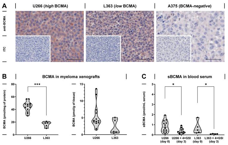
Prior to in vivo testing of radiocopper-labeled anti-BCMA mAbs in mice bearing human myeloma xenografts, BCMA concentrations in subcutaneous tumors as well as sBCMA concentrations in the blood serum were characterized. Immunohistochemistry showed positive staining for BCMA in tissue sections from U266 and L363 myeloma xenografts, but not in A375 melanoma xenografts serving as negative controls (Figure 1 A). Quantitative analysis of BCMA in tissue lysates by ELISA showed 2.7-fold higher concentrations in U266 compared to L363 tumors (44.4 ± 8.08 versus 16.5 ± 4.35 pmol/mg of protein, respectively; Figure 1 B). Serum samples obtained from mice bearing U266 and L363 myeloma xenografts showed the release of sBCMA into the blood (0.85 ± 0.55 and 0.68 ± 0.62 pmol/mL, respectively; Figure 1 C). Treatment with GSI for 4 d effectively reduced the concentration of sBCMA in the blood serum obtained from both U266 and L363 tumor-bearing mice (to 0.38 ± 0.26 and 0.09 ± 0.02 pmol/mL, respectively). These results provided the rationale to only include GSI-treated myeloma xenograft mice in the in vivo testing of radiocopper-labeled anti-BCMA mAbs in order to minimize their interaction with sBCMA in the blood.
BCMA and sBCMA in mice bearing subcutaneous human myeloma xenografts; (A) Immunohistochemical staining of U266, L363 and A375 tumor microsections; insets: negative controls incubated with a mAb isotype control; scale bars: 50 µm; (B) Concentrations of BCMA in myeloma homogenates measured by ELISA; (left) concentrations normalized to protein content, (right) concentrations normalized to tissue mass; (C) Concentrations of tumor-derived sBCMA in the blood serum measured by ELISA; reduction of the initial concentrations (day 0) after four daily treatments (day 0-3) with a γ-secretase inhibitor (GSI); significance of differences determined using Mann-Whitney test: * p < 0.05*** p < 0.001.
Distribution of copper-64-labeled anti-BCMA mAbs in tumor-bearing mice
The distribution of the radiocopper-labeled bispidine-mAb conjugates [64Cu]Cu-N2py4-MAB193 and [64Cu]Cu-N2py4-Vicky-1 was monitored by PET imaging for up to 48 h after injection in mice bearing subcutaneous BCMA-positive myeloma (U266 and L363) and BCMA-negative melanoma (A375) xenografts. Of note, [64Cu]Cu-N2py4-Vicky-1 was excluded from further investigations in vivo due to its low tumor uptake and high liver background (Supporting information, Figure S 7 and Figure S 8).
In PET images, [64Cu]Cu-N2py4-MAB193 enabled specific visual detection of both U266 and L363 tumors, unlike the corresponding isotype control [64Cu]Cu-N2py4-IgG2A-ITC, indicating that the accumulation of [64Cu]Cu-N2py4-MAB193 in the myeloma xenografts was BCMA-specific (Figure 2 A, B). In A375 melanoma xenografts, both [64Cu]Cu-N2py4-MAB193 and its corresponding isotype control [64Cu]Cu-N2py4-IgG2A-ITC showed low but comparable uptake, indicating that the accumulation of the two radiolabeled mAbs in the BCMA-negative tumor model was due to non-specific effects (Figure 2 C).
PET images and image-derived uptake values showing the distribution and tumor-specific accumulation of [64Cu]Cu-N2py4-MAB193 depending on the different BCMA levels of subcutaneous U266, L363, and A375 tumor xenografts in nude mice; corresponding isotype control: [64Cu]Cu-N2py4-IgG2A-ITC; (A-C) PET maximum intensity projections; a signals from both vena cava and aorta; (D-E) Uptake values extracted from PET images; (F) Total activity contents and excretion: b extrapolated from uptake values using a dose model for 30 g-mice, c calculated through balancing activity contents in intestine, urinary bladder, and total body; see materials and methods for details; (SUVmean) region-averaged standardized uptake value, reference values measured ex vivo in tissue explants are included in the Supporting information, Figure S 8; means ± standard deviation; significance of differences: ** p < 0.01, *** p < 0.001.
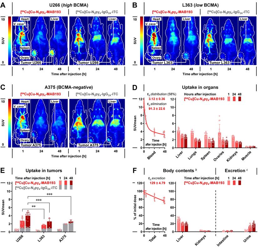
Quantitative analysis of PET images provided time courses of region-averaged standardized uptake values (SUVmean) for [64Cu]Cu-N2py4-MAB193 in normal tissues and tumors (Figure 2 D, E). The radiolabeled mAb showed a biphasic pharmacokinetic profile in the blood with half-lives of 3.12 h for distribution (58% of the initial dose) and 87.8 h for elimination. Of note, blood kinetics of [64Cu]Cu-N2py4-MAB193 were the same in γ-secretase inhibitor-treated myeloma-bearing mice and naïve A375 melanoma-bearing mice, ruling out effects from binding towards circulating sBCMA. After 48 h, 29% of the initial antibody concentration remained detectable in the blood.
Within 48 h, the uptake values decreased continuously in normal tissues of the liver, lungs, spleen, ovaries, kidneys, and muscles, while increasing to 4.01 and 2.59 in U266 and L363 tumors, respectively. The tumor-specific uptake of [64Cu]Cu-N2py4-MAB193 corresponded to the higher BCMA levels of U266 compared to L363 cells. Consequently, the tumor-to-background ratios were higher in U266 compared to L363 myeloma-bearing mice (tumor-to-blood: 1.28 versus 0.68 [p < 0.001]; tumor-to-muscle: 14.9 versus 9.53 [p < 0.05], respectively). Radioluminographic analysis of cryosections prepared from tumor explants 24 h after [64Cu]Cu-N2py4-MAB193 injection confirmed this trend (Supporting information, Figure S 9).
The distribution of the isotype control [64Cu]Cu-N2py4-IgG2A-ITC differed partially from that of [64Cu]Cu-N2py4-MAB193, most notably in liver uptake, remaining higher after 48 h (5.01 versus 2.72 [p < 0.001], respectively). Nevertheless, the U266 tumor-to-background ratios for [64Cu]Cu-N2py4-IgG2A-ITC remained well below those of [64Cu]Cu-N2py4-MAB193 (tumor-to-blood: 0.45 versus 1.28 [p < 0.001]; tumor-to-muscle: 3.82 versus 14.9 [p < 0.001], respectively). A similar trend was observed in L363 tumor-bearing mice, indicating that the tumor contrast provided by [64Cu]Cu-N2py4-MAB193 in the two myeloma xenografts models was BCMA-specific. SUVmean, tissue-to-background ratios, and statistical analyses are summarized in the Supporting information (Table S 2 and Table S 3).
The initially administered activity dose of [64Cu]Cu-N2py4-MAB193 was excreted from the animals with a biological half-life of 129 h, predominantly via the renal pathway (Figure 2 F). After 48 h, 19% and 2.9% of the initial dose remained detectable in liver and kidneys, respectively.
The radiation doses predicted for immunoPET in humans using [64Cu]Cu-N2py4-MAB193 were estimated based on quantitative PET data from U266 myeloma-bearing mice (Supporting information, Table S 4). Organs expected to receive the highest absorbed doses in the range between 66.2 and 138 µGy/MBq were spleen < ovaries < liver < heart wall. The predicted human effective doses were 35.3 µSv/MBq for women and 25.3 µSv/MBq for men, without taking tumor-specific accumulation into account.
BCMA-specific binding of [64Cu]Cu-N2py4-MAB193 on tumor explants
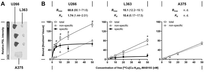
Whether the tumor-specific uptake of [64Cu]Cu-N2py4-MAB193 in U266, L363, and A375 tumor xenografts in vivo corresponds to tumor-specific BCMA levels was further investigated by saturation binding on tumor cryosections in vitro. After incubation of the cryosections with [64Cu]Cu-N2py4-MAB193, quantitative analysis of radioluminograms showed equilibrium dissociation constants (Kd) in the low nanomolar range and binding capacities (Bmax) that were five times higher on U266 compared to L363 tumor sections (Figure 3 A, B). Since the differences between total and non-specific binding reached statistical significance only for U266 and L363 tumor sections, binding towards A375 tumor sections was ascribed to non-specific effects. The in vitro binding capacities of U266, L363, and A375 cryosections for [64Cu]Cu-N2py4-MAB193 are therefore consistent with the trends in tumor-specific uptake in vivo, indicating that the radiolabeled mAb is a BCMA-specific radiotracer.
Binding of [64Cu]Cu-N2py4-MAB193 to U266, L363, and A375 tumor cryosections in vitro; (A) Exemplary radioluminograms of tissue sections incubated with the radiolabeled mAb at a concentration of 5 nM; photostimulated luminescence (PSL) intensities; (B) Tumor-specific BCMA binding constants determined by quantitative analyses of radioluminograms (n = 3); non-specific binding was determined in presence of excess anti-human BCMA sdAb 269A37948; binding constants were calculated using the non-linear regression model ‘one-site specific binding’; Bmax values given in fmol/mm3 of tissue; Kd values given in nM; significance of differences compared to non-specific binding (ANOVA with Fisher’s LSD test): * p > 0.05, # p > 0.001; means with 68% confidence interval; (n.d.) not determined.
SPECT imaging and dose delivery of [67Cu]Cu-N2py4-MAB193 in tumor-bearing mice
The suitability of the copper-67-labeled mAb [67Cu]Cu-N2py4-MAB193 for SPECT image-based estimations of therapeutic doses was tested in three mice bearing subcutaneous U266 myeloma xenografts, and treatment effects were documented in the setting of a pilot study. Each of the three animals received a single treatment with an individual total activity of 25, 50, and 80 MBq, respectively. Quantitative SPECT imaging showed that both distribution and tumor accumulation of [67Cu]Cu-N2py4-MAB193 were consistent with those of its copper-64-labeled counterpart (Figure 4 A and Supporting information, Table S 2 and Table S 5).
SPECT imaging, time-activity profiles, and a pilot study on treatment effects of [67Cu]Cu-N2py4-MAB193 in mice bearing subcutaneous U266 myeloma xenografts; (A) SPECT images after treatment with 50 MBq; a signals from both vena cava and aorta; (B) Normalized time-activity curves extracted from SPECT images; (C) Normalized cumulated volume activity in tissues, (n = 3); (D) Total excretion excluding activity in tumors; dashed black lines: decay-corrected courses fitted with exponential regression models: b ‘two-phase decay’, c ‘two-phase association’, d ‘one-phase decay’; continuous red lines: effective courses accounting for radionuclide decay; (E-F) Changes in tumor volume and body weight comparing untreated controls (w/o, n = 4) with radioimmunotherapy at individual initial activities (n = 1); dotted black line: treatment start, means ± standard deviation.
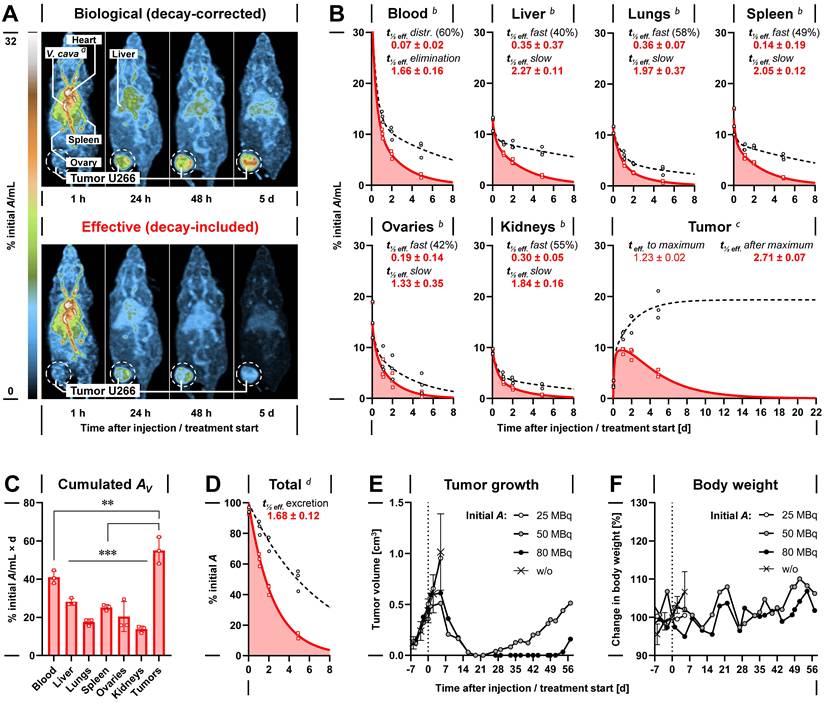
The pharmacokinetic profiles of [67Cu]Cu-N2py4-MAB193 in the liver, lungs, spleen, ovaries, and kidneys showed similar shapes as in the blood (effective half-lives: distribution 1.68 h, elimination 39.8 h), indicating that the retention of the radiolabeled mAb in the vasculature is the primary contributor to the cumulated activity in normal tissues (Figure 4 B). Compared to normal tissues, BCMA-specific uptake of [67Cu]Cu-N2py4-MAB193 in U266 tumors provided a significantly higher time-cumulated tissue activity (Figure 4 C). The initially administered activity was excreted with an effective half-life of 40.3 h (Figure 4 D). An increase in the total molar amount of [67Cu]Cu-N2py4-MAB193 from 0.36 to 1.14 nmol had no effect on relative uptake and pharmacokinetic profiles.
Single treatment with [67Cu]Cu-N2py4-MAB193 provided absorbed doses between 0.98 and 1.26 Gy/MBq in U266 tumors. By increasing the administered total activity from 25 to 80 MBq, the total absorbed doses in the tumors increased from 32.3 to 89.0 Gy. In two animals, total absorbed doses of >52 Gy were associated with a reduction in tumor volume and extension of progression-free survival, up from 0 d in the untreated control group to 29-55 d (Figure 4 E, Table 2). The relative changes in body weight following treatment with [67Cu]Cu-N2py4-MAB193 remained below 10% (Figure 4 F).
Absorbed doses in U266 myeloma xenografts and survival of tumor-bearing mice after treatment with [67Cu]Cu-N2py4-MAB193; Conditions at start of observation/treatment: (BW) body weight, (VStart, Tumor) tumor volume, (IA) initial activity dose, (ID molar) initial molar dose; (AUC Tumor) area under normalized time-activity curves also referred to as normalized cumulated volume activity in tumors; (DT, Tumor) absorbed tissue dose in tumors calculated using the ‘Sphere’ model implemented in Olinda 2.2.3, (PFS) progression-free survival defined as < 50 mm3 increase in tumor volume compared to minimum; (w/o, n = 4) untreated controls; means ± standard deviation
| Animal | BWStart | VStart, Tumor | Treatment | IA | ID molar | AUC Tumor | DT, Tumor per MBq | DT, Tumor | PFS |
|---|---|---|---|---|---|---|---|---|---|
| # | [g] | [cm3] | [MBq] | [nmol] | [% IA/mL×d] | [Gy/MBq] | [Gy] | [d] | |
| 1-4 | 29.6 ± 2.99 | 0.44 ± 0.12 | w/o | - | - | - | - | - | 0 |
| 5 | 29.2 | 0.50 | [67Cu]Cu-N2py4-MAB193 | 25 | 0.36 | 62.1 | 1.26 | 32.3 | 0 |
| 6 | 30.4 | 0.36 | [67Cu]Cu-N2py4-MAB193 | 50 | 0.71 | 48.1 | 0.98 | 52.0 | 29 |
| 7 | 31.4 | 0.47 | [67Cu]Cu-N2py4-MAB193 | 80 | 1.14 | 54.9 | 1.12 | 89.0 | 55 |
The expected radiation exposure for humans undergoing radioimmunotherapy with [67Cu]Cu-N2py4-MAB193 were predicted based on quantitative SPECT imaging of U266 myeloma-bearing mice (Supporting information, Table S 6 A). Organs that received the highest absorbed doses, ranging from 416 to 612 µGy/MBq, were spleen < ovaries < liver < heart wall. The predicted human effective doses were 162 µSv/MBq for women and 112 µSv/MBq for men, without taking tumor-specific accumulation into account. In comparison, the mean difference in dose estimates for [67Cu]Cu-N2py4-MAB193 extrapolated from PET imaging using its corresponding copper-64-labeled counterpart was below 6% (Table S 6 B).
Discussion
This preclinical study demonstrates the theranostic potential and practical applicability of the radionuclide pair copper-64 and copper-67 in BCMA-targeted immunotheranostics of MM, as indicated by the theranostic capabilities of experimental radiocopper-labeled bispidine-mAb conjugates directed against BCMA in models of human myeloma.
The starting point for this study was the pre-selection of three recombinant anti-BCMA mAbs MAB193, Vicky-1, and E6D7B as candidates for conversion into radiocopper-labeled bispidine-mAb conjugates, based on published results demonstrating their antigen-specific immunoreactivity [48-50], different antibody subtypes, and availability in sufficient quantities from commercial vendors.
With Kd values in the picomolar to nanomolar range (0.33-22 nM), the two BCMA-directed mAbs, MAB193 and Vicky-1, meet the binding affinity requirements that warrant their conversion into radioimmunotheranostic agents. Their binding affinity is comparable to other biological vectors that have been recently converted into BCMA radiotracers [27, 30-32]. Despite the reported BCMA-specific immunoreactivity of the third mAb candidate, E6D7B, which has been demonstrated in Western blot experiments and immunohistochemistry using formalin-fixed, paraffin-embedded tissue [50, 51], the antibody was excluded from further investigations due to insufficient binding to native human BCMA.
For labeling with radiocopper, MAB193 and Vicky-1 were modified using the bifunctional bispidine N2py4-Bn-NCS based on the following considerations: First, the NCS group enables straightforward lysine residue-directed bioconjugation of antibodies resulting in covalent thiourea bonds with desirable stability in buffers and human blood serum [52]. Second, N2Py4 offers rapid and efficient complex formation with CuII under mild conditions (room temperature and neutral pH), particularly favorable for mAbs-based radioconjugates, as they prevent detrimental effects on immunoreactivity and pharmacokinetic properties, potentially resulting from harsh labeling conditions which are often required for radiometal complex formation with other chelating moieties [53]. Third, the Cu-N2py4 complex exhibits high kinetic inertness which is especially important for radiocopper-labeled conjugates, due to the high abundance of copper-avid proteins in vivo [38, 54].
To characterize the pharmacokinetic properties of [64Cu]Cu-N2py4-MAB193 and [64Cu]Cu-N2py4-Vicky-1 in biological settings, U266 and L363 cells were used for generating complementary myeloma models that differ in their natural BCMA concentration. Additionally, A375 melanoma cells were included as a BCMA-negative control model to verify the antigen-specificity of tumor cell immunotargeting. The observation that U266 cells exhibit higher BCMA levels than L363 cells is consistent with previous reports [13, 55]. The present results further demonstrate that the intrinsically different BCMA levels of U266 and L363 myeloma cells are maintained in the corresponding subcutaneous tumor xenografts in mice. In fact, subcutaneous tumor xenograft models were chosen because, at this early stage of radiotracer characterization, they allow for straightforward in vivo pharmacokinetic monitoring - with clearly identifiable tumor sites being a favorable prerequisite for extracting meaningful tumor uptake values from quantitative PET and SPECT images.
Comparative investigations that include both U266 and L363 tumors offer a valuable opportunity to test the BCMA-dependent tumor specificity of theranostic agents in vivo across different biological variants of the same tumor entity, i.e., requiring no genetic manipulation of BCMA gene expression or blockade experiments in presence of a BCMA-specific competitor. The higher non-specific uptake of [64Cu]Cu-N2py4-MAB193 in the BCMA-negative A375 melanoma xenografts, compared to the L363 myeloma xenografts, exemplifies that characterizing the off-target uptake of radioimmunoconjugates in tumor models of a different entity can occasionally lead to false conclusions.
BCMA is known to be cleaved from the myeloma cell surface by γ-secretase, resulting in the release of sBCMA into the bloodstream [19]. In myeloma-bearing mice, the effective reduction in sBCMA levels in the peripheral blood by systematic short-term treatment with GSI (maintained until 48 h after radiotracer injection) largely rules out any effects of circulating sBCMA on the blood kinetics of the radiocopper-labeled mAbs. This is further supported by the observation that, e.g., blood kinetics of [64Cu]Cu-N2py4-MAB193 were the same in both GSI-treated myeloma-bearing mice and naïve A375 melanoma-bearing mice. However, it is known that GSI treatment with crenigacestat used in the present study affects several signaling pathways, including Notch signaling, and can alter tumor biology and microenvironment [56]. For example, short-term GSI treatment in mice has been reported to increase cell surface levels of BCMA in MM.1R myeloma xenografts [57]. Since this was not systematically investigated in the present study, BCMA-stimulatory and other biological effects of GSI treatment on myeloma xenografts cannot be ruled out. For this reason, GSI treatment was applied to all myeloma-bearing mice to ensure consistent experimental conditions across in vivo experiments.
Results from PET imaging in U266 myeloma-bearing mice and ex vivo analysis of tissue explants demonstrate that [64Cu]Cu-N2py4-MAB193 is a suitable PET radiotracer for the specific detection of BCMA-positive myeloma xenografts, with encouragingly low background in normal tissues. On the other hand, insufficient tumor uptake and extensive liver background in the U266 myeloma xenograft model led to the exclusion of [64Cu]Cu-N2py4-Vicky-1 from further investigations. It can be assumed that the almost instantaneous trapping of [64Cu]Cu-N2py4-Vicky-1 in the liver, which results in rapid clearance from the blood, is the main reason for its low tumor uptake. However, the exact mechanisms underlying the vastly different liver accumulation of the two radiocopper-labeled bispidine-mAb conjugates remain elusive at this stage.
Taking into account both the concentration of a tissue-specific molecular target and the binding affinity of a target-specific vector, a Bmax/Kd ratio ≥ 10 has been proposed to identify target/vector combinations that promise sufficient tissue contrast in molecular in vivo imaging [58]. The binding constants measured on tumor cryosections demonstrate that the BCMA-specific recognition of U266 myeloma xenografts by [64Cu]Cu-N2py4-MAB193 exceeds this benchmark, providing a Bmax/Kd ratio of approximately 40.
[64Cu]Cu-N2py4-MAB193 showed no binding to BCMA-negative A375 melanoma cryosections in vitro, indicating that its non-specific tumor uptake in vivo is likely not driven by cross reactivity with non-BCMA antigens or other specific molecular interactions. This is further supported by results from flow cytometry demonstrating no relevant expression of potentially antibody-capturing Fc receptors in A375 melanoma cells, nor in U266 and L363 myeloma cells. Therefore, non-specific uptake of [64Cu]Cu-N2py4-MAB193 in both melanoma and myeloma xenografts in vivo is most likely attributable to target-independent mechanisms, such as ‘enhanced permeability and retention’ (EPR). Especially subcutaneous tumor models are prone to EPR-driven effects [59]. This suggests, for example, that EPR-driven effects may account, for 26-33% of the total uptake in U266 myeloma xenografts.
As a PET radiotracer, [64Cu]Cu-N2py4-MAB193 showed no accumulation in most normal mouse tissues. However, a moderate uptake in the liver followed by slower clearance from the hepatic tissues compared to the blood suggests metabolic processing in the liver. Since its metabolic profile was not investigated in this study, no definitive conclusions can be drawn at this stage regarding the potential implications of radiometabolites for accumulation in the liver. However, referring to previous studies, it seems unlikely that a release of [64Cu]Cu2+ has any major role in liver accumulation, since the intact Cu-N2py4 complex has shown high resistance to demetalation in blood serum and to transchelation by physiological competing ligands present in the murine liver [38, 60]. Furthermore, there is no evidence from our previous investigations that might raise serious concerns about the stability of the thiourea bond in chelator-mAb conjugates [52].
The tumor uptake of [64Cu]Cu-N2py4-MAB193, detected in PET images 24 h after injection in U266 myeloma-bearing mice (SUVmean 2.74; equiv. to ~ 9.13% initial A/mL), is within the range reported for other radiolabeled mAbs targeting BCMA in subcutaneous myeloma xenograft models, for example, [89Zr]Zr-DFO-BCMAh230430 in MM.1S tumors (~5% initial A/mL) [30] and [89Zr]Zr-DFO-PFBH0L0 in H929 tumors (~10% initial A/mL) [31]. In PET imaging based on copper-64-based radiotracers, the further accumulation of mAbs in tumors – as also observed for [64Cu]Cu-N2py4-MAB193 reaching tumor-to-muscle ratios up to 14.9 in U266 myeloma-bearing mice after 48 h - may be of limited relevance in current clinical settings due to the progressive decay of copper-64 and the resulting decrease in signal intensity. On the other hand, this limitation can be overcome by use of modern long axial field of view (LAFOV) PET scanners, which offer higher sensitivity compared to conventional PET scanners [61].
As an advantage over to the zirconium-89-labeld mAbs, both of which exhibit high liver background as well as a significant release and bone accumulation of [89Zr]Zr4+ in vivo [30, 31], the lower normal tissue background of [64Cu]Cu-N2py4-MAB193 and its non-bone-avid radiolabel suggests that BCMA-targeted immunoPET using a copper-64-labeled bispidine-mAb conjugate holds promise for improved lesion detection, particularly in bones. This requires further examination in future studies. Furthermore, the predicted human effective dose for immunoPET using [64Cu]Cu-N2py4-MAB193 (25 and 35 µSv/MBq in adult women and men, respectively) corresponds to only 4-10% of the radiation exposure predicted for the use of zirconium-89-labeled mAbs (344 and 680 µSv/MBq) [30, 31].
Besides mAb-based approaches, other BCMA-specific vector molecules have been converted into gallium-68- and fluorine-18-containing PET radiotracers, including peptides [29] and sdAbs [27, 32, 33]. However, using these PET radiotracers for prospective therapeutic dose planning is challenging because their therapeutic counterparts are usually not chemically identical and contain radionuclides with a considerably longer physical half-life, as exemplified by the theranostic pair of sdAbs [18F]FPy-BCMA-Nb and [131I]I-BCMA-Nb [32]. Other PET radiotracers, such as the peptide-based [68Ga]Ga-DOTA-BP1 [29] and sdAb-based [68Ga]Ga-NOTA-MMBC derivatives [27], have shown uptake values below 0.6% initial A/mL in myeloma xenografts and fast washout from the tissue, presumably limiting the anti-tumor efficacy of their radiotherapeutic counterparts.
Compared to the existing BCMA-targeted peptide- and sdAb-based PET radiotracers containing short-lived radionuclides [27, 29, 32], copper-64-labeled bispidine-mAb conjugates unfold their advantage in light of existing chemically identical, copper-67-labeled counterparts, for which copper-64 provides a sufficiently long imaging window to enable prospective therapeutic dose planning. This is supported by the fact that the dose estimates derived directly from treatment of mice with [67Cu]Cu-N2py4-MAB193 are consistent with those extrapolated from PET imaging using its copper-64-labeled diagnostic counterpart. Furthermore, lower renal accumulation of [64Cu/67Cu]Cu-N2py4-MAB193 points towards lower radiation exposure to kidneys associated with BCMA-targeted immunotheranostics based on radiocopper-labeled bispidine-mAb conjugates. On the other hand, their longer blood retention and slower tumor accumulation point towards higher radiation exposure to blood and bone marrow cells, as well as slower build-up of the desired tumor-to-background contrast in PET imaging.
As an advantage over copper-64, the longer physical half-life of copper-67 allowed for monitoring [67Cu]Cu-N2py4-MAB193 in U266 tumor-bearing mice for 5 d using quantitative small animal SPECT imaging. Its extended traceability increased the accuracy of the extrapolated time-activity profiles for normal tissues and tumors, and thus dose estimates. This suggests that BCMA-targeted therapies using copper-67-labeled bispidine-mAb conjugates offer sufficiently long imaging windows to allow for precise SPECT-based dose monitoring. Given the high abundance of γ photons emitted during nuclear conversion of copper-67 [5, 6], a low-activity use of copper-67-labeled bispidine-mAb conjugates may also be considered for prospective SPECT-based dose planning with a favorable imaging window.
In mice, radiolabeled mAbs of the IgG type directed against tumor-specific molecular targets typically show biological half-lives in the blood between 4 and 10 d [62, 63], and reach their peak concentrations in solid tumors between 1 and 4 d, occasionally within up to 7 d after injection [64-67]. [67Cu]Cu-N2py4-MAB193 is well within these ranges, reaching its highest pharmacological concentration in U266 myeloma xenografts between 2 and 5 d after injection. However, less than 58% of the initially possible radiation dose per pharmacological amount of [67Cu]Cu-N2py4-MAB193 remain to be delivered to the tumor tissue from 2 d onwards (within another 2 weeks), due to the already advanced decay of copper-67 at the time point when the antibody approaches its highest pharmacologic concentration in U266 tumors.
Whereas the accumulation of IgG type antibodies in solid tumors frequently follows similar time courses in both mice [62-66] and humans [64, 68-71], their elimination from the blood is usually slower in humans, with biological half-lives between 20 and 24 d [72]. Consequently, it can be assumed that the ratio of radiation exposure between tumor and blood will be lower in a clinical setting of BCMA-targeted therapy using copper-67-labeled bispidine-mAb conjugates compared to the preclinical setting.
Despite the relatively slow accumulation of [67Cu]Cu-N2py4-MAB193 in U266 myeloma xenografts, single-dose treatment provided absorbed tissue doses between 0.96 and 1.27 Gy/MBq in the tumors. This is comparable to the absorbed tumor doses delivered by clinically approved β‾ particle-emitting radiopharmaceuticals addressing other critical cancer targets in preclinical settings, for example by [177Lu]Lu-PSMA-617 in PSMA-positive prostate cancer models (0.22-0.68 Gy/MBq) [73], and by [177Lu]Lu-DOTA-TATE in SSTR2-positive neuroendocrine tumor models (0.27-0.82 Gy/MBq) [5, 74-76].
Single-dose treatments with [67Cu]Cu-N2py4-MAB193 that delivered total absorbed doses >52 Gy in U266 myeloma xenografts reduced the tumor mass and prolonged the progression-free survival of mice. This suggests that BCMA-targeted radioimmunotherapy using copper-67-labeled bispidine-mAb conjugates can be effective against MM, although no conclusions on dose-response relationships can be drawn from the pilot study at this stage. Nevertheless, the sensitivity of U266 myeloma xenografts to treatment with [67Cu]Cu-N2py4-MAB193 is in line with other reports demonstrating therapeutic efficacy of β‾ particle-emitting immunoconjugates targeting BCMA in myeloma xenograft models, for example sdAb- and mAb-based agents labeled with iodine-131 and lutetium-177, respectively [30, 32].
Short-term GSI treatment has been reported to exert no growth-reducing effects on MM.1R myeloma xenografts in mice [57]. It can therefore be assumed that this treatment regimen has no effect on U266 tumor growth in the present study either. Furthermore, it has been reported that an unconjugated anti-BCMA mAb, J6M0 (Kd = 1 nM), slowed the growth of subcutaneous H929 myeloma xenografts in mice but did not reduce tumor mass, even when up to four doses of 4 mg/kg (~0.8 nmol/30-g-animal) were administered twice weekly (total cumulated dose of ~3 nmol/animal) [77]. Therefore, it can be assumed that the efficient reduction of the U266 tumor mass following treatment with [67Cu]Cu-N2py4-MAB193 at single molar doses of 0.72 and 1.12 nmol/animal, is predominantly attributable to the radiotoxic effects of copper-67. However, the anti-tumor effects of the unconjugated MAB193 were not investigated in the pilot study herein.
The observation that tumors regressed while the animals maintained their body weight and remained in good general health, even in response to treatment with [67Cu]Cu-N2py4-MAB193 at an initial activity dose of up to 80 MBq, provides a solid foundation for the future therapeutic evaluation of BCMA-targeted therapies using copper-67-labeled radioimmunoconjugates; however, it does not yet allow for definitive conclusions regarding safety of this approach.
The effective doses predicted for BCMA-targeted radioimmunotherapy in humans using [67Cu]Cu-N2py4-MAB193 (less than 161µSv/MBq) are lower compared to other BCMA-directed mAbs, such as [89Zr]Zr-DFO-BCMAh230430 (344 µSv/MBq) [30] and [89Zr]Zr-DFO-PFBH0L0 (680 µSv/MBq) [78], but align more closely with the effective doses predicted for mAbs that target other critical cancer antigens and have yet been included in clinical trials, for example, anti-PSMA [177Lu]Lu-DOTA-rosopatamab (210 µSv/MBq) [79] and anti-CD20 [177Lu]Lu-DOTA-rituximab (220 µSv/MBq) [80]. Of note, the effective dose predicted for the copper-67-labeled anti-HER2 [67Cu]Cu-NOTA-trastuzumab (17 µSv/MBq) [9] may have underestimated the potential radiation exposure in humans, although its activity concentrations in mouse tissues are comparable to those of [67Cu]Cu-N2py4-MAB193 in the preclinical setting.
The predicted radiation exposure in humans from treatment with [67Cu]Cu-N2py4-MAB193 is more than 2.3-fold higher compared to clinically approved peptide-based radiopharmaceuticals such as [177Lu]Lu-PSMA-617 (50 µSv/MBq) [81] and [177Lu]Lu-DOTA-TATE (70 µSv/MBq) [82], which are used to treat prostate cancer and neuroendocrine tumors, respectively. This suggests that the further development of BCMA-targeted radioimmunotherapies based on copper-67 requires immunoconjugates offering both faster uptake in tumors and faster elimination from the blood.
This preclinical study has limitations. Besides their usefulness for early-stage radiopharmaceutical characterization, subcutaneous myeloma models do not reflect the typical clinical manifestation of MM. Clinically, MM is a disseminated systemic malignancy characterized by diffuse bone marrow infiltration, where vascular permeability, antigen accessibility, and microenvironmental interactions may differ substantially from the subcutaneous tissue. Subcutaneous models are prone to EPR-driven effects, which may artificially inflate tumor accumulation of large molecules such as mAbs. Therefore, tumor uptake and pharmacokinetics of radiolabeled mAbs, as well as therapeutic responses may differ between subcutaneous and disseminated bone marrow disease. However, in one recent study, a fluorine-18-labeled BCMA-specific sdAb has shown comparable uptake in both subcutaneous and disseminated myeloma xenografts [32]. On the other hand, alpha-radioimmunotherapy using an astatine-211-labeled mAb directed against CD38 has been reported to be less effective in subcutaneous compared to disseminated models of MM [83]. Furthermore, radiation doses predicted based on the distribution of BCMA-targeted radioimmunoconjugates in mice bearing subcutaneous myeloma xenografts may underestimate bone marrow exposure in humans potentially resulting from uptake in diffuse and intermedullary lesions.
Due to the species selectivity of [64Cu/67Cu]Cu-N2py4-MAB193 for human BCMA, whose occurrence in myeloma xenograft mice is restricted to the human tumor tissue (as well as to tumor-derived human sBCMA in peripheral blood), the potential contributions of physiologic BCMA to the uptake in normal mouse tissues remained undetectable in the present study.
It should also be noted that [64Cu/67Cu]Cu-N2py4-MAB193 itself does not meet the molecular requirements for direct medical use. Efforts towards clinical translation require molecular engineering of its Fc region (rat IgG2A) to generate a humanized chimeric mAb which is compatible with the human immune system and bypasses human Fcγ receptor-mediated uptake in normal tissues.
To more precisely tailor the pharmacokinetic properties of BCMA-targeted immunotherapeutic agents for their use with radiocopper - particularly with regard to faster blood kinetics - various strategies are known to be effective and may therefore be considered in future developments, including pretargeting [84], the use of antibody-nanoparticle conjugates [28], or antibody fragments with smaller molecular weights such as sdAbs [27, 32, 33, 40], minibodies and diabodies [85], or Fab fragments [86].
Conclusion
The fundamental efficacy of copper-64 and copper-67 in BCMA-targeted immunotheranostics of MM promises both precise PET-based dose planning and radioimmunotherapy in combination with highly sensitive SPECT-based dose monitoring, as demonstrated by the theranostic capabilities of the chemically identical bispidine-mAb conjugates [64Cu]Cu-N2py4-MAB193 and [67Cu]Cu-N2py4-MAB193 in tumor-bearing mice. The results provide a strong incentive for incorporating the CopperNostics approach into the further development of BCMA-targeted radioimmunotheranostic agents, including precise tailoring of their pharmacokinetic properties to the physical half-life of copper-67.
Abbreviations
Am: molar activity; BCMA: B cell maturation antigen; CT: computed tomography; EDTA: ethylenediaminetetraacetic acid; GSI: gamma secretase inhibitor; IgG: immunoglobulin G; ITC: isotype control; mAb: monoclonal antibody; MALDI-TOF: matrix-assisted laser desorption/ionization time-of-flight mass spectrometry; MM: multiple myeloma; PET: positron emission tomography; sBCMA: soluble B cell maturation antigen; sdAb: single-domain antibody; SPECT: single-photon emission computed tomography; SPR: surface plasmon resonance; SUVmean: region-averaged standardized uptake value; TLC: thin layer chromatography.
Supplementary Material
Supplementary methodological details and additional results (images, tables, graphs).

Acknowledgements
The expert technical assistance of Julia Aldinger, Karl Anger, Mareike Barth, Cornelius Donat, PhD, Juliane Meyer, Aline Morgenegg, Andrea Suhr, Johanna Wodtke, and Adrian Wünsche is greatly acknowledged. The authors would also like to thank Lena Richter for her support in carrying out the animal experiments. Furthermore, we would like to thank the team at the animal facility, Katrin Baumgart, Norman Fielko, and Helge Gläser, for the excellent preparation of the experiments, as well as Martin Kreller, PhD, Santiago Brühlmann, PhD, and the cyclotron team for providing radiocopper. We would also like to thank Christin Neuber, PhD, for her comments and stimulating discussions.
The authors disclose that no AI tools were used for data collection, data analysis, or image generation. German text passages from the draft manuscript were translated into English using DeepL Translator (https://www.deepl.com) and manually reviewed for content correctness, stylistic flow, and grammatical accuracy.
Funding
Funded by the Deutsche Forschungsgemeinschaft (DFG, German Research Foundation) – Project number PI 304/5-1 “Antibody-based BCMA-directed theranostics of multiple myeloma” (J.P. and B.B.).
Contributions
Conceptualization, J.P., B.B.; methodology, M.U., K.Z., M.K., M.L., B.B.; investigation, M.U., K.Z., M.K., M.L., B.B.; visualization, M.U., K.Z., M.K., M.L., B.B.; resources, J.P.; project administration, J.P.; funding acquisition, J.P., B.B.; writing—original draft preparation, M.U., K.Z., M.K., M.L., J.P., B.B.; writing—review and editing, M.U., K.Z., M.K., M.L., J.P., B.B. All authors have read and agreed to the published version of the manuscript.
Data availability
Beyond the supplementary material, additional raw data can be provided upon request; please contact the corresponding author.
Competing Interests
The authors have declared that no competing interest exists.
References
1. Bruhlmann SA, Walther M, Kopka K, Kreller M, Kiss OC. CopperNostics-Here we are now, entertain us!. Pharmaceuticals (Basel). 2026;19:321
2. Zimmermann R. Are 61Cu, 64Cu, and 67Cu really happening? J Nucl Med. 2026: jnumed.126.272416.
3. Badier L, Quelven I. Zirconium-89 and copper-64 for immunoPET: From antibody bioconjugation and radiolabeling to molecular imaging. Pharmaceutics. 2024;16:882
4. ICRP 2008. Nuclear decay data for dosimetric calculations. ICRP Publication 107. Ann. ICRP 38 (3).
5. Ullrich M, Wodtke R, Brandt F, Freudenberg R, Kotzerke J, Richter S. et al. The heterobivalent (SSTR2/albumin) radioligand [67Cu]Cu-NODAGA-cLAB4-TATE enables efficient somatostatin receptor radionuclide theranostics. Theranostics. 2024;14:5371-87
6. Hao G, Mastren T, Silvers W, Hassan G, Oz OK, Sun X. Copper-67 radioimmunotheranostics for simultaneous immunotherapy and immuno-SPECT. Sci Rep. 2021;11:3622
7. Krasnovskaya OO, Abramchuck D, Erofeev A, Gorelkin P, Kuznetsov A, Shemukhin A. et al. Recent advances in 64Cu/67Cu-based radiopharmaceuticals. Int J Mol Sci. 2023;24:9154
8. O'Donnell RT, DeNardo GL, Kukis DL, Lamborn KR, Shen S, Yuan A. et al. 67Copper-2-iminothiolane-6-[p-(bromoacetamido)benzyl-TETA-Lym-1 for radioimmunotherapy of non-Hodgkin's lymphoma. Clin Cancer Res. 1999;5:3330s-36s
9. Pougoue Ketchemen J, Njotu FN, Babeker H, Ahenkorah S, Tikum AF, Nwangele E. et al. Effectiveness of [67Cu]Cu-trastuzumab as a theranostic against HER2-positive breast cancer. Eur J Nucl Med Mol Imaging. 2024;51:2070-84
10. Rudd SE, Van Zuylekom J, Cullinane C, Blyth BJ, Donnelly PS. Potential theranostics of breast cancer with copper-64/67 sarcophagine-trastuzumab. Chem Sci. 2025;16:3998-4005
11. Pineau J, Ollier C, Mothes C, Belderbos S, Le Bris N, Tripier R. Advanced 64Cu/67Cu-based theranostics: Application of the copper chelator TE1PA to a murine model of HER2-positive gastric cancer. Inorg Chem. 2025;64:12189-97
12. Hughes OD, Bishop MC, Perkins AC, Wastie ML, Denton G, Price MR. et al. Targeting superficial bladder cancer by the intravesical administration of copper-67-labeled anti-MUC1 mucin monoclonal antibody C595. J Clin Oncol. 2000;18:363-70
13. Carpenter RO, Evbuomwan MO, Pittaluga S, Rose JJ, Raffeld M, Yang S. et al. B-cell maturation antigen is a promising target for adoptive T-cell therapy of multiple myeloma. Clin Cancer Res. 2013;19:2048-60
14. Hu Y, Xie Y, Wang X, Yang L, Geng H, Yi Z. et al. Targeting BCMA in multiple myeloma: Designs, challenges, and future directions. Cancer Immunol Immunother. 2025;74:77
15. Avery DT, Kalled SL, Ellyard JI, Ambrose C, Bixler SA, Thien M. et al. BAFF selectively enhances the survival of plasmablasts generated from human memory B cells. J Clin Invest. 2003;112:286-97
16. Chiu A, Xu W, He B, Dillon SR, Gross JA, Sievers E. et al. Hodgkin lymphoma cells express TACI and BCMA receptors and generate survival and proliferation signals in response to BAFF and APRIL. Blood. 2007;109:729-39
17. Thompson JS, Schneider P, Kalled SL, Wang L, Lefevre EA, Cachero TG. et al. BAFF binds to the tumor necrosis factor receptor-like molecule B cell maturation antigen and is important for maintaining the peripheral B cell population. J Exp Med. 2000;192:129-35
18. Solimando AG, Krebs M, Desantis V, Marziliano D, Caradonna IC, Morizio A. et al. Breaking through multiple myeloma: A paradigm for a comprehensive tumor ecosystem targeting. Biomedicines. 2023;11:2087
19. Laurent SA, Hoffmann FS, Kuhn PH, Cheng Q, Chu Y, Schmidt-Supprian M. et al. Gamma-secretase directly sheds the survival receptor BCMA from plasma cells. Nat Commun. 2015;6:7333
20. Ghermezi M, Li M, Vardanyan S, Harutyunyan NM, Gottlieb J, Berenson A. et al. Serum B-cell maturation antigen: a novel biomarker to predict outcomes for multiple myeloma patients. Haematologica. 2017;102:785-95
21. Sanchez L, Dardac A, Madduri D, Richard S, Richter J. B-cell maturation antigen (BCMA) in multiple myeloma: The new frontier of targeted therapies. Ther Adv Hematol. 2021;12:2040620721989585
22. Sheykhhasan M, Ahmadieh-Yazdi A, Vicidomini R, Poondla N, Tanzadehpanah H, Dirbaziyan A. et al. CAR T therapies in multiple myeloma: Unleashing the future. Cancer Gene Ther. 2024;31:667-86
23. Yue T, Sun Y, Dai Y, Jin F. Mechanisms for resistance to BCMA-targeted immunotherapies in multiple myeloma. Blood Rev. 2025;70:101256
24. Hu K, Yahalom J. Radiotherapy in the management of plasma cell tumors. Oncology (Williston Park). 2000;14:101-8 11; discussion 11-2, 15
25. Herrmann K, Schottelius M, Lapa C, Osl T, Poschenrieder A, Hanscheid H. et al. First-in-human experience of CXCR4-directed endoradiotherapy with 177Lu- and 90Y-labeled pentixather in advanced-stage multiple myeloma with extensive intra- and extramedullary disease. J Nucl Med. 2016;57:248-51
26. Lapa C, Herrmann K, Schirbel A, Hanscheid H, Luckerath K, Schottelius M. et al. CXCR4-directed endoradiotherapy induces high response rates in extramedullary relapsed multiple myeloma. Theranostics. 2017;7:1589-97
27. Wei W, Zhang Y, Zhang D, Liu Q, An S, Chen Y. et al. Annotating BCMA expression in multiple myelomas. Mol Pharm. 2022;19:3492-501
28. Thomas E, Mathieu C, Moreno-Gaona P, Mittelheisser V, Lux F, Tillement O. et al. Anti-BCMA immuno-nanoPET radiotracers for improved detection of multiple myeloma. Adv Healthc Mater. 2022;11:e2101565
29. Song L, Jiang S, Yang Q, Huang W, Qiu Y, Chen Z. et al. Development of a novel peptide-based PET tracer [68Ga]Ga-DOTA-BP1 for BCMA detection in multiple myeloma. J Med Chem. 2024;67:15118-30
30. Ma J, Zhang S, Yang N, Shang J, Gao X, Chen J. et al. Discovery of a highly specific radiolabeled antibody targeting B-cell maturation antigen: Applications in PET imaging of multiple myeloma. Eur J Nucl Med Mol Imaging. 2025;52:599-610
31. Wang T, Yang Q, Huang W, Sun X, Lei Y, Xu H. et al. Development and preclinical validation of a novel 89Zr-labeled BCMA-targeting antibody for immunoPET imaging: Application in multiple myeloma models and first-in-nonhuman-primates. J Med Chem. 2025;68:14843-58
32. Ghaemi B, Olkowski CP, Basuli F, Shi J, Young R, Kazandjian D. et al. Precision radiolabeled B-cell maturation nanobody for targeted PET imaging and radioligand therapy of disseminated multiple myeloma. Theranostics. 2026;16:2748-63
33. Gu T, Chen Z, Wang T, Dong Y, Kang L. B-cell maturation antigen targeted PET/CT imaging in multiple myeloma: a first-in-human study. J Hematol Oncol. 2025;18:101
34. Bleiholder C, Borzel H, Comba P, Ferrari R, Heydt M, Kerscher M. et al. Coordination chemistry of a new rigid, hexadentate bispidine-based bis(amine)tetrakis(pyridine) ligand. Inorg Chem. 2005;44:8145-55
35. Juran S, Walther M, Stephan H, Bergmann R, Steinbach J, Kraus W. et al. Hexadentate bispidine derivatives as versatile bifunctional chelate agents for copper(II) radioisotopes. Bioconjug Chem. 2009;20:347-59
36. Comba P, Kerscher M, Ruck K, Starke M. Bispidines for radiopharmaceuticals. Dalton Trans. 2018;47:9202-20
37. Singh G, Zarschler K, Hunoldt S, Martinez IIS, Ruehl CL, Matterna M. et al. Versatile bispidine-based bifunctional chelators for 64Cu(II)-labelling of biomolecules. Chemistry. 2020;26:1989-2001
38. Kubeil M, Neuber C, Starke M, Arndt C, Rodrigues Loureiro L, Hoffmann L. et al. 64Cu tumor labeling with hexadentate picolinic acid-based bispidine immunoconjugates. Chemistry. 2024;30:e202400366
39. Fan X, Zhuang Q, Wang P, Wang L, Yang L, Hao J. et al. Chimeric antigen receptors targeting BCMA and methods of use thereof (patent number: WO-2018028647-A1). Nanjing Legend Biotechnology Co Ltd; Legend Biotech Ireland Ltd; Legend Biotech USA Inc. 2018
40. Trommer J, Ullrich M, Reissig F, Bruhlmann SA, Nitt-Weber AK, Novy Z. et al. It's a match: use of the radionuclide theranostic pair 133La/225Ac for the radiopharmacological characterization of EGFR-targeted single-domain antibodies. EJNMMI Radiopharm Chem. 2025;10:31
41. Saleh HA, Mitwasi N, Ullrich M, Kubeil M, Toussaint M, Deuther-Conrad W. et al. Specific and safe targeting of glioblastoma using switchable and logic-gated RevCAR T cells. Front Immunol. 2023;14:1166169
42. Thieme S, Walther M, Pietzsch HJ, Henniger J, Preusche S, Mading P. et al. Module-assisted preparation of 64Cu with high specific activity. Appl Radiat Isot. 2012;70:602-8
43. Kreller M, Pietzsch HJ, Walther M, Tietze H, Kaever P, Knieß T. et al. Introduction of the new Center for Radiopharmaceutical Cancer Research at Helmholtz-Zentrum Dresden-Rossendorf. Instruments. 2019;3:9
44. Bruhlmann SA, Walther M, Kreller M, Reissig F, Pietzsch HJ, Kniess T. et al. Cyclotron-based production of 67Cu for radionuclide theranostics via the 70Zn(p,alpha)67Cu reaction. Pharmaceuticals (Basel). 2023;16:314
45. Ullrich M, Bergmann R, Peitzsch M, Zenker EF, Cartellieri M, Bachmann M. et al. Multimodal somatostatin receptor theranostics using [64Cu]Cu-/[177Lu]Lu-DOTA-(Tyr3)octreotate and AN-238 in a mouse pheochromocytoma model. Theranostics. 2016;6:650-65
46. Keenan MA, Stabin MG, Segars WP, Fernald MJ. RADAR realistic animal model series for dose assessment. J Nucl Med. 2010;51:471-6
47. Kranz M, Sattler B, Tiepolt S, Wilke S, Deuther-Conrad W, Donat CK. et al. Radiation dosimetry of the alpha4beta2 nicotinic receptor ligand (+)-[18F]flubatine, comparing preclinical PET/MRI and PET/CT to first-in-human PET/CT results. EJNMMI Phys. 2016;3:25
48. Chapellier M, Pena-Martinez P, Ramakrishnan R, Eriksson M, Talkhoncheh MS, Orsmark-Pietras C. et al. Arrayed molecular barcoding identifies TNFSF13 as a positive regulator of acute myeloid leukemia-initiating cells. Haematologica. 2019;104:2006-16
49. Notas G, Alexaki VI, Kampa M, Pelekanou V, Charalampopoulos I, Sabour-Alaoui S. et al. APRIL binding to BCMA activates a JNK2-FOXO3-GADD45 pathway and induces a G2/M cell growth arrest in liver cells. J Immunol. 2012;189:4748-58
50. Ran Y, Cheng W, Yi X, Yang S, Zhang W, Xu J. Limited expression of B-cell maturation antigen in acute myeloid leukemia. J Immunother Cancer. 2026 14
51. Garfall AL, Cohen AD, Susanibar-Adaniya SP, Hwang WT, Vogl DT, Waxman AJ. et al. Anti-BCMA/CD19 CAR T cells with early immunomodulatory maintenance for multiple myeloma responding to initial or later-line therapy. Blood Cancer Discov. 2023;4:118-33
52. Striese F, Neuber C, Grassel S, Arndt C, Ullrich M, Steinbach J. et al. Preclinical characterization of the 177Lu-labeled prostate stem cell antigen (PSCA)-specific monoclonal antibody 7F5. Int J Mol Sci. 2023;24:9420
53. Khandave PY, Gudavalekar PU, Das T, Pande AH. Radioligand-antibody conjugates: Integrating antibody engineering, chelator chemistry, and radioligands for precision theranosis. Mol Diagn Ther. 2026;30:259-80
54. Cai Z, Anderson CJ. Chelators for copper radionuclides in positron emission tomography radiopharmaceuticals. J Labelled Comp Radiopharm. 2014;57:224-30
55. Kimman T, Cuenca M, Tieland RG, Rockx-Brouwer D, Janssen J, Motais B. et al. Engineering anti-BCMA CAR T cells for enhancing myeloma killing efficacy via apoptosis regulation. Nat Commun. 2025;16:4638
56. Olsauskas-Kuprys R, Zlobin A, Osipo C. Gamma secretase inhibitors of Notch signaling. Onco Targets Ther. 2013;6:943-55
57. Pont MJ, Hill T, Cole GO, Abbott JJ, Kelliher J, Salter AI. et al. gamma-Secretase inhibition increases efficacy of BCMA-specific chimeric antigen receptor T cells in multiple myeloma. Blood. 2019;134:1585-97
58. Patel S, Gibson R. In vivo site-directed radiotracers: A mini-review. Nucl Med Biol. 2008;35:805-15
59. Kim J, Cho H, Lim DK, Joo MK, Kim K. Perspectives for improving the tumor targeting of nanomedicine via the EPR effect in clinical tumors. Int J Mol Sci. 2023;24:10082
60. Bass LA, Wang M, Welch MJ, Anderson CJ. In vivo transchelation of copper-64 from TETA-octreotide to superoxide dismutase in rat liver. Bioconjug Chem. 2000;11:527-32
61. Cherry SR, Jones T, Karp JS, Qi J, Moses WW, Badawi RD. Total-body PET: Maximizing sensitivity to create new opportunities for clinical research and patient care. J Nucl Med. 2018;59:3-12
62. Vieira P, Rajewsky K. The half-lives of serum immunoglobulins in adult mice. Eur J Immunol. 1988;18:313-6
63. Gurbaxani B, Dela Cruz LL, Chintalacharuvu K, Morrison SL. Analysis of a family of antibodies with different half-lives in mice fails to find a correlation between affinity for FcRn and serum half-life. Mol Immunol. 2006;43:1462-73
64. Guo X, Zhou N, Chen Z, Liu T, Xu X, Lei X. et al. Construction of 124I-trastuzumab for noninvasive PET imaging of HER2 expression: From patient-derived xenograft models to gastric cancer patients. Gastric Cancer. 2020;23:614-26
65. Aerts HJ, Dubois L, Perk L, Vermaelen P, van Dongen GA, Wouters BG. et al. Disparity between in vivo EGFR expression and 89Zr-labeled cetuximab uptake assessed with PET. J Nucl Med. 2009;50:123-31
66. Woo SK, Jang SJ, Seo MJ, Park JH, Kim BS, Kim EJ. et al. Development of 64Cu-NOTA-trastuzumab for HER2 targeting: A radiopharmaceutical with improved pharmacokinetics for human studies. J Nucl Med. 2019;60:26-33
67. Omweri JM, Saini S, Houson HA, Tekin V, Pyles JM, Parker CC. et al. Development of 52Mn labeled trastuzumab for extended time point PET imaging of HER2. Mol Imaging Biol. 2024;26:858-68
68. Kurdziel K, Mena E, Adler S, Paik C, Brechbiel M, McKinney Y. et al. Human dosimetry and tumor kinetics of 111In CHX-A” DTPA trastuzumab in solid tumors using gamma-camera imaging. Journal of Nuclear Medicine. 2011;52:1469-69
69. Dijkers EC, Oude Munnink TH, Kosterink JG, Brouwers AH, Jager PL, de Jong JR. et al. Biodistribution of 89Zr-trastuzumab and PET imaging of HER2-positive lesions in patients with metastatic breast cancer. Clin Pharmacol Ther. 2010;87:586-92
70. Even AJ, Hamming-Vrieze O, van Elmpt W, Winnepenninckx VJ, Heukelom J, Tesselaar ME. et al. Quantitative assessment of zirconium-89 labeled cetuximab using PET/CT imaging in patients with advanced head and neck cancer: A theragnostic approach. Oncotarget. 2017;8:3870-80
71. Menke-van der Houven van Oordt CW, Gootjes EC, Huisman MC, Vugts DJ, Roth C, Luik AM. et al. 89Zr-cetuximab PET imaging in patients with advanced colorectal cancer. Oncotarget. 2015;6:30384-93
72. Cruse JM, Lewis RE. Atlas of immunology. 3 ed: CRC Press. 2010
73. Arbuznikova D, Klotsotyra A, Uhlmann L, Domogalla LC, Steinacker N, Mix M. et al. Exploring the role of combined external beam radiotherapy and targeted radioligand therapy with [(177)Lu]Lu-PSMA-617 for prostate cancer - from bench to bedside. Theranostics. 2024;14:2560-72
74. Dalmo J, Spetz J, Montelius M, Langen B, Arvidsson Y, Johansson H. et al. Priming increases the anti-tumor effect and therapeutic window of 177Lu-octreotate in nude mice bearing human small intestine neuroendocrine tumor GOT1. EJNMMI Res. 2017;7:6
75. Dalm SU, Nonnekens J, Doeswijk GN, de Blois E, van Gent DC, Konijnenberg MW. et al. Comparison of the therapeutic response to treatment with a 177Lu-labeled somatostatin receptor agonist and antagonist in preclinical models. J Nucl Med. 2016;57:260-5
76. Feijtel D, Doeswijk GN, Verkaik NS, Haeck JC, Chicco D, Angotti C. et al. Inter and intra-tumor somatostatin receptor 2 heterogeneity influences peptide receptor radionuclide therapy response. Theranostics. 2021;11:491-505
77. Tai YT, Mayes PA, Acharya C, Zhong MY, Cea M, Cagnetta A. et al. Novel anti-B-cell maturation antigen antibody-drug conjugate (GSK2857916) selectively induces killing of multiple myeloma. Blood. 2014;123:3128-38
78. Wang L, Ma J, Zhang S, Hu K. Discovery of a highly specific radiolabeled antibody targeting B-cell maturation antigen: Applications in PET imaging and radiotherapy of multiple myeloma. Journal of Nuclear Medicine. 2024;65:242574-74
79. Lenzo N, O'Byrne K, Ngai S, Meyrick D, Patel N, Hayward C. 177Lu-TLX591 safety, biodistribution and dosimetry study (ProstACT SELECT). Journal of Nuclear Medicine. 2023;64:P1145-P45
80. Yadav MP, Singla S, Thakral P, Ballal S, Bal C. Dosimetric analysis of 177Lu-DOTA-rituximab in patients with relapsed/refractory non-Hodgkin's lymphoma. Nucl Med Commun. 2016;37:735-42
81. Kamaldeep Wanage G, Sahu SK Maletha P, Adnan A Suman S. et al. Examining absorbed doses of indigenously developed 177Lu-PSMA-617 in metastatic castration-resistant prostate cancer patients at baseline and during course of peptide receptor radioligand therapy. Cancer Biother Radiopharm. 2021;36:292-304
82. Kamaldeep Loharkar S, Das T Basu S, Banerjee S. Estimation of absorbed doses of indigenously produced "direct-route" lutetium-177-[177Lu]Lu-DOTA-TATE PRRT in normal organs and tumor lesions in patients of metastatic neuroendocrine tumors: Comparison with no-carrier-added [177Lu]Lu-DOTA-TATE and the trend with multiple cycles. Cancer Biother Radiopharm. 2022;37:214-25
83. O'Steen S, Comstock ML, Orozco JJ, Hamlin DK, Wilbur DS, Jones JC. et al. The alpha-emitter astatine-211 targeted to CD38 can eradicate multiple myeloma in a disseminated disease model. Blood. 2019;134:1247-56
84. Frampas E, Rousseau C, Bodet-Milin C, Barbet J, Chatal JF, Kraeber-Bodere F. Improvement of radioimmunotherapy using pretargeting. Front Oncol. 2013;3:159
85. Jain M, Venkatraman G, Batra SK. Optimization of radioimmunotherapy of solid tumors: Biological impediments and their modulation. Clin Cancer Res. 2007;13:1374-82
86. Chakravarty R, Goel S, Valdovinos HF, Hernandez R, Hong H, Nickles RJ. et al. Matching the decay half-life with the biological half-life: ImmunoPET imaging with 44Sc-labeled cetuximab Fab fragment. Bioconjug Chem. 2014;25:2197-204
87. Karges J, Jakubaszek M, Mari C, Zarschler K, Goud B, Stephan H. et al. Synthesis and characterization of an epidermal growth factor receptor-selective Ru(II) polypyridyl-nanobody conjugate as a photosensitizer for photodynamic therapy. Chembiochem. 2020;21:531-42
88. Laemmli UK. Cleavage of structural proteins during the assembly of the head of bacteriophage T4. Nature. 1970;227:680-5
89. Cieslik P, Kubeil M, Zarschler K, Ullrich M, Brandt F, Anger K. et al. Toward personalized medicine: One chelator for imaging and therapy with lutetium-177 and actinium-225. J Am Chem Soc. 2022;144:21555-67
Author contact
Corresponding author: Martin Ullrich, Ph.D., Helmholtz-Zentrum Dresden-Rossendorf, Institute of Radiopharmaceutical Cancer Research, Department of Radiopharmaceutical and Chemical Biology, Bautzner Landstraße 400, 01328 Dresden, Germany, Phone: +49-351-260 4046, Fax: +49-351-260 12622, E-mail: m.ullrichde.