Impact Factor




Theranostics 2026; 16(15):8860-8879. doi:10.7150/thno.133456 This issue Cite
Research Paper
Colon-targeted oral delivery of melatonin for regulating macrophage activity and intestinal barrier in ulcerative colitis
Nhu-Nam Nguyen1,†, Yoojin Seo2,†, Tiep Tien Nguyen1, Kim-Phuong Phan1, Rabyya Kausar1, Hu-Lin Jiang1,3, Jae Young Lee4, Seho Kweon5, Nguyen-Thach Tung6, Hyung-Sik Kim2,  , Jee-Heon Jeong1,
1. Department of Precision Medicine, School of Medicine, Sungkyunkwan University, Suwon, Gyeonggi, Republic of Korea.
2. Department of Oral Biochemistry, Dental and Life Science Institute, School of Dentistry, Pusan National University, Yangsan, Republic of Korea.
3. State Key Laboratory of Natural Medicines, Department of Pharmaceutics, China Pharmaceutical University, Nanjing, China.
4. College of Pharmacy and Research Institute of Pharmaceutical Sciences, Seoul National University, Seoul, Republic of Korea.
5. College of Pharmacy, Chonnam National University, Gwangju, Republic of Korea.
6. Department of Pharmaceutics, Hanoi University of Pharmacy, Vietnam.
†These authors made equal contributions to this work.
Received 2026-2-24; Accepted 2026-7-28; Published 2026-8-24
Abstract
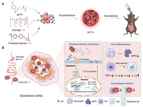
Background: Despite its promising therapeutic potential, inefficient site-specific targeting and complex fabrication processes hinder the clinical translation of oral melatonin therapy for ulcerative colitis (UC). Herein, we present Melatonin-Eudragit-Thioketal polymer-based Assembly (META), a dual pH/ROS-responsive oral microsphere for precise melatonin delivery to the inflamed colon and enhanced therapeutic outcomes.
Methods: META was fabricated via a scalable emulsification process using Eudragit® FS 30 D and a thioketal polymer to enable dual pH- and ROS-responsive release. Its targeting delivery and therapeutic effects were then evaluated in simulated gastrointestinal (GI) fluids, 2D cell models, intestinal organoids, and a murine colitis model.
Results: Using cell and animal models, we showed the important role of melatonin in GI health, supporting our strategy for UC treatment. META exhibited controlled melatonin release and better accumulation in the inflamed colon. The targeted delivery helped promote disease recovery by modulating macrophages and improving the intestinal barrier.
Conclusion: META delivers melatonin precisely to the colon, which reprograms colonic macrophages and reinforces the epithelial barrier. This study offers a practical, clinically relevant approach for improving the use of endogenous therapeutic molecules in UC and other inflammatory GI diseases.
Keywords: ulcerative colitis, melatonin, colon-targeted oral delivery, macrophage modulation, intestinal barrier
Introduction
Ulcerative colitis (UC) is a chronic inflammatory bowel disease (IBD) that mainly affects the large intestine and leads to persistent inflammation and ulceration of the colonic mucosa [1, 2]. Its incidence has continued to rise worldwide, making it an increasing long-term burden for millions of people [2-4]. The disease develops through a complex interplay of genetic susceptibility, impaired epithelial barrier function, dysregulated immune responses, and environmental factors [5, 6]. Macrophages are central to this process. They arise from circulating monocytes and respond to inflammatory signals by differentiating into cells [7] that help maintain intestinal homeostasis. In UC, however, this balance is disrupted, and a shift toward pro-inflammatory M1 macrophages rather than anti-inflammatory M2 macrophages is thought to drive disease progression [8, 9]. Current treatment options mainly include anti-inflammatory drugs, immunomodulators, and biologics such as 5-aminosalicylic acid (5-ASA) agents, corticosteroids, immunosuppressants, and anti-TNF-α therapies [1, 2]. Although these treatments can help control symptoms, they often act on only one part of the inflammatory process rather than the whole disease mechanism. As a result, many patients do not achieve complete remission, and systemic side effects remain a serious concern [10, 11]. Immunosuppressive drugs can weaken normal immune function and increase the risk of infection and organ toxicity, while biologics are often limited by poor stability, short half-life, high cost, and the need for repeated intravenous administration, which can reduce compliance and overall treatment effectiveness [10, 12, 13]. Together, these limitations highlight the need for safer and more effective therapies with better delivery systems [14, 15].
Melatonin (N-acetyl-5-methoxytryptamine) is best known for its role in regulating circadian rhythms, but it also acts as a potent immunomodulator and antioxidant [16]. In addition to being secreted by the pineal gland, melatonin is produced locally by enterochromaffin cells in the gastrointestinal (GI) tract [17, 18]. It exerts antioxidant and anti-inflammatory effects that may be beneficial in a range of GI disorders [19, 20]. As an endogenous molecule, melatonin has the advantage of good biocompatibility, which may improve both efficacy and tolerability compared with conventional therapeutic agents [21]. Although preclinical and clinical studies have suggested that melatonin can reduce intestinal damage and alleviate UC symptoms, its therapeutic potential remains largely underexplored [22-25]. Because melatonin is stable in the acidic environment of the stomach [26], it has been considered for oral delivery, a route favored by patients for its convenience and potential access to the colon [19]. However, oral melatonin delivery is still limited by poor pharmacokinetics, insufficient accumulation in inflamed colonic tissue, limited retention at disease sites, and the hostile inflammatory microenvironment that reduces local drug activity [27-30]. In many cases, current formulations are absorbed too early in the small intestine, resulting in poor colonic accumulation and unwanted systemic effects [31, 32]. Advanced oral delivery systems for melatonin in UC have not yet reached clinical trials, and most studies still rely on free melatonin (Table S1). This gap underscores the need for precisely responsive delivery systems tailored to the unique pathophysiology of UC to improve therapeutic outcomes.
Although many advanced oral delivery platforms have been developed for UC, poor targeting to diseased tissue and complicated fabrication processes continue to limit their clinical translation [33-36]. For melatonin, a simple but effective platform that improves site-specific delivery could help unlock its full therapeutic potential and support clinical use. An effective oral delivery system for melatonin should consider disease-related changes in GI physiology, including altered pH and elevated reactive oxygen species (ROS) levels, to achieve precise targeting and consistent therapeutic effects [37, 38]. ROS levels in intestinal lesions from patients with IBD have been reported to be 10- to 100-fold higher than those in healthy individuals and play an important role in colitis progression [38, 39]. For this reason, ROS-responsive delivery systems are considered promising for UC treatment [39-45]. However, the spatial variability of ROS along the GI tract raises the risks of premature or off-target activation, which can reduce reliability [40, 46]. Similarly, the distinct pH profile along the GI tract has inspired many colon-targeted formulations based on pH-dependent release [47-49]. Yet variation in luminal pH among patients, as well as differences between healthy and diseased states, can reduce the consistency and effectiveness of these systems [43, 50, 51]. Given these challenges, monotherapies based on either ROS or pH responsiveness alone are often not sufficient, especially for melatonin, which requires highly precise delivery. By combining both triggers, a synergistic pH/ROS-responsive strategy can better navigate the dynamic GI microenvironment, allowing more controlled oral release of melatonin and improving therapeutic outcomes in UC.
In this study, we developed an oral delivery system for melatonin, Melatonin-Eudragit-Thioketal polymer-based Assembly (META), designed to respond to both pH and ROS. By taking advantage of these two disease-related triggers, the system can more selectively target inflamed colonic tissue and improve therapeutic efficacy in a murine colitis model. META incorporates biocompatible and functional materials, including Eudragit® FS 30 D, an FDA-approved excipient that responds to high pH [37, 52], and thioketal polymers that are sensitive to elevated ROS levels [43]. These components form a polymeric matrix that encapsulates melatonin through a simple and scalable emulsification process (Figure 1). Compared with more complex delivery platforms, this approach is easier to prepare and may be more practical for future translation. Beyond developing a delivery system, we also sought to better understand the role of melatonin in GI physiology and its mechanism in colonic repair via macrophage modulation and intestinal barrier reinforcement. Using single-cell analysis, intestinal organoid models, and in vivo colitis mouse models, we showed that META achieved effective colonic targeting and meaningful therapeutic benefit, with macrophages playing an important role in the response. Collectively, these findings may advance the understanding of melatonin-based therapies and offer a promising, clinically translatable strategy for treating UC.
Schematic illustration of META preparation and its mechanism of action in UC treatment. A, META fabrication and administration. B, Melatonin is released and accumulates specifically at injured colon sites, where it exerts therapeutic effects by modulating macrophage polarization and enhancing intestinal barrier integrity. MT: melatonin, TJ: tight junction, AJ: adherens junction.
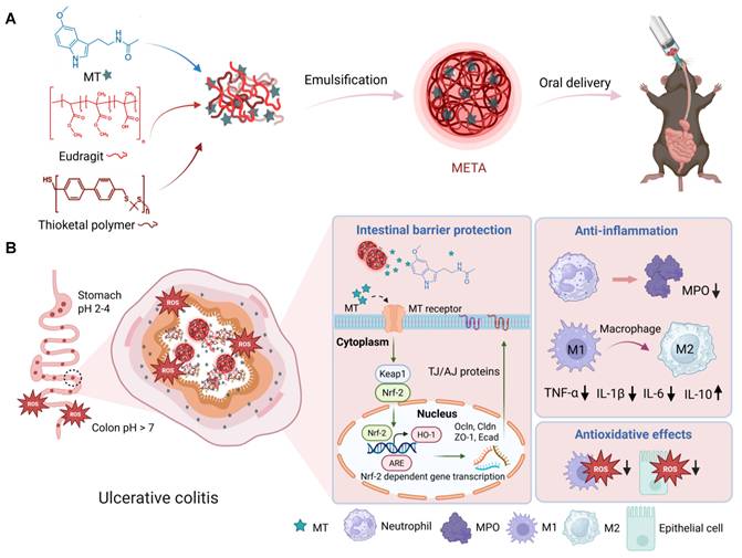
Relationship between melatonin and colonic inflammation. A, Induction of the mouse colitis model through oral dextran sulfate sodium (DSS) treatment. B, Body weight change measurement (n = 4-7 mice). C, Survival rate of mice (n = 4-7 mice). D, Representative histological images of colon tissue were obtained with H&E staining. Scale bar, 250 µm. E, Quantification of melatonin levels in colon tissue by ELISA (n = 4-5 mice). F, Relative mRNA expression of Aanat, Tph1, and Hiomt derived from the colon tissue using RT-PCR (n = 4-5 mice). G, Heat map depicting mRNA expression in RAW 264.7 macrophages (n = 3). H, Melatonin’s mechanism of action underlying macrophage modulation. I, Heat map depicting mRNA expression in Caco-2 (n = 3). J, Melatonin’s mechanism of action underlying protective effects. Data in (B) are presented as mean ± s.e.m. Data in (E, F) are presented as mean ± s.d. Data in (B, E, F) are analyzed using an unpaired two-tailed t-test. In (B) *p < 0.05, **p < 0.01, ***p < 0.001, ****p < 0.0001. In (E, F), significant p values are shown. Figures 2A, H, J were created with BioRender.com. MT: melatonin, TJ: tight junction, AJ: adherens junction, LPS: lipopolysaccharide, si-Scr: scrambled negative control siRNA.
Results
Relationship between melatonin and colonic inflammation
Melatonin (MT), which is abundantly secreted by enterochromaffin cells in the GI tract, plays an important role in maintaining gut integrity [53]. To investigate whether melatonin is involved in UC, we established a dextran sulfate sodium (DSS)-induced colitis model in C57BL/6 mice (Figure 2A). The model closely reproduces the major clinical and histological features of human UC [54]. Compared with healthy mice, DSS-treated mice showed marked body weight loss, reduced survival, and higher disease activity index (DAI) scores, indicating more severe disease (Figures 2B, C, and Figure S1). Histological examination also revealed crypt damage, epithelial injury, and inflammatory cell infiltration (Figure 2D). We then evaluated melatonin levels in the colon of both healthy and diseased mice. Mice with colitis showed significantly lower colonic melatonin and downregulated melatonin biosynthetic genes such as Aanat, Tph1, and Hiomt (Figure 2E, F). Taken together, these results suggest an association between lowered melatonin levels and intestinal damage in UC, and they also imply that melatonin deficiency may drive disease progression.
Based on the known anti-inflammatory and cytoprotective effects of melatonin in intestinal disorders, we further examined its impact on macrophage polarization, which is a key process in UC pathogenesis. Because STAT3 is an important regulator of inflammation [55], we used siRNA to knock down STAT3 in RAW 264.7 macrophages, with a scrambled negative control siRNA (si-Scr), to determine whether STAT3 signaling is required for melatonin’s anti-inflammatory effects. We confirmed efficient suppression of STAT3 at both the mRNA and protein levels (Figure S2A, B). Lipopolysaccharide (LPS) stimulation promoted M1 polarization, as shown by increased NF-κB p65 expression and upregulation of inflammatory genes. In contrast, melatonin treatment strongly suppressed these responses while increasing IL-10 expression, a marker associated with M2 macrophages (Figure 2G). Importantly, STAT3 silencing reduced these anti-inflammatory effects of melatonin, indicating that STAT3 signaling is necessary for melatonin activity in RAW 264.7 cells (Figures 2G, H). Melatonin increased p-STAT3 expression in a concentration-dependent manner (Figure S2C), suggesting activation of the STAT3 pathway. In addition, the STAT3 inhibitor stattic weakened the effects of melatonin on macrophage polarization (Figure S2D-G). Taken together, these findings suggest that melatonin regulates macrophage polarization largely through STAT3 signaling.
Melatonin also protected Caco-2 colonic epithelial cells from H2O2-induced injury and increased tight and adherens junction factors (Figure 2I). Mechanistic analysis suggested that this protective effect was mediated through the NRF-2/HO-1 pathway (Figures 2I, J). In addition, melatonin showed clear antioxidant activity (Figure S3), further supporting its ability to counter colitis-like stress. Together, these findings highlight the important role of melatonin in UC and suggest that melatonin supplementation may be a promising therapeutic option for UC.
Fabrication and characterization of META
We developed a dual pH- and ROS-responsive microsphere platform, namely META, designed to address the limitations of previous systems and precisely deliver melatonin to colonic inflammation sites via synergistic responsiveness.
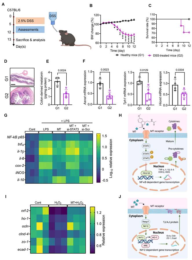
META was fabricated by a simple and scalable emulsification method as illustrated in Figure 3A. Eudragit® FS 30 D is an FDA-approved excipient for drug development with high pH-responsive properties (above pH 7) [37, 52]. The thioketal polymer was developed with highly biocompatible properties and introduced in our previous study [43]. The confirmed chemical structure showed that the ROS-sensitive linker enables this polymer to respond effectively in an enriched ROS environment (Figure S4). Individual polymer-based microspheres were also prepared and characterized following the same method for comparative purposes (Figure S5A-F). The melatonin loading capacity and entrapment efficiency of META were 6.41 ± 0.09% and 64.15 ± 0.91%, respectively (Figure 3B). META exhibited a spherical shape with a uniform size of approximately 3 µm (Figures 3C, E-initial timepoint) and a negative zeta potential, indicating favorable dispersibility and colloidal stability (Figure S5D, G). To better mimic disease-relevant conditions, luminal pH was first measured between healthy and colitis mice (Figure S6). We then comprehensively evaluated melatonin release from different formulations under simulated gastric fluid (SGF), simulated intestinal fluid (SIF), and simulated colonic fluid (SCF) corresponding to predetermined UC-mimicking conditions. The incubation times were selected to simulate the transit time of a single-unit dosage form in the gastrointestinal tract, specifically 2 h for SGF, 4 h for SIF, and an extended period for SCF [56, 57]. Interestingly, META showed minimal release in SGF and SIF but a pronounced, controlled release in SCF with elevated pH and ROS, indicating strong site-specific responsiveness (Figure 3D). Consistently, SEM images also revealed stimulus-triggered degradation of META with significant morphology changes in different conditions, especially in the SCF (Figure 3E). Besides, Melatonin-Eudragit microsphere (ME-MS) showed rapid melatonin release (with a similarity factor f2 between META and ME-MS of 34.2) from SIF to SCF due to extensive ionization of Eudragit in high pH conditions (Figure 3D). In contrast, Melatonin-Thioketal microsphere (MTK-MS) showed a delayed release (with a similarity factor f2 between META and MTK-MS of 27.5), reflecting the slower ROS-mediated cleavage of thioketal linkers (Figure 3D), as observed in a previous study [43]. These data indicated that a rational combination of functional polymers, including pH-responsive Eudragit and ROS-responsive thioketal polymer, effectively controls melatonin release and enhances precise delivery to the inflamed colon (Figure 3F).
META exhibits anti-inflammatory effects and intestinal barrier protection
After confirming the cytotoxicity profile of the microsphere (Figure S7), we evaluated multiple effects of META on single-cell models.
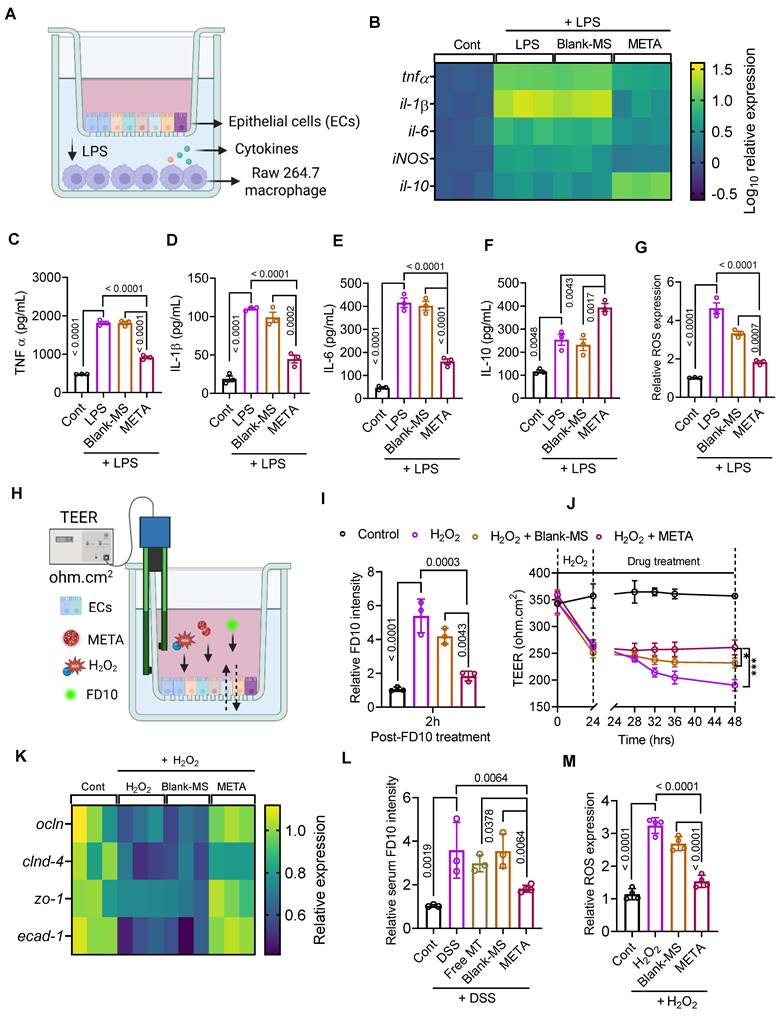
To evaluate the anti-inflammatory effects of META, we used a Caco-2/RAW 264.7 co-culture model (Figure 4A). RAW 264.7 macrophages were first driven into an inflammatory state with LPS, then treated with different formulations. As expected, inflammatory genes were upregulated in the LPS and Blank-MS groups, but META strongly dampened their expression (Figure 4B). Additionally, IL-10 mRNA was notably upregulated in RAW cells treated with META compared with the LPS or Blank-MS groups (Figure 4B). Consistent with these gene changes, cytokine measurements in the conditioned medium showed that META lowered pro-inflammatory cytokines, including TNF-α, IL-1β, and IL-6 (Figures 4C-E), while boosting IL-10 (Figure 4F). Altogether, these findings indicate that META suppresses inflammation by decreasing M1 macrophage polarization and promoting M2 activation. Furthermore, META also reduced oxidative stress with reduced ROS levels in treated RAW cells (Figure 4G), which supports its wider protective profile.
To determine whether META could protect the intestinal barrier from ROS-induced damage, we established a transwell-based epithelial cell model for permeability assessments (Figure 4H and Figure S8). Epithelial disruption was induced by H2O2 to mimic intestinal injury. META effectively preserved epithelial integrity with a lower FITC-Dextran 10 kDa (FD10) signal in the basolateral chamber compared to the H2O2 and Blank-MS groups (Figure 4I). Transepithelial electrical resistance (TEER) measurement further confirmed the protective effects of META, with significantly higher TEER values than in the H2O2 and Blank-MS groups (Figure 4J). In addition, META increased the expression of tight junction and adherens junction factors, indicating improved epithelial barrier function (Figure 4K). Importantly, in a murine colitis model, META showed better protective effects than other groups with lower serum FD10 levels (Figure 4L). Although free melatonin showed protective effects in vitro (Figure 2I, J), orally delivered melatonin had little impact on the colitis model, underscoring the need for targeted delivery to achieve therapeutic benefit (Figure 4L). Besides, Caco-2 cells treated with META also showed lower ROS levels, supporting the antioxidant capacity of this promising therapy (Figure 4M). Together, these results suggest META provides broad protection in colitis by combining antioxidant effects with reinforcement of the intestinal barrier, which may inhibit disease progression.
META notably preserves intestinal organoid integrity
META demonstrated excellent intestinal barrier protection, as evidenced by both the 2D cell model in vitro and the mouse colitis model (Figure 4). However, to further explore and comprehensively characterize META’s protective therapeutic effects, an intestinal organoid-based model was further employed (Figure 5A).
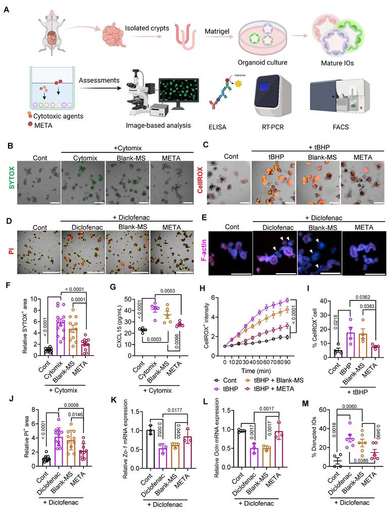
Upon confirming that neither META nor Blank-MS adversely affected organoid growth or viability (Figures S9A, B), we investigated the protective role of META in intestinal organoids (IOs) challenged with various cytotoxic agents (Figure 5A). To stimulate inflammation-mediated injury, IOs were exposed to a combination of pro-inflammatory cytokines, including IFN-γ, TNF-α, and IL-1β (Cytomix), for 24 h. Following treatment, the proportion of dead cells (SYTOX+) within the organoids increased significantly (Figures 5B, F). This tendency was mitigated by co-treatment with META in a dose-dependent manner, whereas Blank-MS showed no effect (Figures 5B, F). META treatment also significantly attenuated the secretion of the important immune mediator CXCL15/IL-8 in IOs in response to cytomix exposure (Figure 5G), indicating its ability to interrupt the inflammatory cascade in epithelial cells.
Building on the antioxidant effects of META observed in 2D epithelial cell models (Figure 4), we further examined its protective capacity against oxidative stress in a more physiologically relevant IO model. Real-time imaging showed that intracellular ROS levels rose quickly after tBHP treatment, confirming that oxidative stress had been successfully triggered (Figures 5C, H, and Figure S9C). The Blank-MS group showed similarly high ROS levels, almost the same as the tBHP group, while META treatment significantly attenuated tBHP-induced ROS accumulation (Figures 5C, H, and Figure S9C). Consistent with these findings, flow cytometric analysis further confirmed that META reduced the fraction of ROS-high cells to less than half compared to the tBHP-treated group (Figure 5I and Figure S9D). Overall, these data reinforce that META offers strong protection against ROS-induced damage to the intestinal barrier.
Next, we modeled NSAID-induced enteropathy by treating organoids with diclofenac, which disrupts epithelial tight junctions [58]. In the PI staining-based permeability assay, diclofenac-treated organoids showed higher PI penetration than the control group with intact tight junctions, indicating compromised barrier integrity (Figures 5D, J). Additionally, SYTOX staining also revealed reduced cell viability after diclofenac exposure (Figure S9E). These detrimental effects of diclofenac were markedly reduced when META was co-administered (Figures 5D, J, and Figure S9E). Prompted by these findings, we then examined the expression of tight junction marker genes. META effectively prevented diclofenac-induced down-regulation of tight junction-associated transcripts, including Zo-1 and Ocln, compared to the diclofenac and Blank-MS groups (Figures 5K, L). Because tight junction integrity is essential for preserving organoid structure, we next evaluated epithelial polarity. Under normal conditions, intact IOs typically exhibit basal-out polarity, with F-actin staining outlining the apical surface that faces the lumen (Figure S9F). In contrast, diclofenac treatment disrupted epithelial integrity and polarity, leading to structural disorganization and loss of the typical organoid morphology (Figures 5E, M). Interestingly, META largely preserved epithelial polarity of IOs, whereas Blank-MS showed severe structural disruption similar to that seen with the diclofenac group (Figures 5E, M). Collectively, these findings indicate that META effectively protects IOs against diverse cytotoxic agents, highlighting its therapeutic potential in preserving epithelial viability and barrier integrity under stress.
Dual pH/ROS-response of META enhances drug delivery to the inflamed colon
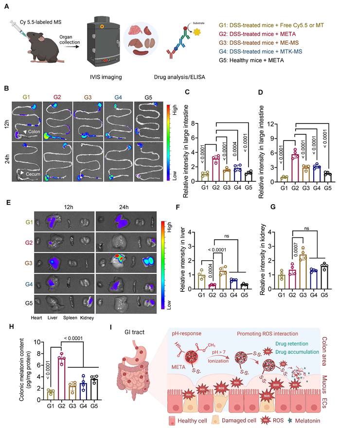
Resistance to the harsh conditions of the GI tract and prolonged retention in the colon are critical attributes for orally administered microspheres used in colitis treatment. In vitro evidence indicated controlled melatonin release from META under GI tract-mimicking conditions in colitis, as previously demonstrated (Figure 3). To further confirm the precise drug delivery of META, we investigated its fate following oral administration in healthy mice and DSS-induced colitis mice. Fluorescence images of Cy5.5-labeled microspheres were obtained using an in vivo imaging system (IVIS) at 12 and 24 h post-treatment (Figure 6A). Free Cy5.5 and individual polymer-based microspheres were included for comparative purposes.
Fabrication and characterization of META. A, Fabrication of the META platform. B, Melatonin loading capacity and entrapment efficiency of META (n = 3). C, META size distribution. D, Melatonin release profile of melatonin-loaded microspheres (n = 3). E, SEM images of META in GI tract-mimicking conditions in UC. F, Drug release mechanism of META in the GI tract. Figures 3A, F were created with BioRender.com. MT: melatonin, PVA: polyvinyl alcohol, SGF: simulated gastric fluid, SIF: simulated intestinal fluid, SCF: simulated colonic fluid.
META exhibits anti-inflammatory effects and protects the intestinal barrier. A, Co-culture model of the Caco-2 epithelial cells (ECs) and RAW 264.7 macrophages. B, Gene expression of inflammatory and anti-inflammatory factors in RAW cells (n = 3). C-F, Enzyme-linked immunosorbent assay (ELISA) quantification of extracellular cytokines, including TNF-α, IL-1β, IL-6, and IL-10, respectively (n = 3). G, ROS assessments in RAW cells (n = 3). H, Epithelial Caco-2 cell monolayer model for in vitro permeability assessment. I, FD10 intensity measurement in the basolateral chamber (n = 3). J, TEER values indicating epithelial barrier integrity, measured between apical and basolateral chambers (n = 3). K, Gene expression in the tight and adherens junctions in Caco-2 cells (n = 3). L, FD10 intensity measurement in the serum from mice after treatment (n = 3-4 mice). M, ROS assessments in Caco-2 cells (n = 4). Data are presented as mean ± s.d. Data are analyzed using one-way ANOVA with Tukey’s multiple comparisons test (significant p values are shown). Figures 4A, H were created with BioRender.com. TEER: trans-epithelial electrical resistance, FD10: FITC-Dextran 10kDa.
META notably preserves intestinal organoid integrity. A, Intestinal organoid (IO)-based assessments are shown. B, Representative images of Cytomix-induced IOs damage stained with SYTOX. Scale bar: 300 µm. C, CellROX staining showing oxidative stress-induced damage in IOs treated with tBHP. Scale bar: 300 µm. D, Propidium iodide (PI) staining depicts diclofenac-induced IO damage. Scale bar: 1000 µm. E, F-actin staining shows changes in organoid morphology and epithelial integrity after diclofenac treatment. Arrows indicate the disrupted structure. Scale bar: 300 µm. F, SYTOX-positive area of IO measurement (n = 11-12). G, CXCL15 concentration secreted from IOs (n = 5). H, Time course of CellROX intensity in IOs (n = 3-8). I, Percentage of CellROX-positive cells by flow cytometry after 60 min of treatment (n = 4). J, PI-positive area of IO measurement (n = 10-11). K-L, Relative mRNA expression of Zo-1 and Ocln in IOs (n = 3). M, Percentage of disrupted IOs (n = 4-6). In (F-M), data are presented as mean ± s.e.m. Data in (F-M) are analyzed using one-way ANOVA with the two-stage linear step-up method of Benjamini, Krieger, and Yekutieli (significant q values are shown). Figure 5A was created with BioRender.com. IOs: intestinal organoids, PI: propidium iodide, SYTOX: dead-cell nucleic acid stain, CellROX: ROS detection dye.
Dual pH/ROS-response of META enhances drug delivery to the inflamed colon. A, Illustration of biodistribution after oral treatment in mice using the in vivo imaging system (IVIS) and ELISA measurement. G1: DSS-treated mice + Free Cy5.5 or Free Melatonin (MT), G2: DSS-treated mice + META, G3: DSS-treated mice + ME-MS, G4: DSS-treated mice + MTK-MS, G5: Healthy mice + META. B, Fluorescence imaging shows the distribution of Cy5.5 in the GI tract. C-D, Relative fluorescence intensity in the large intestine 12 h (C) and 24 h (D) post-administration (n = 4 mice). E, Fluorescence distribution of Cy5.5 in major organs following different treatments. F-G, Relative fluorescence intensity 24 h post-administration in liver (F) and kidney (G) (n = 4 mice). H, Quantification of colonic melatonin content at 12 h post-administration by ELISA (n = 4 mice). I, pH- and ROS-interaction of META with the GI tract enhances drug retention and drug accumulation in the inflamed colon. Data in (C, D, F, G, H) are presented as mean ± s.d. Data in (C, D, F, G, H) are analyzed using one-way ANOVA with Tukey’s multiple comparisons test (significant p values are shown). ns, no significance. Figures 6A, I were created with BioRender.com. MS: microsphere, MT: melatonin, ECs: epithelial cells.
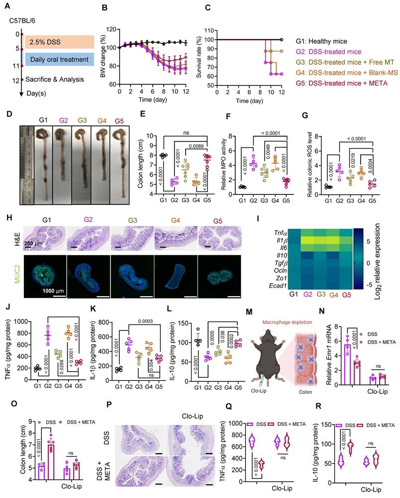
META therapy effectively ameliorates UC through macrophage modulation. A, Induction of the mouse colitis model through oral DSS delivery and subsequent drug treatment. G1: Healthy mice, G2: DSS-treated mice, G3: DSS-treated mice + free melatonin (MT), G4: DSS-treated mice + Blank-MS, G5: DSS-treated mice + META. B, Body weight change measurement (n = 5-8 mice). C, Survival rate of mice (n = 8 mice). D, Representative images of colon tissues. E, Colon length measurement (n = 5-8 mice). F, Myeloperoxidase (MPO) activity in colon tissues analysis (n = 5-8 mice). G, Colonic ROS measurement (n = 5-8 mice). H, Representative images of colon sections stained with H&E, scale bar: 250 µm (above panel) and representative immunofluorescence staining for MUC2 (green), scale bar: 1000 µm (below panel). I, Heat map of gene expression in colon tissues (n = 3-5 mice). J-L, ELISA measurement of colonic TNF-α (J), IL-1β (K), and IL-10 (L) (n = 5 mice). M, Macrophage depletion in mice via intraperitoneal injection of clodronate liposome (Clo-Lip). N, Emr1 expression in colon tissue (n = 4-6 mice). O, Colon length measurement (n = 4-6 mice). P, Representative images of colon sections stained with H&E. Scale bar, 250 µm. Q-R, ELISA measurement of colonic TNF-α (Q) and IL-10 (R) (n = 4-6 mice). Data are presented as mean ± s.d. Data in (E, F, G, J, K, L) are analyzed using one-way ANOVA with Tukey’s multiple comparisons test (significant p values are shown). Data in (N, O, Q, R) are analyzed using two-way ANOVA with Tukey’s multiple comparisons test (significant p values are shown). Figure 7M was created with BioRender.com. MS: microsphere, MT: melatonin.
Orally administered free drug or microsphere exhibited distinct distribution patterns in the large intestine (Figure 6B). In colitis mice, free Cy5.5 treatment showed low intensity at 12 h and was nearly undetectable at 24 h. Meanwhile, microsphere-based treatment revealed a stronger intensity in the colon area at both time points than that in the free Cy5.5 group. Notably, META demonstrated high efficiency of drug delivery to the inflamed colon with robust intensity of Cy5.5 compared to the individual polymer-based formulations (Figures 6B-D).
These differences can be attributed to the interactive properties between each formulation and the altered GI environment in colitis. Free Cy5.5, lacking a protective carrier, is vulnerable to enzymatic degradation, premature systemic absorption in the small intestine, and poor mucosal adherence, which are all factors reducing its stability and retention in the GI tract [59, 60]. Similarly, orally administered free melatonin faces comparable challenges, potentially limiting its therapeutic effectiveness. In contrast, META, a rationally designed delivery platform inspired by the physiological properties of the GI tract in colitis, could tune the interactions within this platform and effectively regulate drug release along with transit time in the colon. This design avoids either premature release due to high pH in the small intestine observed in ME-MS formulations or delayed release due to ROS-induced time-dependent response observed in MTK-MS formulations, both leading to diminished intensity due to rapid absorption or elimination from the colon before release. In healthy mice, META treatment exhibited a significantly lower colonic fluorescence intensity than that in the colitis mice (Figures 6B-D). The lower ROS levels and intact mucosal barrier in healthy animals reduce META-mucus interactions, facilitating rapid clearance and significantly decreased Cy5.5 intensity (Figures 6B-D). Thus, META enhanced drug retention and drug accumulation in the inflamed area, thereby potentially accelerating therapeutic response in colitis.
Major organs were also collected for fluorescence imaging to explore the fate of orally administered drugs. Notably, strong fluorescence signals were detected in the liver and kidney of the ME-MS group at both 12- and 24-h post-administration (Figures 6E-G). This finding indicates that premature ME-MS release, triggered by the elevated pH in the small intestine, leads to systemic absorption and accumulation in off-target organs, resulting in off-target delivery and possible side effects. Similarly, free Cy5.5 showed substantial hepatic accumulation at both time points (Figures 6E-G). As mentioned above, free Cy5.5 is unstable in the GI tract, undergoing rapid systemic absorption and degradation, which leads to poor intestinal retention and pronounced distribution to non-target tissues. In contrast, META and MTK-MS showed limited distribution in distant organs, attributable to controlled intestinal release profiles (Figures 6E-G). However, delayed colonic release by the MTK-MS platform could reduce therapeutic efficacy due to elimination by peristaltic movement in the GI tract [61].
Melatonin in the colon was quantified in a mouse model administered oral free melatonin or microsphere formulations, following the same treatment protocol as that employed for Cy5.5 imaging, to confirm the targeted delivery capacity of META. The META group exhibited significantly higher colonic melatonin levels than both free melatonin and individual polymer-based microsphere groups in the colitis mice and also exceeded that observed in healthy mice treated with the same META platform (Figure 6H). Collectively, these results demonstrate that the rationally designed META, featuring dual pH/ROS-responsiveness, enables precise and localized melatonin delivery to the inflamed colon, enhances drug retention, and promotes colonic drug accumulation via optimized interactions with GI tract physiology (Figure 6I).
META therapy effectively ameliorates UC through macrophage modulation
The DSS-induced colitis mouse model was used to evaluate the therapeutic effects of META in UC (Figure 7A). After 5 days of DSS treatment, mice then received daily doses of various treatments [19, 25, 27]. As expected, the DSS-only and Blank-MS group exhibited the most severe decline in body weight, followed by the free melatonin groups. In contrast, mice treated with META lost less weight and began to recover earlier than the other colitis groups (Figure 7B). Remarkably, META provided complete survival protection in DSS-treated mice, whereas other groups showed varying degrees of mortality. The free melatonin group had a survival rate of 87.5%, while the lowest survival rate of 62.5% was observed in colitis mice receiving no treatment or Blank-MS treatment (Figure 7C).
Among all treatment groups, META-treated colitis mice exhibited the most pronounced recovery, with colon length restored to nearly that of healthy mice. Mice receiving free melatonin also had longer colons than untreated or Blank-MS-treated colitis mice, although the effect was less marked (Figures 7D, E). In parallel, both myeloperoxidase (MPO) activity and ROS levels in the colon were significantly reduced in the META group compared with DSS group (Figures 7F, G). H&E staining further showed that META provided the strongest protection, preserving crypt architecture and epithelial barrier integrity while reducing inflammatory cell infiltration more effectively than free melatonin, Blank-MS, or no treatment (Figure 7H, above panel). Immunofluorescence staining also revealed higher mucin 2 (MUC2) expression in META-treated mice, indicating better maintenance of the intestinal barrier (Figure 7H, below panel). Consistent with these findings, gene analysis showed that META inhibited M1 markers and increased M2 markers. META also promoted the expression of tight junction and adherens junction markers, supporting improved barrier integrity (Figure 7I and Figure S10). These results were confirmed by ELISA, which showed that META reduced TNF-α and IL-1β levels while increasing IL-10 (Figures 7J-L). To further assess macrophage polarization in colon tissue, we examined CD86 and CD206, which are surface markers associated with M1 and M2 macrophages, respectively. PCR results showed that META markedly suppressed CD86 expression and increased CD206 expression in the colons of colitis mice, further supporting its immunomodulatory effect (Figure S11).
We also examined changes in the gut microbiota after META oral administration (Figure S12). The results showed that DSS greatly altered the microbial profile compared with healthy controls. META partially restored a healthier microbial balance while reducing dysbiosis-related changes. These findings suggest that META may also work, at least in part, by modulating the gut microbiota, potentially through a melatonin-microbiota-intestinal barrier/macrophage axis.
Moreover, META therapy was safe in vivo and did not cause systemic toxicity after treatment (Figure S13). In addition, we evaluated the therapeutic stability of META to further support its practical applicability. The results showed that META effectively protected melatonin and controlled its release under SGF-mimicking conditions for at least 12 h (Figure S14A). After storage at room temperature for 3 months, META retained its spherical morphology, an average size of approximately 3 µm, a negative zeta potential, and melatonin loading capacity comparable to that of fresh META (Figure S14B-D). Furthermore, in a DSS-induced mouse colitis model, stored META showed therapeutic efficacy similar to fresh META, as demonstrated by diverse assessments (Figure S14E-M). Collectively, these findings highlight that META effectively ameliorated colonic inflammation in UC and exhibits strong potential for practical application.
Macrophages as key mediators of META therapeutic activity in UC
To discover the functional role of tissue macrophages in META therapeutic efficacy, we depleted intestinal macrophages by intraperitoneal administration of clodronate liposomes (Clo-Lip) before and during the DSS-induced colitis model (Figure 7M). Macrophage depletion was confirmed with the significant reduction in the mRNA of Emr1, the gene encoding F4/80, which is a key marker of macrophages, in the Clo-Lip-treated groups (Figure 7N). Interestingly, in Clo-Lip-untreated groups, META treatment exhibited better recovery in colon length compared to DSS groups, whereas there was no significant difference between Clo-Lip-treated groups, and both resembled the Clo-Lip-untreated DSS group (Figure 7O). Histological assessments showed the severe condition of colon tissue with damaged crypt structure and epithelial barriers in Clo-Lip-treated groups, including DSS and META groups (Figure 7P). Furthermore, clodronate liposome treatment reversed META’s suppressive effect on the inflammatory cytokine TNF-α compared to the macrophage-intact META group (Figure 7Q). Macrophage depletion also abolished IL-10 production in both META and DSS groups (Figure 7R). Collectively, these findings indicate that the therapeutic effects of META are largely dependent on the presence of colonic macrophages.
Dual pH/ROS-response in META is critical for outstanding therapeutics against UC
The individual polymer-based microspheres, including ME-MS and MTK-MS, also showed potential therapeutic effects under in vitro conditions (Figure S15 and Figure S16). This is consistent with the fact that, in vitro, melatonin can be readily released and directly interacts with cells in a controlled environment. However, in vivo delivery is much more challenging. After oral administration, microspheres must pass through several barriers in the GI tract before reaching the inflamed colon. The acidic gastric environment and the risk of premature release in the small intestine may limit the ability of single-polymer systems to achieve efficient colonic delivery and sustained therapeutic effects. As shown above, the dual pH/ROS-responsive META platform improved drug delivery to the inflamed colon more effectively than the single platforms. We next evaluated whether META could also provide better therapeutic outcomes in the DSS-induced colitis model than the individual polymer-based microspheres, ME-MS and MTK-MS (Figure S17A). All treatment groups improved body weight compared with untreated DSS mice, but META induced earlier and more pronounced recovery (Figure S17B). Survival rates were also improved in all treatment groups, with META and ME-MS completely preventing mortality in colitis mice (Figure S17C). In addition, META restored colon length more effectively than DSS alone, while ME-MS and MTK-MS showed intermediate improvement (Figures S17D, E). MPO activity further indicated that META suppressed neutrophil infiltration and inflammation more effectively than either ME-MS or MTK-MS (Figure S17F). At the molecular level, META produced the strongest therapeutic response, with clear downregulation of pro-inflammatory markers (Tnfα and Il1β), upregulation of anti-inflammatory (Il10) and tight junction genes (Ocln and Zo1) compared to single polymer-based formulations (Figure S17G). Overall, these findings show that the rational dual pH/ROS-responsive design of META enables more precise melatonin delivery in the GI tract and leads to better therapeutic efficacy than the individual polymer-based systems.
Discussion
Among the existing treatments for UC, the highest patient compliance is observed with oral therapies, including 5-ASA, corticosteroids, immunosuppressants, and biologics. However, these often encounter limited colon-specific targeting, systemic side effects, and suboptimal efficacy due to the complex pathophysiology of UC [2, 29, 30]. These treatments primarily address downstream symptoms or isolated pathways, frequently failing to interrupt the disease’s vicious cycle or to restore intestinal barrier integrity comprehensively [10, 62].
Melatonin is an endogenous hormone produced in the GI tract and is an attractive therapeutic candidate because of its intrinsic multiple beneficial effects, including anti-inflammatory activity, antioxidant effects, and protection of the intestinal barrier, as shown in this study. Unlike many small-molecule drugs that do not directly match the underlying mechanisms of UC, melatonin acts on key upstream drivers such as immune imbalance and oxidative stress. Because it is an endogenous molecule, melatonin may be highly favorable to the GI tract condition, supporting tissue repair and recovery. However, the therapeutic potential of oral melatonin is limited by its early absorption and poor accumulation at inflamed colonic sites, which reduces its effectiveness and may also increase systemic side effects [27, 29, 31, 32].
In this study, we indicated that melatonin plays an important role in colonic inflammation. We also addressed the main limitation of oral melatonin delivery by using the META platform. This allows melatonin to be delivered more precisely, helping regulate immune responses and protect the intestinal barrier. However, to better assess the targeting performance of META, future studies will need more advanced methods such as LC-MS to more accurately evaluate the pharmacokinetics of the drug and microspheres.
Mechanistically, META could modulate macrophage polarization and reinforce the intestinal epithelial barrier. Deletion of intestinal macrophages reduces META’s therapeutic benefit, underscoring the pivotal role of macrophage-melatonin interactions in disease amelioration. Further studies should clarify the interaction between melatonin, an endogenous agent, and macrophages, an important immune cell type implicated in UC. Deep mechanistic insights into these interactions could inform drug selection and the design of delivery systems such as META, which promote native therapeutic effects at the site of inflammation. This integrated approach may yield superior therapies combining precision delivery with biological rationality, minimizing off-target effects, and maximizing efficacy.
In addition to the mechanisms assessed in this study, melatonin has been reported to regulate gut microbiota composition in experimental colitis models, and this action is increasingly recognized as critical to its therapeutic effect [23, 63]. Melatonin-mediated improvements in barrier function, mucin production, and antimicrobial peptide induction contribute to shaping a healthy microbial environment, while shifts in microbiota further modulate host immune responses [64]. Our PCR-based microbiota abundance analysis showed that DSS caused a pronounced dysbiotic shift, whereas META partially reversed these changes and moved the microbial profile toward that of healthy mice. In particular, the recovery of beneficial taxa together with the attenuation of DSS-associated microbial alterations supports a potential role for the gut microbiota in the therapeutic action of META. However, these analyses were based on limited information regarding the gut microbiota and therefore cannot fully elucidate the underlying mechanisms of META. In future studies, more comprehensive 16S rRNA sequencing analyses, including microbiota composition, α/β diversity, and key taxonomic shifts, will be of great interest for characterizing the “melatonin-microbiota-intestinal barrier/macrophage” axis, and may reveal further synergistic mechanisms underlying META therapeutic benefit in UC.
A notable advancement of this study is the use of IOs as an advanced in vitro model to evaluate META’s protective effects on the intestinal barrier under inflammatory and oxidative stress conditions. IOs could closely recapitulate the complex epithelial architecture, cell heterogeneity, and physiological responses of the intestine [65]. Our META significantly preserved organoid integrity from different damage factors. This organoid-based assessment strongly supports the translational potential of META from in vitro experiments to in vivo outcomes.
The DSS-induced colitis model reflects many key features of human UC, which supports the relevance of our findings. However, mouse models still have clear limitations, because immune responses and disease progression can vary between species. This is especially important for META, since it is designed to respond to the altered GI environment during colitis. Before clinical translation, future studies in larger animal models and non-human primates will be needed to confirm its efficacy and safety.
Other than its biological success, the META drug could also be produced using an easy and scalable emulsification technique, which would increase its translational capability. Through the advancement of technologies such as microfluidic technology, the META system could be improved to increase its capacity to load drugs and its encapsulation effectiveness, thus making large-scale production cost-effective. The easy production technique will solve one of the key problems facing the translational capability of pharmaceuticals [66], bridging the gap from the lab to the bedside. Additionally, since the META drug delivery system is modular, it could be modified to deliver a wide variety of therapeutic drugs other than melatonin for other conditions. Besides, META is generated from biocompatible and safe materials with solid evidence. Together, these features give META both useful functionality and good translational potential.
In conclusion, this study provides mechanistic insight with practical designs to develop an orally effective delivery system. META improves melatonin delivery, which regulates macrophage activity and the intestinal barrier in the inflamed colon. Its simple preparation and consistent performance suggest that it could be a promising approach for UC and other inflammatory GI diseases.
Materials and Methods
Animals and cell culture
Male C57BL/6 mice (8–10-week-old) were purchased from Orient Bio (Republic of Korea). All animal experiments were performed following the national ethical guidelines and were approved by the Institutional Animal Care and Use Committee at Sungkyunkwan University (Suwon-si, Gyeonggi-do, Republic of Korea) (Approval no. SKKU IACUC 2023-09-28-1).
The RAW 264.7 murine macrophage cell line (ATCC) and the human intestinal epithelial cell line Caco-2 (ATCC) were cultured in Dulbecco’s Modified Eagle’s Medium (DMEM; ByLABS, cat# BY0011) supplemented with 10% fetal bovine serum (FBS; Gibco, cat# 12483020) and 1% penicillin-streptomycin solution (ByLABS, cat# BY1411), at 37 °C and 5% CO2.
Fabrication and characterization of melatonin-loaded microspheres
Melatonin-loaded microspheres were synthesized using an emulsification solvent-evaporation technique. Briefly, 90 mg of polymer, either thioketal polymer, Eudragit® FS 30 D (Evonik, Germany, CAS# 26936-24-3 and Lot# C220165001), or an equal mixture of both, was dissolved with 10 mg of melatonin (Thermo Fisher Scientific, cat# J62452.14) in 1 mL of dichloromethane (DCM). This organic phase was slowly added dropwise to 5 mL of an aqueous 1% (w/v) polyvinyl alcohol (PVA) solution under homogenization at 13,000 rpm for 5 min to generate an oil-in-water emulsion. The emulsion was then transferred to 60 mL of 1% PVA and stirred at RT for 5 h to allow solvent evaporation. The resulting microspheres were then recovered by centrifugation, washed, and freeze-dried. Their morphology was examined by scanning electron microscopy (SEM; JEOL, JSM-IT800), particle size distribution was determined using laser diffraction (Beckman Coulter LS 13320, CA, USA), and zeta potential was measured with a Zetasizer (Malvern, UK). Additionally, we prepared a blank microsphere (Blank-MS), used as a control of META, containing Eudragit® FS 30 D and thioketal polymer without melatonin, for further functional assessments.
The amount of melatonin was analyzed by high-performance liquid chromatography (HPLC; Waters, USA) using a C18 column (5 µm, 150 × 4.6 mm; GL Science). Mobile phases for HPLC were prepared using acetonitrile and water (ratio 70:30, v/v) with a flow rate of 1 mL/min. Melatonin detection was done using a photodiode array detector at a wavelength of 220 nm. The loading capacity and entrapment efficiency were calculated using the formulas given in Table S2.
To assess the pH/ROS-responsive degradation of META, microspheres were sequentially incubated in simulated gastric fluid (SGF; pH 2.5) for 2 h, simulated intestinal fluid (SIF; PBS pH 7.2) for 4 h, and simulated colonic fluid (SCF; PBS pH 7.4) containing 1 mM H2O2 to mimic elevated ROS. Changes in microsphere surface morphology were examined by SEM. Melatonin release under these simulated GI conditions was quantified by HPLC. The release profiles of different formulations were compared using the similarity factor f2 (Table S2) with f2 values below 50 indicating dissimilar dissolution profiles [67].
In vitro ROS assay
RAW 264.7 macrophage and Caco-2 epithelial cells were seeded in 96-well plates at a density of 1 × 10⁴ cells per well. RAW 264.7 cells were activated with lipopolysaccharide (LPS, 500 ng/mL, cat# 2880-25MG), while Caco-2 cells were exposed to 100 µM H2O2. After 12 h of stimulation, cells were incubated with either free melatonin, Blank-MS, or the META for 24 h. Subsequently, the cells were stained with DCFH-DA (Sigma-Aldrich, cat# 35845-1G) for 15 min, followed by three washes with PBS. Fluorescence imaging was performed using a fluorescent microscope (Nikon, Japan), while quantitative analysis was conducted with a microplate reader (TECAN, Austria) at an excitation wavelength of 485 nm and emission wavelengths of 535 nm [68, 69].
In vitro anti-inflammatory assay
A co-culture model was established for Caco-2 and RAW 264.7 macrophages using 6-transwell insert plates (0.4 µm pore size, SPL), following a previously described method [70]. RAW 264.7 macrophages were then activated with LPS for 12 h. Subsequently, Blank-MS or META were treated to the basolateral chamber and incubated for 24 h. Supernatants were withdrawn for cytokine analysis by ELISA, including TNF-α (BioLegend, cat# 430904), IL-1β (BioLegend, cat# 432601), IL-6 (BioLegend, cat# 431304), and IL-10 (BioLegend, cat# 431414). Cells were harvested to evaluate inflammatory gene expression using RT-PCR.
In vitro permeability assessment
For in vitro cellular permeability studies, Caco-2 cells (1 × 10⁵ cells) were seeded in the apical chamber with a pore size of 0.4 µm of 24-transwell plates (Corning, USA). Upon reaching confluence and forming a monolayer, cells were treated with 100 µM H2O2 for 24 h. The monolayer cells were then washed twice with PBS and incubated with either Blank-MS or META for an additional 24 h. After treatment, monolayer cells were washed twice with PBS, and 200 µL of fluorescein isothiocyanate-dextran 10 kDa (FD10, Sigma-Aldrich, cat# FD10S-100MG) at 1 mg/mL in HBSS was added to the apical chamber. Samples from the basolateral chamber were collected after 2 h, and FD10 fluorescence was determined using a microplate reader (TECAN, Austria) at excitation/emission wavelengths of 485/525 nm [71, 72]. Additionally, TEER (Table S2) was measured at predefined time points using an EVOM2 epithelial voltohmmeter (World Precision Instruments) to monitor monolayer integrity throughout the assay [73].
Culturing IOs and image-based analysis
IOs were derived from mouse small intestine and cultured following established protocols [74]. Only organoids from passage 4 exhibiting stable growth and typical morphology were selected for experiments. Organoids were treated with varying concentrations (5-40 µg/mL) of Blank-MS or META, as well as with inflammatory stimuli including interferon-γ (IFN-γ; 10 ng/mL, Peprotech, Rockyhill, NJ), tumor necrosis factor-α (TNF-α; 10 ng/mL, Peprotech), interleukin-1β (IL-1β; 10 ng/mL, Peprotech), diclofenac (100 or 300 µM, MedChem Express, Monmouth Junction, NJ), and tert-butyl hydroperoxide (tBHP; 200 µM, Sigma-Aldrich, St. Louis, MO). Organoid morphology and viability were assessed by imaging with a BioTek Cytation 5 Cell Imaging Multi-Mode Reader (Agilent, Santa Clara, CA), and images were processed using Gen5 software with the image analysis module.
Assessments of IO viability and permeability
For growth assays, 50 organoids per well were cultured into 96-well plates for 3 days. Bright-field images were captured at the time of seeding (day 0) and on day 3. Relative growth was quantified by calculating the ratio of the organoid-covered area on day 3 to that on day 0. SYTOX Green (Thermo Fisher Scientific, Waltham, MA) staining was performed to assess organoid viability. A final concentration of 500 nM SYTOX Green was added to the culture medium and incubated for 15 min. Fluorescent images were then acquired to quantify the proportion of dead organoids, defined by SYTOX Green-positive staining. To evaluate cell-cell junctional integrity following damage, a permeability assay using propidium iodide (PI) was conducted [75]. IOs were cultured in 96-well plates (50 organoids per well) for 48 h and then treated with diclofenac, with or without META. After 24 h, PI (5 µg/mL; Thermo Fisher Scientific) was added to each well and incubated for 20 min at 37 °C. Bright-field and red fluorescence images were sequentially captured to visualize total organoid structures and PI-positive (permeable) regions, respectively.
Oxidative stress induction in IOs and ROS assessments
For real-time viability assessment, 5 µM CellROX Orange Reagent (Thermo Fisher Scientific) was added to organoid cultures on day 3 and incubated at 37 °C for 20 min. Following this, organoids were treated with t-BHP in the presence of either Blank-MS or META. Organoid images were acquired at 10-min intervals for a total duration of 90 min and analyzed to calculate the intensity of CellROX-positive organoid area over time. Meanwhile, the ROS levels in the IOs were assessed using flow cytometry. After 1-h exposure to tBHP in the presence of microspheres, organoids were incubated with 5 µM CellROX Deep Red Reagent (Thermo Fisher Scientific) for 20 min at 37 °C. Organoids were then harvested, dissociated into single cells via trypsinization, and subjected to flow cytometric analysis using an Accuri C6 Plus flow cytometer (BD Biosciences, Ashland, OR, USA).
DSS-induced acute colitis model and treatment
Acute UC was induced in male C57BL/6 mice (8–10-week-old) by administering 2.5% (w/v) DSS (36-50 kDa, MP Biomedicals, cat# 160110) for 5 days [54, 76]. Following DSS treatment, mice received daily oral gavage of either free melatonin (5 mg/kg/day) [25], melatonin-loaded microspheres (equivalent to 5 mg/kg/day of free melatonin), or Blank-MS for 7 days. Body weight and clinical symptoms were recorded daily until mice were sacrificed. The disease activity index (DAI) was measured following Table S3. On day 12, mice were euthanized, and colon tissues were collected for cytokine quantification, gene expression analysis, and histological assessments. In addition, colonic contents were harvested, and total bacterial DNA was extracted using a QIAamp FAST DNA Stool Mini Kit (Qiagen, Germany, Cat #51604) for qRT-PCR analysis to evaluate the abundance of disease-associated bacterial candidates (Table S4).
In vivo intestinal permeability assay and MPO activity assay
Gut barrier function was assessed in C57BL/6 mice using the FD10 permeability assay. Acute colitis was induced, and treatments were administered as described previously. Mice were fasted for 6 h prior to the assay and then orally gavaged with 150 µL of 80 mg/mL FD10 solution. Four hours post-gavage, blood samples were collected, and serum FD10 levels were quantified by fluorescence measurement at excitation/emission wavelengths of 485/525 nm [54, 71, 77]. The myeloperoxidase (MPO) activity was evaluated using a previously described method [43, 74].
Colonic ROS measurement
Colon tissues were rinsed twice with PBS (pH 7.4) and cut into small fragments. The tissue pieces were then digested in serum-free RPMI-1640 medium containing 0.1% collagenase P (Roche, cat# 11213857001) and 0.01% DNase I (Roche, cat# 11284932001) at 37 °C for 30-45 min with gentle shaking. The resulting suspension was passed through a 40-µm strainer to obtain single-cell suspensions. The cells were then washed, resuspended in culture medium, and incubated with 20 µM DCFH-DA at 37 °C for 15 min in the dark. After removing the excess dye by washing, intracellular ROS levels were quantified by a microplate reader at excitation and emission wavelengths of 485 and 535 nm, respectively. Fluorescence intensity was normalized against cell number.
Quantitative real-time polymerase chain reaction
Total RNA was extracted from Caco-2 cells, RAW 264.7 cells, or colon tissues using TRIzol reagent (Invitrogen, cat# 15596018). RNA concentration and purity were quantified using a NanoDrop 2000 spectrophotometer (Thermo Fisher Scientific). cDNA was synthesized using the RevertAid First Strand cDNA Synthesis Kit (Thermo Fisher Scientific, cat# K1622). qRT-PCR was then performed on a QuantStudio real-time PCR system using SYBR Green Master Mix (Applied Biosystems, cat# 4309155). Relative gene expression was calculated using the ΔΔCt method, with Gapdh or Actb serving as internal reference genes. Primer sequences were obtained from Bioneer Corporation (Daejeon, Republic of Korea) and are listed in Table S5.
Enzyme-linked immunosorbent assay
Cytokine and melatonin collected from cells and colon tissues were quantified using ELISA kits (for melatonin, mouse ELISA kit, ELK Biotechnology, cat# ELK9480) following the instructions from the manufacturer. Cells or tissues were lysed in RIPA buffer containing 1% protease inhibitor and 1% EDTA (Thermo Fisher Scientific). Absorbance was then read at 450 nm.
In vivo imaging analysis
To evaluate the targeted delivery capability of the META platform, Cy5.5-labeled microspheres and free Cy5.5 were orally administered to healthy and DSS-induced colitis mice at equivalent Cy5.5 doses. For labeling, Cy5.5 dye was incorporated into the microsphere formulation during the preparation process under the same conditions used for the corresponding blank or drug-loaded microspheres. After preparation, the Cy5.5-labeled microspheres were collected and washed to remove unbound dye. The Cy5.5 content in each microsphere formulation was quantified by fluorescence measurement to ensure consistent labeling and to allow dose adjustment for equivalent Cy5.5 administration across all groups. Mice were sacrificed at 12 and 24 h post-administration. The GI tract and major organs were harvested for fluorescence imaging using an In Vivo Imaging System (IVIS, Caliper Life Sciences) equipped with a Cy5.5 filter.
Depletion of intestinal macrophages
In brief, 200 µL of clodronate-liposome (Clo-Lip) (Encapsula NanoSciences LLC, Brentwood, USA) was intraperitoneally administered to mice 4 days before initiating DSS-induced colitis to deplete resident macrophages, and subsequently every 3 days throughout the protocol to eliminate infiltrating macrophages [78, 79].
AI tool
Perplexity was used to improve the language and readability of the Materials and methods section. Specifically, we first drafted this section ourselves and then used Perplexity to refine it for the above purposes. After using this tool/service, the authors carefully reviewed and edited the content as needed and take full responsibility for the content of the published article.
Statistical analysis
GraphPad Prism was used for statistical analyses. Unpaired two-tailed t-tests assessed differences between two groups, while comparisons involving three or more groups were conducted using one-way ANOVA or two-way ANOVA followed by post hoc tests (Tukey’s multiple comparison or false discovery rate, FDR correction). FDR-controlled tests report q values, whereas other tests present p values. Statistical significance was considered as p or q < 0.05.
Abbreviations
AJ: adherens junction; Clo-Lip: clodronate liposome; DSS: dextran sulfate sodium; EC: epithelial cell; ELISA: enzyme-linked immunosorbent assay; FD10: FITC-Dextran 10 kDa; GI: gastrointestinal; H&E: hematoxylin and eosin; IO: intestinal organoid; IVIS: in vivo imaging system; LPS: lipopolysaccharide; ME-MS: Melatonin-Eudragit microsphere; META: Melatonin-Eudragit-Thioketal polymer-based Assembly; MTK-MS: Melatonin-Thioketal microsphere; MT: melatonin; PI: propidium iodide; ROS: reactive oxygen species; SCF: simulated colonic fluid; SEM: scanning electron microscopy; SGF: simulated gastric fluid; SIF: simulated intestinal fluid; TEER: transepithelial electrical resistance; TJ: tight junction; UC: ulcerative colitis.
Supplementary Material
Supplementary methods, figures and tables.

Supplementary figure 9, movie.
Acknowledgements
The authors would like to acknowledge Evonik, South Korea, for kindly providing Eudragit® FS 30 D materials used in this study.
Funding
This research was supported by the National Research Foundation of Korea (NRF), funded by the Ministry of Science and ICT (grant No. 2021K1A3A1A20002609 and No. RS-2023-00272815), by the Bio & Medical Technology Development Program of the NRF funded by the Korean government (MSIT) (grant No. 2022M3A9G8017220 and RS-2023-00223591), and by a Korean Fund for Regenerative Medicine (KFRM) grant funded by the Ministry of Science and ICT and the Ministry of Health & Welfare (grant No. 22A0205L1 and No. 26B0106L1). In addition, this paper was supported by the 63 Research Fund, Sungkyunkwan University, 2025.
Author contributions
N.-N.N., H.-S.K., and J.-H.J. conceived the study. N.-N.N. and Y.S. designed and performed experiments, analyzed data, and wrote the manuscript. T.T.N., K.-P.P., and R.K. supported the experimental works. H.-L.J., J.Y.L., S.K., and N.-T.T. interpreted data and revised the manuscript. J.-H.J. and H.-S.K. revised the manuscript and supervised the project.
Data availability statement
The data supporting the findings of this study are available from the corresponding author upon reasonable request.
Declaration of generative AI and AI-assisted technologies in the manuscript preparation process
During the preparation of this work, the authors used Perplexity to improve the language and readability of the Materials and methods section. Specifically, we first drafted this section ourselves and then used Perplexity to refine it for the above purposes. After using this tool/service, the authors carefully reviewed and edited the content as needed and take full responsibility for the content of the published article.
Competing Interests
J.-H.J., N.-N.N., and H.-S.K. are submitting a patent application that includes work in this manuscript. The other authors have no conflicts of interest to declare.
References
1. Taku K, Britta S, Chen WS, Ferrante M, Shen B, Bernstein CN. et al. Ulcerative colitis (primer). Nat Rev Dis Primers. 2020;6:74
2. Berre CL, Honap S, Peyrin-Biroulet L. Ulcerative colitis. Lancet. 2023;402:571-84
3. Kaplan GG. The global burden of IBD: from 2015 to 2025. Nat Rev Gastroenterol Hepatol. 2015;12:720-7
4. Alatab S, Sepanlou SG, Ikuta K, Vahedi H, Bisignano C, Safiri S. et al. The global, regional, and national burden of inflammatory bowel disease in 195 countries and territories, 1990-2017: a systematic analysis for the Global Burden of Disease Study 2017. Lancet Gastroenterol Hepatol. 2020;5:17-30
5. Neurath MF. Targeting immune cell circuits and trafficking in inflammatory bowel disease. Nat Immunol. 2019;20:970-9
6. Danese S, Fiocchi C. Ulcerative colitis. N Engl J Med. 2011;365:1713-25
7. Zhao M, Yu S, Zhang M, Huang Y, Dou Z, Tian B. et al. Macrophages in Ulcerative Colitis: Immunomodulatory Roles, Phenotypic Switching, and Therapeutic Targeting. J Innate Immun. 2026;18:85-103
8. Chen S, Qin Z, Lin X, Zhou S, Xu Y, Zhu Y. Macrophages: emerging targets for ulcerative colitis. Front Immunol. 2025;16:1623491
9. Chen S, Saeed A, Liu Q, Jiang Q, Xu H, Xiao GG. et al. Macrophages in immunoregulation and therapeutics. Signal Transduct Target Ther. 2023;8:207
10. Xu M, Xin W, Xu J, Wang A, Ma S, Dai D. et al. Biosilicification-mimicking chiral nanostructures for targeted treatment of inflammatory bowel disease. Nat Commun. 2025;16:2551
11. Taylor KM, Irving PM. Optimization of conventional therapy in patients with IBD. Nat Rev Gastroenterol Hepatol. 2011;8:646-56
12. Tabas I, Glass CK. Anti-inflammatory therapy in chronic disease: challenges and opportunities. Science. 2013;339:166-72
13. Farrell RJ. Biologics beyond anti-TNF agents for ulcerative colitis - efficacy, safety, and cost? N Engl J Med. 2019;381:1279-81
14. Ungaro R, Mehandru S, Allen PB, Peyrin-Biroulet L, Colombel JF. Ulcerative colitis. Lancet. 2017;389:1756-70
15. Honap S, Jairath V, Danese S, Peyrin-Biroulet L. Navigating the complexities of drug development for inflammatory bowel disease. Nat Rev Drug Discov. 2024;23:546-62
16. Tarocco A, Caroccia N, Morciano G, Wieckowski MR, Ancora G, Garani G. et al. Melatonin as a master regulator of cell death and inflammation: molecular mechanisms and clinical implications for newborn care. Cell Death Dis. 2019;10:317
17. Zheng J, Zhou Y, Zhang D, Ma K, Gong Y, Luo X. et al. Intestinal melatonin levels and gut microbiota homeostasis are independent of the pineal gland in pigs. Front Microbiol. 2024;15:1352586
18. Wang B, Zhu S, Liu Z, Wei H, Zhang L, He M. et al. Increased Expression of Colonic Mucosal Melatonin in Patients with Irritable Bowel Syndrome Correlated with Gut Dysbiosis. Genomics Proteomics Bioinformatics. 2020;18:708-20
19. Asemani Y, Heidari R, Ezzatifar F, Mehrzadi S, Mosaed R, Karami E. et al. Melatonin supplementation in preclinical colitis models: A systematic review and dose-response meta-analysis on inflammation, oxidative stress, and colon repair. PharmaNutrition. 2024;30:100414
20. Gao T, Wang T, Wang Z, Cao J, Dong Y, Chen Y. Melatonin-mediated MT2 attenuates colitis induced by dextran sodium sulfate via PI3K/AKT/Nrf2/SIRT1/RORalpha/NF-kappaB signaling pathways. Int Immunopharmacol. 2021;96:107779
21. Kamfar WW, Khraiwesh HM, Ibrahim MO, Qadhi AH, Azhar WF, Ghafouri KJ. et al. Comprehensive review of melatonin as a promising nutritional and nutraceutical supplement. Heliyon. 2024;10:e24266
22. Mannino G, Caradonna F, Cruciata I, Lauria A, Perrone A, Gentile C. Melatonin reduces inflammatory response in human intestinal epithelial cells stimulated by interleukin-1beta. J Pineal Res. 2019;67:e12598
23. Zhao ZX, Yuan X, Cui YY, Liu J, Shen J, Jin BY. et al. Melatonin Mitigates Oxazolone-Induced Colitis in Microbiota-Dependent Manner. Front Immunol. 2021;12:783806
24. Cuzzocrea S, Mazzon E, Serraino I, Lepore V, Terranova ML, Ciccolo A. et al. Melatonin reduces dinitrobenzene sulfonic acid-induced colitis. J Pineal Res. 2001;30:1-12
25. Ahmed O, Farid A, Elamir A. Dual role of melatonin as an anti-colitis and anti-extra intestinal alterations against acetic acid-induced colitis model in rats. Sci Rep. 2022;12:6344
26. Pranil T, Moongngarm A, Loypimai P. Influence of pH, temperature, and light on the stability of melatonin in aqueous solutions and fruit juices. Heliyon. 2020;6:e03648
27. Mohanbhai SJ, Sardoiwala MN, Gupta S, Shrimali N, Choudhury SR, Sharma SS. et al. Colon targeted chitosan-melatonin nanotherapy for preclinical Inflammatory Bowel Disease. Biomater Adv. 2022;136:212796
28. Harpsoe NG, Andersen LP, Gogenur I, Rosenberg J. Clinical pharmacokinetics of melatonin: a systematic review. Eur J Clin Pharmacol. 2015;71:901-9
29. Shivaji UN, Nardone OM, Cannatelli R, Smith SC, Ghosh S, Iacucci M. Small molecule oral targeted therapies in ulcerative colitis. Lancet Gastroenterol Hepatol. 2020;5:850-61
30. Duran-Lobato M, Niu Z, Alonso MJ. Oral Delivery of Biologics for Precision Medicine. Adv Mater. 2020;32:e1901935
31. Larki RA, Iranmanesh A, Gholami D, Manzouri L. The effect of oral melatonin on the quality of life, sleep and blood pressure of hemodialysis patients: a randomized clinical trial. BMC Nephrol. 2025;26:451
32. Yener S, Akbulut KG, Karakuş R, Erdoğan D, Acartürk F. Development of melatonin loaded pectin nanoparticles for the treatment of inflammatory bowel disease: In vitro and in vivo studies. J Drug Deliv Sci Technol. 2022;67:102861
33. Kotla NG, Rana S, Sivaraman G, Sunnapu O, Vemula PK, Pandit A. et al. Bioresponsive drug delivery systems in intestinal inflammation: State-of-the-art and future perspectives. Adv Drug Deliv Rev. 2019;146:248-66
34. Tibbitt MW, Dahlman JE, Langer R. Emerging Frontiers in Drug Delivery. J Am Chem Soc. 2016;138:704-17
35. Ahadian S, Finbloom JA, Mofidfar M, Diltemiz SE, Nasrollahi F, Davoodi E. et al. Micro and nanoscale technologies in oral drug delivery. Adv Drug Deliv Rev. 2020;157:37-62
36. Zhao P, Xia X, Xu X, Leung KKC, Rai A, Deng Y. et al. Nanoparticle-assembled bioadhesive coacervate coating with prolonged gastrointestinal retention for inflammatory bowel disease therapy. Nat Commun. 2021;12:7162
37. Zhang Y, Wang Y, Lu Y, Quan H, Wang Y, Song S. et al. Advanced oral drug delivery systems for gastrointestinal targeted delivery: the design principles and foundations. J Nanobiotechnology. 2025;23:400
38. Li X, Lu C, Yang Y, Yu C, Rao Y. Site-specific targeted drug delivery systems for the treatment of inflammatory bowel disease. Biomed Pharmacother. 2020;129:110486
39. Zhang X, Zhao X, Hua Z, Xing S, Li J, Fei S. et al. ROS-triggered self-disintegrating and pH-responsive astaxanthin nanoparticles for regulating the intestinal barrier and colitis. Biomaterials. 2023;292:121937
40. Xiong T, Xu H, Nie Q, Jia B, Bao H, Zhang H. et al. Reactive oxygen species triggered cleavage of thioketal-containing supramolecular nanoparticles for inflammation-targeted oral therapy in ulcerative colitis. Adv Funct Mater. 2025;35:2411979
41. Sun Q, Luan L, Arif M, Li J, Dong QJ, Gao Y. et al. Redox-sensitive nanoparticles based on 4-aminothiophenol-carboxymethyl inulin conjugate for budesonide delivery in inflammatory bowel diseases. Carbohydr Polym. 2018;189:352-9
42. Vong LB, Tomita T, Yoshitomi T, Matsui H, Nagasaki Y. An orally administered redox nanoparticle that accumulates in the colonic mucosa and reduces colitis in mice. Gastroenterology. 2012;143:1027-36 e3
43. Regmi S, Pathak S, Nepal MR, Shrestha P, Park J, Kim JO. et al. Inflammation-triggered local drug release ameliorates colitis by inhibiting dendritic cell migration and Th1/Th17 differentiation. J Control Release. 2019;316:138-49
44. Liang J, Liu B. ROS-responsive drug delivery systems. Bioeng Transl Med. 2016;1:239-51
45. Huang Y, Canup BSB, Gou S, Chen N, Dai F, Xiao B. et al. Oral nanotherapeutics with enhanced mucus penetration and ROS-responsive drug release capacities for delivery of curcumin to colitis tissues. J Mater Chem B. 2021;9:1604-15
46. Aviello G, Knaus UG. ROS in gastrointestinal inflammation: Rescue Or Sabotage? Br J Pharmacol. 2017;174:1704-18
47. Gao W, Chan JM, Farokhzad OC. pH-Responsive nanoparticles for drug delivery. Mol Pharm. 2010;7:1913-20
48. Singh J, Nayak P. pH-responsive polymers for drug delivery: trends and opportunities. J Polym Sci. 2023;61:2828-50
49. Fan S, Zhao Y, Yao Y, Shen X, Chai X, Li J. et al. Oral colon-targeted pH-responsive polymeric nanoparticles loading naringin for enhanced ulcerative colitis therapy. J Transl Med. 2024;22:878
50. Liu L, Yao W, Rao Y, Lu X, Gao J. pH-Responsive carriers for oral drug delivery: challenges and opportunities of current platforms. Drug Deliv. 2017;24:569-81
51. Lou J, Duan H, Qin Q, Teng Z, Gan F, Zhou X. et al. Advances in Oral Drug Delivery Systems: Challenges and Opportunities. Pharmaceutics. 2023;15:484
52. Rabito MF, Reis AV, Freitas Ados R, Tambourgi EB, Cavalcanti OA. A pH/enzyme-responsive polymer film consisting of Eudragit FS 30 D and arabinoxylane as a potential material formulation for colon-specific drug delivery system. Pharm Dev Technol. 2012;17:429-36
53. Ahmadi S, Taghizadieh M, Mehdizadehfar E, Hasani A, Khalili Fard J, Feizi H. et al. Gut microbiota in neurological diseases: Melatonin plays an important regulatory role. Biomed Pharmacother. 2024;174:116487
54. Chassaing B, Aitken JD, Malleshappa M, Vijay-Kumar M. Dextran sulfate sodium (DSS)-induced colitis in mice. Curr Protoc Immunol. 2014;104:15 25 1-15 25 14
55. Samad MA, Ahmad I, Hasan A, Alhashmi MH, Ayub A, Al-Abbasi FA. et al. STAT3 Signaling Pathway in Health and Disease. MedComm (2020). 2025;6:e70152
56. Kali G, Knoll P, Bernkop-Schnurch A. Emerging technologies to increase gastrointestinal transit times of drug delivery systems. J Control Release. 2022;346:289-99
57. Feng K, Wei Y-s, Hu T-g, Linhardt RJ, Zong M-h, Wu H. Colon-targeted delivery systems for nutraceuticals: A review of current vehicles, evaluation methods and future prospects. Trends Food Sci Technol. 2020;102:203-22
58. Bhatt AP, Gunasekara DB, Speer J, Reed MI, Peña AN, Midkiff BR. et al. NSAID-induced leaky gut modeled using polarized monolayers of primary human intestinal epithelial cells. ACS Infect Dis. 2017;4:46
59. He J, Zhu T, Yu L, Mao N, Lu X, Shi X. et al. A based Cistanche deserticola polysaccharide functional-nanoparticle delivery system for effective oral vaccine to facilitate both systemic and mucosal immunity through enhancing oral delivery. Mater Today Bio. 2025;32:101939
60. He H, Qin Q, Xu F, Chen Y, Rao S, Wang C. et al. Oral polyphenol-armored nanomedicine for targeted modulation of gut microbiota-brain interactions in colitis. Sci Adv. 2023;9:eadf3887
61. Patel KS, Thavamani A. Physiology, Peristalsis. StatPearls. Treasure Island (FL): StatPearls Publishing. 2026
62. Yin L, Jiang X, Wang M, Yang Y, He Z, Sun J. et al. Phytoconstituent-derived nano-medicines/vesicles providing a promising dawn for inflammatory bowel disease. Chin Chem Lett. 2025;36:110224
63. Yi X, Cai R, Shaoyong W, Wang G, Yan W, He Z. et al. Melatonin promotes gut anti-oxidative status in perinatal rat by remodeling the gut microbiome. Redox Biol. 2023;65:102829
64. Ma N, Zhang J, Reiter RJ, Ma X. Melatonin mediates mucosal immune cells, microbial metabolism, and rhythm crosstalk: A therapeutic target to reduce intestinal inflammation. Med Res Rev. 2020;40:606-32
65. Xiang T, Wang J, Li H. Current applications of intestinal organoids: a review. Stem Cell Res Ther. 2024;15:155
66. Gazzi R, Gelli R, Aleandri S, Carone M, Luciani P. Bioinspired and bioderived nanomedicine for inflammatory bowel disease. Wiley Interdiscip Rev Nanomed Nanobiotechnol. 2024;16:e1986
67. Xie F, Ji S, Cheng Z. In vitro dissolution similarity factor (f2) and in vivo bioequivalence criteria, how and when do they match? Using a BCS class II drug as a simulation example. Eur J Pharm Sci. 2015;66:163-72
68. Shi C, Dawulieti J, Shi F, Yang C, Qin Q, Shi T. et al. A nanoparticulate dual scavenger for targeted therapy of inflammatory bowel disease. Sci Adv. 2022;8:eabj2372
69. Liu Y, Cheng Y, Zhang H, Zhou M, Yu Y, Lin S. et al. Integrated cascade nanozyme catalyzes in vivo ROS scavenging for anti-inflammatory therapy. Sci Adv. 2020;6:eabb2695
70. Mu K, Kitts DD. Gallic acid mitigates intestinal inflammation and loss of tight junction protein expression using a 2D-Caco-2 and RAW 264.7 co-culture model. Arch Biochem Biophys. 2024;756:109978
71. Singh R, Chandrashekharappa S, Bodduluri SR, Baby BV, Hegde B, Kotla NG. et al. Enhancement of the gut barrier integrity by a microbial metabolite through the Nrf2 pathway. Nat Commun. 2019;10:89
72. Choi SH, Eom JY, Kim HJ, Seo W, Kwun HJ, Kim DK. et al. Aloe-derived nanovesicles attenuate inflammation and enhance tight junction proteins for acute colitis treatment. Biomater Sci. 2023;11:5490-501
73. Tong L, Zhang S, Liu Q, Huang C, Hao H, Tan MS. et al. Milk-derived extracellular vesicles protect intestinal barrier integrity in the gut-liver axis. Sci Adv. 2023;9:eade5041
74. Regmi S, Seo Y, Ahn JS, Pathak S, Acharya S, Nguyen TT. et al. Heterospheroid formation improves therapeutic efficacy of mesenchymal stem cells in murine colitis through immunomodulation and epithelial regeneration. Biomaterials. 2021;271:120752
75. den Daas SA, Soffientini U, Chokshi S, Mehta G. A permeability assay for mouse intestinal organoids. STAR Protoc. 2022;3:101365
76. Kotla NG, Singh R, Baby BV, Rasala S, Rasool J, Hynes SO. et al. Inflammation-specific targeted carriers for local drug delivery to inflammatory bowel disease. Biomaterials. 2022;281:121364
77. Berg S, Suljovic D, Karrberg L, Englund M, Bonisch H, Karlberg I. et al. Intestinal Absorption of FITC-Dextrans and Macromolecular Model Drugs in the Rat Intestinal Instillation Model. Mol Pharm. 2022;19:2564-72
78. Bader JE, Enos RT, Velazquez KT, Carson MS, Nagarkatti M, Nagarkatti PS. et al. Macrophage depletion using clodronate liposomes decreases tumorigenesis and alters gut microbiota in the AOM/DSS mouse model of colon cancer. Am J Physiol Gastrointest Liver Physiol. 2018;314:G22-G31
79. Weisser SB, van Rooijen N, Sly LM. Depletion and reconstitution of macrophages in mice. J Vis Exp. 2012: 4105.
Author contact
Corresponding author: Jee-Heon Jeong, Ph.D., Department of Precision Medicine, School of Medicine, Sungkyunkwan University, Suwon 16419, Republic of Korea, Tel: +82-31-299-6165, E-mail: jeeheonedu. Hyung-Sik Kim, Ph.D., Department of Oral Biochemistry, School of Dentistry, Pusan National University, 49, Busandaehak-ro, Mulgeum-eup, Yangsan, 50612, Korea, Tel: +82-51-510-8231, E-mail: hskimcellac.kr.