Impact Factor




Theranostics 2026; 16(16):9200-9220. doi:10.7150/thno.133733 This issue Cite
Review
Cell-derived biomimetic nanomedicines: Emerging theranostic platforms for precision management of atherosclerosis
Xingji Liu1#, Zhiyue Wang2#, Jiaqi Lu3#, Jie Sheng2  , Longjiang Zhang1,2,3
1. Department of Radiology, Jinling Hospital, The First School of Clinical Medicine, Southern Medical University, Nanjing, Jiangsu 210008, China.
2. Department of Radiology, Jinling Hospital, Affiliated Hospital of Medical School, Nanjing University, Nanjing, Jiangsu 210008, China.
3. Department of Radiology, Jinling Clinical Medical College, Nanjing University of Chinese Medicine, Nanjing, Jiangsu 210008, China.
#Xingji Liu, Zhiyue Wang and Jiaqi Lu contributed equally to this work.
Received 2026-3-1; Accepted 2026-8-18; Published 2026-9-9
Abstract
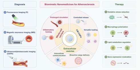
Atherosclerosis (AS), driven by complex cellular interactions, remains a leading cause of mortality worldwide. Conventional therapies often lack diseased blood vessels specificity, limiting therapy efficacy while increasing off-target toxicity. Boasting the biological advantages of cell-derived camouflages, these biomimetic nanomedicines outperform traditional drugs in intraplaque accumulation, circulation half-life and anti-AS efficacy. Current cell-derived biomimetic nanomedicines include whole-cell carriers, cell membrane-coated nanoparticles (NPs), and extracellular vesicles (EVs), all exploitable as novel platforms for plaque imaging and treatment. This review introduces the cellular mechanisms of AS pathology, examines recent advances in biomimetic strategies for AS theranostics and discusses the major barriers in clinical translation including biomaterial design, manufacturing consistency, safety evaluation, and large-scale production.
Keywords: atherosclerosis, biomimetic nanoparticles, drug delivery systems, clinical translation, theranostics
Introduction
Coronary artery disease (CAD) remains a primary cause of morbidity and mortality worldwide [1-5]. Currently, CAD management has shifted from handling ischemic complications to detecting and treating atherosclerotic plaques before the occurrence of clinical events. Earlier intervention is of vital importance to reduce the global burden of cardiovascular disease [6, 7].
Atherosclerosis (AS), the underlying pathology of CAD, is a chronic inflammatory disease. It involves endothelial dysfunction, lipid accumulation, and fibrous cap development within the arterial wall [8, 9]. Current techniques for plaque diagnosis include computed tomography (CT) angiography, magnetic resonance imaging (MRI), and intravascular imaging such as intravascular ultrasound, optical coherence tomography and near-infrared spectroscopy; nevertheless, none is able to directly reveal plaque phenotype and underlying mechanism [10]. As a result, they have limited sensitivity for detecting early-stage disease [11-16].
Lipid-lowering therapy, along with surgical or catheter-based revascularization, remains the conventional intervention of AS. Statins and proprotein convertase subtilisin/kexin type 9 (PCSK9) inhibitors lower low-density lipoprotein (LDL) cholesterol. Although these drugs significantly reduce cardiovascular risk, they exhibit limited efficiency in the plaque-targeted delivery, leading to insufficient drug accumulation at lesions and potential adverse effects, such as statin-associated myopathy [17-19]. Furthermore, surgical interventions such as coronary stenting and coronary artery bypass grafting can achieve revascularization but are associated with restenosis and graft occlusion [20-22]. Given these drawbacks, precise approaches for AS management are urgently required.
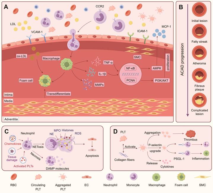
Schematic illustration of the key pathological processes underlying AS initiation, progression, and complications. (A) Endothelial dysfunction initiates atherosclerosis by increasing vascular permeability and upregulating adhesion molecules (VCAM-1 and ICAM-1) and MCP-1. CCR2 positive monocytes are recruited into the intima, where they differentiate into macrophages and take up oxidized low-density lipoprotein (ox-LDL), forming foam cells. Activated macrophages promote inflammation and matrix remodeling. Meanwhile, vascular smooth muscle cells (VSMCs) undergo phenotypic switching, migration, and proliferation through pathways such as NF-κB and PI3K/AKT. (B) Atherosclerotic lesions progress from early lesions to advanced plaques, which may cause cardiovascular complications. (C) During plaque progression, activated neutrophils undergo NETosis and release neutrophil extracellular traps (NETs) containing myeloperoxidase (MPO), histones, neutrophil elastase (NE), and reactive oxygen species (ROS), thereby exacerbating vascular inflammation and cell injury. In addition, necrotic cells release damage-associated molecular patterns (DAMPs), further amplifying inflammatory responses. (D) Plaque rupture or endothelial erosion exposes subendothelial collagen, triggering PLT activation and thrombus formation. Subsequently, activated PLTs increase the surface expression of P-selectin, which binds to P-selectin glycoprotein ligand-1 (PSGL-1) on neutrophils and macrophages. This interaction recruits leukocytes and promotes inflammation. Created by figdraw.com.
Nanomedicine can improve the precision of AS diagnosis and treatment [23-25]. Engineered nanoparticles (NPs) can not only deliver drugs in a controlled manner, but also achieve molecular imaging by targeting specific receptors [26]. However, one of the main challenges in the clinical translation is the immunological clearance of NPs from the circulation by the reticuloendothelial system. This rapid clearance reduces the blood circulation time of NPs, thereby limiting sustained drug delivery and compromising therapeutic efficacy [27-29]. To address this challenge, researchers have incorporated natural cellular delivery systems into nanoparticle designs.
The usage of natural cellular components endows cell-derived biomimetic NPs with extraordinary targeting efficacy to the lesion sites [30, 31]. The preserved membrane proteins and surface molecules provide endogenous recognition signals, such as CD47-mediated “don't eat me” signaling and receptor-mediated targeting interactions, which enhance immune evasion and promote accumulation in diseased tissues [32, 33]. In general, these NPs can be divided into three categories: whole-cell carriers, cell membrane-coated nanoparticles (CM-CNPs), and extracellular vesicles (EVs). Whole-cell carriers conjugate drugs directly to cells in a bid to improve homing ability and half-life in circulation. CM-CNPs combine biomedical materials, such as metal NPs, nanogels, and metal-organic frameworks, with natural cell membranes obtained from various cell types. These nanoplatforms retain functions such as immune evasion and tissue recognition. EVs, including exosomes and microvesicles, transport biological cargo such as proteins, regulatory RNAs, and lipids intercellularly. Upon uptake by target cells, EVs protect these cargo from endosomal degradation and facilitate their entry into the cytoplasm, enabling precise regulation of cellular functions [34, 35]. Collectively, these biomimetic NPs exhibit enhanced delivery and regulatory capabilities, thereby offering a promising avenue to address unmet needs in AS management.
This review explores cell-derived biomimetic nanomedicines for the diagnosis and treatment of AS. We first describe the pathological mechanisms and cellular processes that drive AS progression, then compare whole-cell carriers, CM-CNPs, and EVs and evaluate their applications in plaque imaging and targeted therapy. Moreover, we discuss the potential of clinical translation and the directions for future research.
Pathogenesis of AS
AS pathogenesis involves coordinated cellular and molecular interactions that drive progressive vascular alterations, underscoring the critical roles of diverse cell populations [36, 37]. In this section, we summarize the key cell types involved in AS and their contributions to disease development.
Endothelial cells (ECs)
Endothelial dysfunction is an early hallmark of AS [38]. Atherogenic risk factors, such as disturbed blood flow and oxidative stress, induce endothelial cells (ECs) activation and dysfunction. Activated ECs secrete chemokines, including monocyte chemoattractant protein-1 (MCP-1) and interleukin-8 (IL-8). They also upregulate the expression of vascular cell adhesion molecule-1 (VCAM-1) and intercellular adhesion molecule-1 (ICAM-1), thereby promoting the recruitment and adhesion of circulating immune cells to the vessel wall [9].
Endothelial barrier integrity disruption permits low-density lipoprotein (LDL) to infiltrate the arterial intima. Once retained in the intima, LDL accumulates and undergoes oxidative modification by reactive oxygen species (ROS), forming oxidized LDL (ox-LDL). Ox-LDL further stimulates endothelium and promotes the release of pro-inflammatory mediators, thereby sustaining local inflammation and promoting lesion formation [39-41].
Monocytes and macrophages
Monocytes are key components of the innate immune system and serve as the major contributors of macrophages within plaques [42]. Circulating monocytes are recruited to the activated ECs through coordinated chemokine and adhesion-molecule signals. Monocyte chemoattractant protein-1 (MCP-1) binding to C-C chemokine receptor type 2 (CCR2) drives monocyte chemotaxis, whereas endothelial vascular cell adhesion molecule-1 (VCAM-1) and intercellular adhesion molecule-1 (ICAM-1) mediate firm adhesion. Recruited monocytes then transmigrate across the endothelial layer into the intima, where they differentiate into macrophages and contribute to plaque formation [43].
Following transmigration and differentiation, macrophages internalize oxidized low-density lipoprotein (ox-LDL). Continued lipid uptake transforms the macrophages into foam cells, which is a typical feature of atherosclerotic lesions. Foam cells secrete pro-inflammatory cytokines, including tumor necrosis factor-alpha (TNF-α) and interleukin-1 beta (IL-1β), which increase the endothelial adhesion molecules and enhance the chemokine production [44], thereby amplifying monocyte recruitment and sustaining a feed-forward inflammatory loop [45-47].
Platelets (PLTs)
In response to vascular injury, PLTs adhere to exposed subendothelial collagen, initiating hemostasis and amplifying vascular inflammation [48]. This initial adhesion is mediated by glycoprotein VI (GPVI) and integrin α2β1 (GPIa/IIa). Subsequent platelet activation induces a conformational change in integrin αIIbβ3 (GPIIb/IIIa), enabling fibrinogen to cross-link adjacent PLTs. This interaction promotes PLTs aggregation and stabilizes PLTs adhesion to the injured vessel wall.
Once activated, PLTs translocate P-selectin from α-granules to the cell surface, where it binds to P-selectin glycoprotein ligand-1 (PSGL-1), a high-affinity receptor on circulating monocytes. Through this interaction, activated PLTs support monocyte adhesion and recruitment to atherosclerotic plaques [49-53].
Neutrophils
Neutrophil recruitment to atherosclerotic lesions is driven partly by interleukin-8 (IL-8)-CXCR1/CXCR2 signaling. Once activated, neutrophils produce ROS and release neutrophil elastase, thereby exacerbating endothelial dysfunction and vascular inflammation. Furthermore, neutrophils amplify vascular inflammation through the release of neutrophil extracellular traps (NETs), which activate macrophages and dendritic cells, enhance pro-inflammatory cytokine production and accelerate plaque progression [54-57].
Vascular smooth muscle cells (VSMCs)
VSMCs play both structural and regulatory roles during the development of AS [58]. As plaques develop, VSMCs migrate from the media to the intima, where they produce collagen and extracellular matrix (ECM) components. These components help preserve fibrous cap integrity and contribute to plaque stability.
However, inflammatory signals and oxidative stress can induce VSMC phenotypic switching from contractile state to synthetic state. Synthetic VSMCs lose their contractile function and secrete matrix-degrading enzymes, such as matrix metalloproteinases (MMPs), as well as pro-calcific factors. These secreted factors degrade the ECM, thereby weakening the fibrous cap and increasing plaque vulnerability to rupture [59, 60].
Red blood cells (RBCs)
RBCs contribute to atherosclerotic plaque progression primarily in the context of intraplaque hemorrhage (IPH). Intraplaque hypoxia and oxidative stress promote the formation of neovessels within atherosclerotic lesions. However, these are often immature, fragile and highly permeable, making them prone to rupture and facilitating RBC extravasation into the plaque.
Following IPH, extravasated RBCs undergo hemolysis and release damage-associated molecular patterns (DAMPs), thereby promoting necrotic core expansion, amplifying local inflammation, and increasing plaque vulnerability [61, 62].
Summary
AS is a chronic and complex disease that develops over several decades [63, 64]. Multiple vascular and immune cells play distinct but interconnected roles at different stages of disease. A better understanding of these cellular mechanisms may facilitate the rational design of biomimetic nanomedicines and enable the identification of promising cellular targets for the diagnosis and treatment of AS.
Advances in cell-derived nanotechnology for the theranostics of AS
According to the specific functions of different cells mentioned before, various cell-derived NPs have been developed for the treatment of AS [65]. These NPs retain native cellular functions and modes of intercellular communication, thus achieving improved immune evasion, prolonged circulation, and enhanced plaque accumulation [66-68]. The following section summarizes the major types of cell-derived nanomedicines (Tables 1 and 2) and introduces their benefits and limitations in AS management.
Summary of the main biomimetic nanomedicines for AS diagnosis
| Biomimetic Source | Nanomedicines | Particle Size | Targeting Mechanism | Animal Model | Imaging Modality | Reference |
|---|---|---|---|---|---|---|
| Macrophages | MφMB-Au | 50 nm | Inflammatory chemotaxis | ApoE-/- mouse | US/PA | 75 |
| RBC Membrane | R-Lipo@HDB/CH1055 R-Lipo@Cy7/CH1055 | 136.3 nm/147.6 nm | Passive targeting | ApoE-/- mouse | FI | 85 |
| Macrophage Membrane | MM@HGPBRD | 125.5 nm | Inflammatory chemotaxis | ApoE-/- mouse | MRI | 93 |
| Macrophage Membrane | LC-MM | 35 nm | Integrin α4β1/VCAM-1 recognition | ApoE-/- mouse | FI | 94 |
| PLT Membrane | PM@CBNP | 128.1 nm | GP Ib-IX complex/vWF binding | ApoE-/- mouse | FI | 101 |
| PLT Membrane | PM/Fe3O4@PLGA | 280.33 nm | GP Ib-IX complex/vWF binding | ApoE-/- mouse | MRI | 102 |
| PLT Membrane | PGA@PMP | 185.1 nm | ICAM-1/VCAM-1 recognition | ApoE-/- mouse | PA | 105 |
| PLT Membrane | RBT3-NO-PEG@PM | 130 nm | Glycoprotein receptors interaction | ApoE-/- mouse | FI | 106 |
| Neutrophil Membrane | NNPST | 116.8 nm | LFA-1/ PSGL-1/ TLR4 | ApoE-/- mouse | MRI | 115 |
| Neutrophil Membrane | Neu-Balloons | 8.4 ± 1.2 μm | CD11b/ ICAM-1/ E-selectin | ApoE-/- mouse | FI | 116 |
Summary of the key biomimetic nanomedicines for AS treatment
| Biomimetic Source | Nanomedicines | Particle Size | Mechanisms | Animal Model | Plaque Reduction Percentages | Reference |
|---|---|---|---|---|---|---|
| Neutrophil | MNP_pep | 23.9 nm | ROS ↓ | ApoE-/- mouse | 39.5% | 76 |
| RBC Membrane | Nanoghost | 263.0 nm | M2 polarization ↑ | Rabbit | - | 86 |
| RBC Membrane | miR-10a@H-MNP | 145.0 nm | M2 polarization ↑ | ApoE-/- mouse | 64.0% | 87 |
| RBC Membrane | RBC@P-LVTNPs | 254.0 nm | Lipid deposition ↓ | ApoE-/- mouse | 68.5% | 88 |
| Macrophage Membrane | R@MLP | 116.6 nm | Cholesterol efflux ↑ Macrophage phagocytosis ↑ | ApoE-/- mouse | 69.5% | 95 |
| Macrophage Membrane | (Lipo+M)@E NPs | 183.2 nm | PCSK9 level ↓ | ApoE-/- mouse | 81.2% | 96 |
| Macrophage Membrane | Au-ZnO@MM | 124.8 nm | Cholesterol efflux↑ Macrophage phagocytosis ↑ | ApoE-/- mouse | 58.0% | 97 |
| PLT Membrane | PBP@siR@PM | 140.0 nm | ROS ↓ Macrophage phagocytosis ↑ | ApoE-/- mouse | 82.3% | 107 |
| PLT Membrane | PM@EPPT | 105.3 nm | ROS ↓ Mitophagy ↑ | ApoE-/- mouse | 42.8% | 108 |
| PLT Membrane | Curc@Lignin@TA@PL | 418 nm | M2 polarization ↑ | - | - | 109 |
| Neutrophil Membrane | NeuM@P5c/S/C | 316.9 nm | M2 polarization ↑ Macrophage autophagy ↑ | ApoE-/- mouse | 74.3% | 117 |
| Neutrophil Membrane | Ptdser-NM-Lipo/Fer-1 | 174.9 nm | ROS ↓ | ApoE-/- mouse | - | 118 |
| Neutrophil Membrane | AM@ZIF@NM | 112.5 nm | MicroRNA-155 level ↓ | ApoE-/- mouse | 72.9% | 119 |
| EVs | P-EVs | 129.3 nm | ROS ↓ Lipid deposition ↓ | ApoE-/- mouse | 42.8% | 131 |
| EVs | NVEVs | 97.6 nm | Cholesterol efflux ↑ | SD Rats | - | 132 |
| EVs | P-ENVs | 150.9 nm | Cholesterol efflux ↑ | ApoE-/- mouse | 72.0% | 133 |
Whole-cell-based vehicles
Whole-cell vehicles use intact cells to carry therapeutic agents while preserving their cellular structure and function. Depending on the source cell, these carriers can evade immune clearance, prolong circulation, and migrate toward inflamed tissues. Therefore, these nanomedicines can increase drug accumulation in atherosclerotic plaques and improve therapeutic efficacy [69, 70]. Their prolonged circulation and inflammation-directed homing make whole-cell carriers suitable for targeting intermediate and advanced plaques with higher inflammatory activity [71]. Meanwhile, anti-AS agents encapsulated within cells usually have improved biocompatibility, which is another merit of whole-cell-based vehicles [72]. The intact cell structure shields the therapeutic cargo from immune recognition and complement activation, supporting the use of whole-cell carriers in AS imaging and treatment.
RBC-based vehicles are among the earliest biomimetic drug delivery nanoplatforms. Mature RBCs lack nuclei, facilitating their application as bioengineered carriers [73]. Shen et al. developed a RBC-based nanodrug for simvastatin delivery. The RBC structure reduced clearance by the mononuclear phagocyte system and prolonged the circulation half-life by 4.3-fold compared with the free-drug formulation [74].
Immune cells respond to chemotactic signals and migrate toward inflammatory microenvironments. Zeng et al. exploited this property to develop a dual-modality nanoprobe for early AS diagnosis. They used macrophages to encapsulate gold NPs. Owing to the homing ability of macrophages, this probe can be directed to atherosclerotic plaques and enable prolonged real-time monitoring through ultrasound/photoacoustic (US/PA) imaging [75]. Similarly, neutrophil-based melanin NPs have also been used in AS therapy. In vivo, these nanodrugs delayed disease progression and reduced plaque area by 24.7%, demonstrating that immune cell-biomimetic nanomedicine can effectively deliver therapeutic medicine to inflamed plaques [76].
Cell membrane-coated nanoparticles (CM-CNPs)
Although whole-cell carriers offer great biocompatibility and cell-specific targeting, their application is limited by the large-scale production and long-term storage. The need to maintain structural integrity and functional stability during manufacturing and preservation makes it challenging to meet clinical requirements. These constraints led to the development of CM-CNPs, which preserve cellular functions without requiring intact cells [77].
CM-CNPs are fabricated via coating NP cores with natural cell membranes [78]. Unlike whole-cell carriers, CM-CNPs can encapsulate hydrophobic agents or cytotoxic drugs within their cores and achieve controlled drug release [79-81]. This ability makes them suitable for targeting different stages of AS and obtain both diagnostic and therapeutic purposes. The following sections describe the characteristics and applications of four major types of CM-CNPs in AS.
RBCs
RBC is an ideal source for membrane-coated drug delivery. The anucleate structure of mature RBCs simplifies membrane isolation and modification, while the biconcave shape provides a large surface area for molecular exchange [82]. Compared with 5 to 90 hours of neutrophils and 5 to 7 days of PLTs in circulation, human RBCs circulate for about 120 days, providing a prolonged circulation time that facilitates sustained drug exposure and enhances drug accumulation at atherosclerotic lesions. RBCs are also cleared minimally by immune cells, making them well suited to targeted drug delivery [83].
In AS, the CD47 protein serves as a “do not eat me” signal that confers immune evasion capability upon RBCs, which extends circulation half-life and enhances targeted delivery to atherosclerotic lesions [84]. Based on this property, Liu et al. coated a ROS-responsive fluorescent molecule (CH1055) and a stable fluorescent dye (Cy7) with RBC membranes to construct two biomimetic nanoprobes. The RBC membrane coating prolonged the probe’s in vivo half-life from 1.55 to 2.82 h, enabling dynamic monitoring of inflammatory foci (Figure 2D-F) [85].
RBC membrane-derived nanomedicines for AS theranostics. (A) Schematic illustration of the preparation of miR-10a@H-MNP and its application in the treatment of AS. (B) Representative en face ORO staining images of aortas from ApoE-/- mice following different treatments. (C) Quantification of the plaque areas shown in (B). Adapted with permission from [87], copyright 2025, Springer Nature. (D) Representative ex vivo fluorescence images of aortas collected 24 h after injection of R-Lipo@HDB/CH1055. (E) Schematic illustration of the preparation of R-Lipo@HDB/CH1055 and R-Lipo@Cy7/CH1055. (F) Representative ex vivo fluorescence images of aortas from different treatment groups. Adapted with permission from [85], copyright 2023, Elsevier B.V. Reproduced with permission of the Licensor through PLSclear. All the data mentioned above were illustrated as mean ± SD. *P < 0.05, **P < 0.01, ***P < 0.001 by one-way ANOVA.
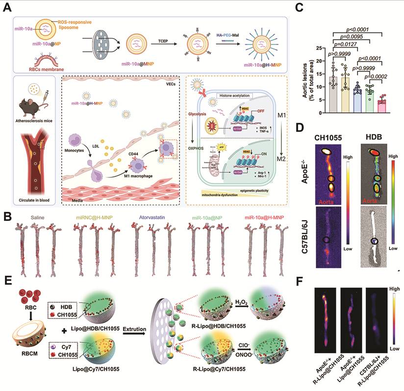
Oxidative stress in the local microenvironment is a key pathogenic factor of AS. Synthesizing anti-ROS agents with RBC membranes shows great potential for AS therapy. Karami et al. synthesized nanoghost (NG) by encapsulating glyburide within PLGA NPs coated with RBC membranes. Histochemical analysis revealed that NG reduced foam cell burden by 1.79-folds compared with un-coated NPs, demonstrating the therapeutic benefit of biomimetic encapsulation [86]. Similarly, an miR-10a-loaded RBC membrane–coated liposomal nanomedicine showed considerable potential for the targeted treatment of AS. This nanodrug considerably inhibited the progression of AS in vivo. This effect was confirmed by Oil Red O staining of mouse aortic sections, which showed an approximately 45% plaque reduction relative to Atorvastatin treatment controls (Figure 2A-C) [87].
Although RBC membrane–based nanomedicines show considerable promise in AS management, their clinical translation is constrained by low production yields. To address this limitation, Cao et al. developed a self-assembled RBC membrane-based nanodrug for AS management. They introduced the PS-binding peptide CLIKKPF onto NPs, enabling specific interactions with PS on RBC membranes. This method drove spontaneous self-assembly and enabled efficient membrane coating without complex processing methods such as electroporation [88].
In summary, RBC membrane-based biomimetic nanomaterials show great potential for AS theranostics. These nanomaterials retain key membrane proteins, such as CD47, which helps them evade immune clearance. This feature prolongs their circulation time and enhances drug stability. However, the lack of intrinsic active targeting limits their accumulation at plaque sites. Future studies should explore active targeting strategies to improve plaque-specific delivery efficiency [89].
Macrophages
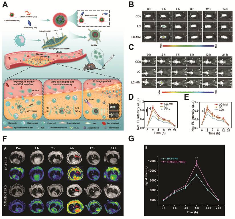
Macrophages coordinate innate immune responses through phagocytosis and immune signaling [90]. During AS progression, activated endothelial cells (ECs) express high levels of vascular cell adhesion molecule-1 (VCAM-1), which interacts with ligands on macrophage membranes, thereby promoting macrophage homing to plaques. As a result, macrophage membrane coating of NPs can facilitate delivery of imaging agents or therapeutics to plaques [91, 92]. For example, Li et al. developed macrophage membrane-coated Prussian blue NPs, termed MM@HGPBRD. In vivo studies showed that MM@HGPBRD produced stronger MRI signal enhancement in inflamed plaques than un-coated NPs (Figure 3F-G) [93]. Similarly, Duan et al. developed a macrophage membrane-cloaked fluorescent nanoprobe (LC-MM). The LC-MM nanoprobe exhibited higher fluorescence intensity in plaque regions than un-coated NPs. These results demonstrated its potential for real-time imaging of vascular inflammation (Figure 3A-E) [94].
Macrophage membrane-derived nanoprobes for AS diagnosis. (A) Schematic illustration of the preparation of LC-MM and its application in AS. (B-C) Representative in vivo fluorescence images of atherosclerotic plaques in ApoE-/- mice following intravenous injection of CDs, LC, or LC-MM, acquired in the prone (B) and supine (C) positions. (D-E) Quantitative analysis of the fluorescence signal intensities corresponding to (B) and (C), respectively. Adapted with permission from [94], copyright 2024, John Wiley and Sons. (F) Representative T1-weighted MR images of aortic plaques (red arrows) in AS model mice at 1, 2, 6, 12, and 24 h after tail vein injection of MM@HGPBRD or HGPBRD. (G) Quantitative analysis of the MR signal intensities of MM@HGPBRD and HGPBRD. Adapted from [93] under the CC BY license, copyright 2025, AAAS. All the data mentioned above were illustrated as mean ± SD. *P < 0.05, **P < 0.01, ***P < 0.001 by one-way ANOVA.
To achieve therapeutic effects, Cai et al. developed macrophage membrane-biomimetic liposomes (R@MLP) with a retinoic acid loaded core. This biomimetic design enabled the targeting and blocking of apoptotic cells with elevated CD47 levels in atherosclerotic plaques. In vivo studies demonstrated an approximately 70% reduction in plaque area compared to controls (Figure 4A-D) [95]. Similarly, Li et al. encapsulated evolocumab, a PCSK9 inhibitor, in macrophage-coated nanoparticles ((Lipo+M)@E NPs). Fluorescence imaging revealed a 1.59-fold higher plaque accumulation of (Lipo+M)@E NPs than un-coated NPs. This enhanced delivery resulted in superior efficacy, as (Lipo+M)@E NPs reduced plaque area to 3.78%, compared with 12.44% in the Lipo@E NP group (Figure 4H) [96].
Macrophage membrane-derived nanomedicines for AS therapy. (A) Schematic illustration of the fabrication and therapeutic mechanism of R@MLP. (B) Schematic illustration of the in vivo experimental design. (C) Representative en face ORO-stained aortas collected from atherosclerotic mice following different treatments. (D) Quantification of the plaque areas in the aortas after R@MLP treatment. Adapted from [95] under the CC BY license, copyright 2025, Elsevier B.V. (E-F) Quantification of the plaque area-to-lumen ratio (E) and necrotic core-to-lumen ratio (F) after treatment. (G) Representative images of aortic arch sections stained with ORO, H&E, and Masson's trichrome, together with immunohistochemical staining for α-SMA and caspase-3 following treatments. Adapted with permission from [97], copyright 2024, John Wiley and Sons. (H) Representative confocal fluorescence images showing Lipo/M@Lipo (red), HUVECs (blue), and ICAM-1 (green) in LPS-treated cells. Adapted from [96] under the CC BY license, copyright 2023, Springer Nature. All the data mentioned above were illustrated as mean ± SD. *P < 0.05, **P < 0.01, ***P < 0.001 by one-way ANOVA.
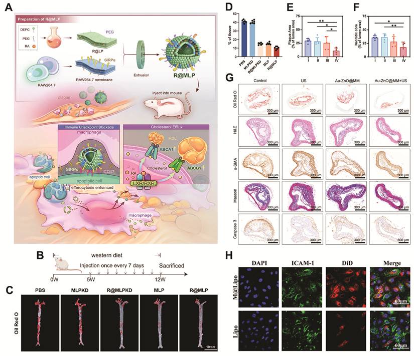
Foam cells are key contributors to atherosclerotic plaque formation, and their apoptosis promotes plaque instability. Cheng et al. developed macrophage membrane-coated Au-ZnO Schottky junctions (Au-ZnO@MM) to enhance foam cell elimination and thereby inhibit necrotic core formation. In vivo experiments showed an approximately 50% reduction in necrotic core area compared with control NPs, demonstrating the strategy’s potential to mitigate AS progression (Figure 4E-G) [97].
In summary, macrophage membrane-based nanomedicines offer substantial promise for AS therapy as an active targeting strategy [98]. Future studies should focus on optimizing macrophage membrane modifications to achieve stable recognition and selective binding to pathological targets, thereby broadening the clinical translation potential of macrophage membrane-based nanoplatforms [99].
PLTs
PLTs play essential roles in vivo, including in hemostasis, wound healing, and inflammatory modulation. Compared to nucleated cells, PLTs have fewer surface antigens and lower immunogenicity, making them attractive candidates for biomimetic drug delivery. Leveraging their targeting capabilities toward injured endothelial cells and thrombi in atherosclerotic lesions, PLT membrane–coated NPs (PLT M–CNPs) can selectively accumulate in the sites of vascular injury [51-53, 100]. This section reviews the application of PLT membrane-based nanotechnology in AS theranostics.
PLT M–CNPs show substantial potential for AS imaging. Zhou et al. designed a PM@CBNP nanoprobe based on the photosensitizer Ce6 for fluorescence imaging. This nanoprobe showed preferential accumulation in atherosclerotic plaques, resulting in 1.5-fold higher fluorescence intensity than bare micelles at 48 h post-injection [101]. To achieve accurate assessment, PM/Fe3O4@PLGA was developed as an MRI contrast agent for AS diagnosis. In vivo, PM/Fe3O4@PLGA showed a specific signal increase in the left carotid artery (LCA) at 24 h post-injection, but the un-coated controls gave almost no signal [102].
To enable real-time assessment of plaque progression, photoacoustic nanoprobes have been developed for AS diagnosis [10, 103, 104]. For example, Ma et al. constructed PGA@PMP by encapsulating the photoacoustic agent PMeDTDPP-THBTD within a PLT membrane. Carotid photoacoustic tomography revealed that PGA@PMP produced strong and specific signals at plaque sites, enabling real-time visualization of lesion progression [105]. Similarly, PLT membranes were used to cloak a near-infrared-II fluorescent dye, enabling real-time plaque imaging, as demonstrated by Chai et al. [106].
During plaque formation, PLTs adhere to the vascular endothelium by binding to exposed fibrinogen through the integrin αIIbβ3. Leveraging this targeting capability, Zhang et al. synthesized PBP@siR@PM NPs by encapsulating black phosphorus nanosheets within PLT membranes. The results demonstrated that these biomimetic NPs exhibited 2-fold greater accumulation at plaque sites than un-coated controls (Figure 5A-B) [107]. Similarly, PLT membrane-coated empagliflozin-loaded PCL-PEG polymeric micelles showed considerable potential for targeted AS treatment. In vivo, PM@EPPT reduced aortic plaque area by 43% compared with free empagliflozin (Figure 5C-E) [108].
PLT membrane-derived nanomedicines for AS therapy. (A) Schematic illustration of PBP@siR@PM for AS therapy. (B) Representative confocal fluorescence images showing the cellular uptake of PBP@siR and PBP@siR@PM by normal macrophages, inflammatory macrophages, and foam cells. Scale bar = 5 μm. Adapted with permission from [107], copyright 2024, Elsevier B.V. Reproduced with permission of the Licensor through PLSclear. (C) Schematic illustration of the in vivo experimental design. (D-E) Representative images of en face ORO-stained aortas from mice treated with PBS, EMP, EPP, EPPT, or PM@EPPT. Adapted from [108] under the CC BY license, copyright 2025, Frontiers Editorial Office. (F) Schematic illustration of Curc@Lignin@TA@PL. (G) Mean fluorescence intensity of CD86 expression in KG-1 cells after 48 h of incubation. Adapted from [109] under the CC BY license, copyright 2024, John Wiley and Sons. All the data mentioned above were illustrated as mean ± SD. *P < 0.05, **P < 0.01, ***P < 0.001 by one-way ANOVA.
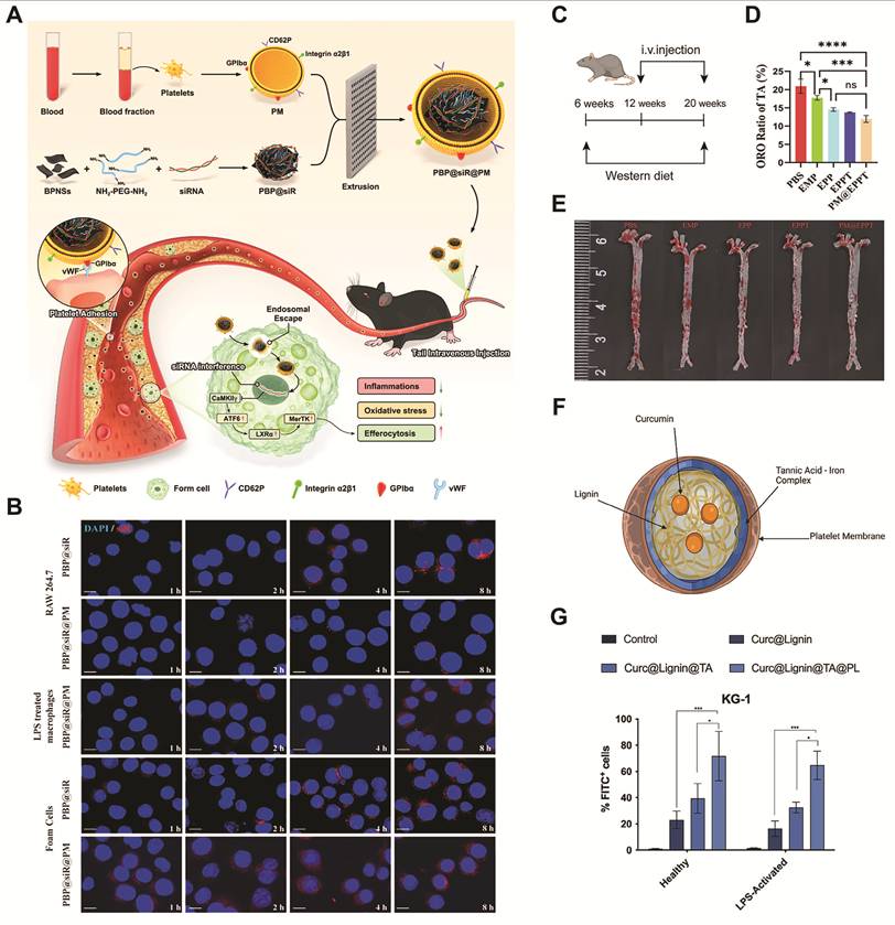
Fontana et al. developed a multilayer-modified nanosystem comprising a curcumin-loaded lignin core coated with tannic acid–iron complexes and an outer layer of PLT membrane fragments. The tannic acid–iron layer increased macrophage uptake from approximately 20% to 30%–40%, while subsequent PLT membrane coating further enhanced uptake to ≥60%. These results demonstrated that membrane modification techniques can enhance cellular targeting and improve plaque-directed drug delivery (Figure 5F-G) [109].
PLT membrane–based nanomedicines offer substantial advantages for AS theranostics, owing to their intrinsic affinity for injured vessels. However, the potential pro-thrombotic risk associated with these nanomaterials remains a key concern for clinical translation. To address this limitation, dynamically reconfigurable nanomaterials have emerged as a promising strategy. Future innovations should focus on smart biointerfaces incorporating stimuli-responsive molecular switches, such as shear stress–activated receptors, to enable self-regulated adhesion to plaques and detachment from healthy endothelium after treatment, thereby improving safety [110-112].
Neutrophils
Neutrophils act as frontline components of innate immunity. They respond to infection and tissue injury through phagocytosis, degranulation, and NET formation [113]. In early AS, activated neutrophils are recruited to damaged ECs through membrane proteins such as CD11b/CD18. This ability to rapidly respond to inflammatory signals makes neutrophil membrane-coated NPs promising candidates for the diagnosis and treatment of AS [56, 114].
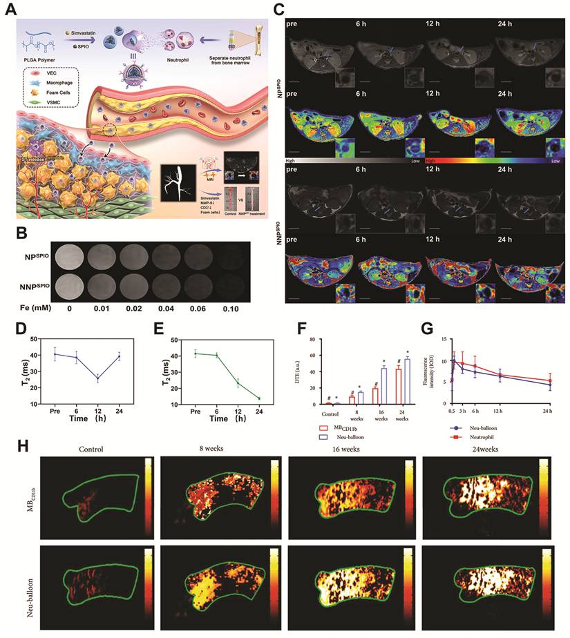
Leveraging this property, Zhang et al. developed neutrophil membrane–based MRI nanoprobes (NNPST), which exhibited a time-dependent reduction in T2 signal intensity within atherosclerotic plaques. At 24 h post-injection, NNPST revealed a 67% signal decrease (Figure 6A-E) [115]. To enhance production efficiency, Liu et al. engineered neutrophil-microbubble complexes (Neu-Balloons). These NPs maintained the same fluorescence intensity with a 60% cut-off of the neutrophils needed, thereby potentially addressing payload limitations in atherosclerotic plaque imaging (Figure 6F-H) [116].
Neutrophil membrane-derived nanoprobes for AS diagnosis. (A) Schematic illustration of the synthesis of NNPST and its application in AS. (B) Representative ex-vivo T2-weighted MR images of NPSPIO and NNPSPIO at different Fe concentrations. (C) Representative T2-weighted MR images and corresponding pseudocolor images of ApoE-/- mice acquired before and at different time points after injection of NPSPIO or NNPSPIO. Scale bar = 5 mm. (D-E) Quantification of the T2 relaxation times of atherosclerotic plaques following injection of NPSPIO (D) and NNPSPIO (E). Adapted from [115] under the CC BY license, copyright 2024, Elsevier. (F) Quantification of the PA signal intensity shown in (H). (G) Quantification of fluorescence intensity within atherosclerotic plaques after probe delivery by Neu-balloons or free neutrophils. (H) Representative PA signals in the Neu-balloon and MBCD11b-treated groups at 8, 16, and 24 weeks. Adapted from [116] under the CC BY license, copyright 2022, AAAS. All the data mentioned above were illustrated as mean ± SD. *P < 0.05, **P < 0.01, ***P < 0.001 by one-way ANOVA.
Neutrophil M–CNPs exhibit substantial anti-inflammatory potential. Liang et al. designed biomimetic NPs to deliver superoxide dismutase and catalase, thereby promoting macrophage polarization from the pro-inflammatory M1 to the anti-inflammatory M2 phenotypes. Treatment with P5c/S/C increased the proportion of M2 macrophages from 0.9% to 4.6% and reduced M1 macrophage proportion from 27.9% to 2.8% (Figure 7D-E) [117]. Similarly, Li et al. developed a neutrophil membrane–fused liposome (PtdSer-NM-Lipo/Fer-1) that achieved a ROS suppression rate of 38.26%, which was substantially higher than the 18.00% suppression observed with free Fer-1 [118].
Neutrophil membrane-derived nanomedicines for AS therapy. (A) Schematic illustration of the biomimetic AM@ZIF@NM NPs for targeted AS therapy. (B) Schematic illustration of the therapeutic protocol for evaluating AM@ZIF@NM NPs in ApoE-/- mice. (C) Representative images of en face ORO-stained aortas from mice treated with different formulations. Adapted with permission from [119], copyright 2023, American Chemical Society. (D) Representative flow cytometry analysis of macrophage subsets in LPS-stimulated RAW264.7 macrophages following 6 h incubation with different formulations. (E) Percentages of M2 (iNOS- and CD206+) and M1 (iNOS+ and CD206-) macrophages shown in (D). Adapted with permission from [117], copyright 2024, John Wiley and Sons. All the data mentioned above were illustrated as mean ± SD. *P < 0.05, **P < 0.01, ***P < 0.001 by one-way ANOVA.
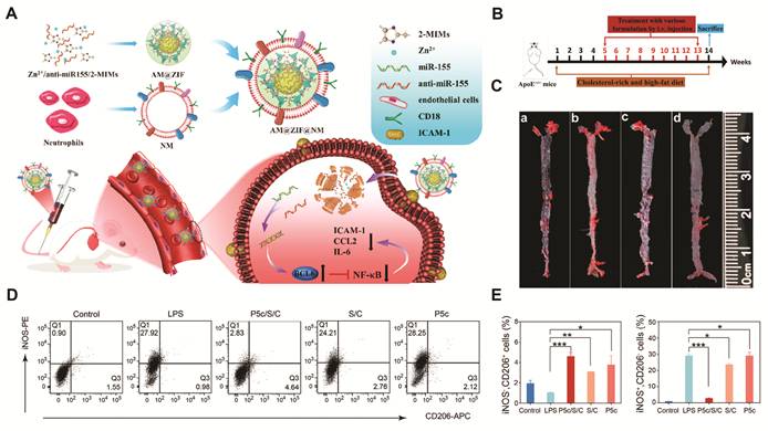
Gene silencing with antisense oligonucleotides (ASOs) is another promising therapeutic strategy for AS. Liu et al. developed neutrophil membrane–camouflaged NPs to deliver an ASO against microRNA-155 to ECs in atherosclerotic lesions. CD18 on the neutrophil membrane bound to ICAM-1 expressed on inflamed endothelium, thereby promoting ASO delivery. The delivered ASO subsequently suppressed the NF-κB pathway, alleviating endothelial inflammation and inhibiting monocyte recruitment. In vivo studies showed that the aortic lesion area decreased from 20% to 6.77%. These results confirmed the high therapeutic efficacy of the treatment (Figure 7A-C) [119].
Neutrophil membrane-coated nanomedicines have attracted increasing attention for AS theranostics because they retain neutrophil-derived chemotactic and adhesive properties, enabling efficient accumulation in inflamed vascular tissues. However, unintended activation by inflammatory signals from non-target tissues may cause off-target accumulation, reduce targeting accuracy, and diminish therapeutic effects, thereby limiting clinical translation [120-122]. Therefore, neutrophil membranes require modifications that limit nonspecific activation while preserving inflammation-directed drug delivery.
Extracellular vesicles (EVs)
EVs mediate intercellular communication by transporting proteins, lipids, nucleic acids, and other bioactive molecules. Based on their size, structure, and origin, EVs can be classified into three types: exosomes, microvesicles, and apoptotic bodies. Among the different EV types, exosomes are particularly useful for biomedical applications because of their nanoscale size of approximately 40 to 200 nm. In particular, mesenchymal stem cell (MSC)-derived exosomes can interact with PLTs and monocytes which are important components of the plaque microenvironment. Moreover, the favorable drug loading efficiency of EVs enables effective drug delivery to atherosclerotic plaques [123-126].
EVs as drug carriers
EVs can transport therapeutic cargo to selected cells and tissues [127]. Zeng et al. developed a delivery platform by camouflaging ginsenoside Rb1/probucol co-assembled NPs with macrophage-derived microvesicles. The structure could retain adhesion proteins, such as VLA-4 and Mac-1, which enhanced targeting to plaques and reduced off-target effects [128].
EVs applied in RNA-based therapeutics
RNA interference (RNAi), including microRNA (miRNA) and small interfering RNA (siRNA), is being investigated as a treatment for AS. Their therapeutic use depends on efficient delivery, because RNA is highly susceptible to degradation in vivo. Thus, RNA cargo needs to reach atherosclerotic lesions, escape endosomal degradation, and enter the cytoplasm to silence its target genes [129, 130]. Li et al. developed platelet membrane-fused EVs loaded with miR-21a-5p, termed P-EVs. Inflammatory macrophages internalized the P-EVs through endocytosis. The vesicles then escaped lysosomal degradation and released the miRNA into the cytoplasm. In vitro and in vivo studies showed that this treatment upregulated the cholesterol efflux transporters ABCA1 and ABCG1 and reduced plaque area from 26.15% to 14.96% (Figure 8A-C) [131].
EV-derived nanomedicines for AS therapy. (A) Schematic illustration of the fabrication and mechanism of P-EVs for the treatment of AS. (B) Representative en face ORO-stained aortas from mice treated with PBS, EVs, or P-EVs. (C) Quantification of the plaque area in the aortas. Adapted with permission from [131], copyright 2023, Elsevier B.V. Reproduced with permission of the Licensor through PLSclear. (D) Representative H&E-stained sections of balloon-injured arteries showing vascular morphology. Adapted with permission from [132], copyright 2025, John Wiley and Sons. (E) Schematic illustration of the synthesis of P-ENVs for AS therapy. (F) Representative ORO- and H&E-stained images of aortic roots from mice treated with PBS, ENVs, or P-ENVs. Scale bar = 200 μm. Adapted with permission from [133], copyright 2024, Informa UK Limited. Reproduced with permission of the Licensor through PLSclear. All the data mentioned above were illustrated as mean ± SD. *P < 0.05, **P < 0.01, ***P < 0.001 by one-way ANOVA.
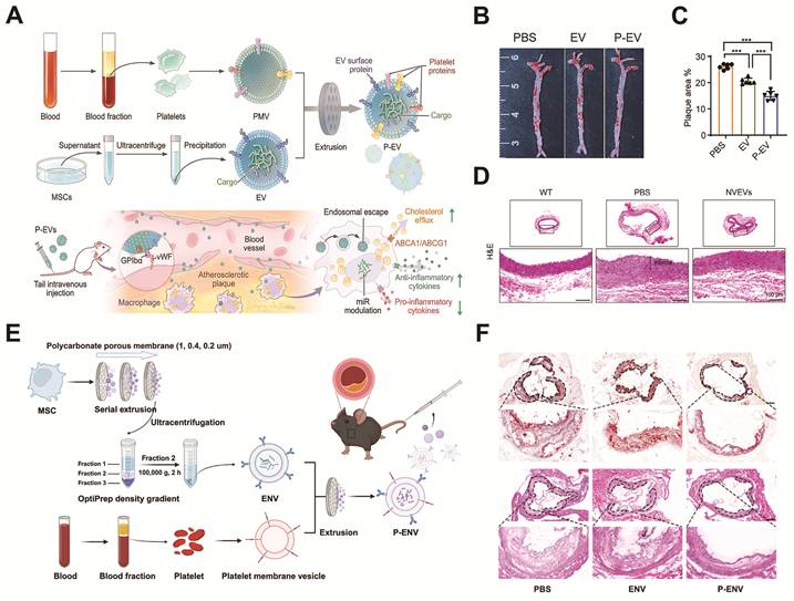
Similarly, neutrophil membrane-engineered EVs (NVEVs) delivering miRNAs such as miR-206-3p have shown promising therapeutic efficacy (Figure 8D) [132]. To enhance scalability, Jiang et al. developed synthetic exosome-mimetic nanovesicles coated with PLT membranes (P-ENVs), achieving a 250-fold increase in yield compared to conventional EVs while maintaining therapeutic performance (Figure 8E-F) [133].
Challenges and future perspectives
Despite these advances, EV-based therapeutics face critical challenges. EV composition and structure are highly heterogeneous, influenced by the cell type of origin, physiological conditions, and production methods. Consequently, the development of standardized isolation and production protocols is imperative for clinical practice [134, 135].
Targeting strategies for distinct atherosclerotic plaque phenotypes
Several microenvironment-targeting CM-CNP formulations have yielded therapeutic effects in experimental models. Their targeting performance, however, may be compromised considering that the plaque environment keeps changing throughout AS progression. Membrane source, therapeutic cargo, and release profile should therefore match the disease stage and plaque phenotype. The following sections introduce strategies tailored to specific stages and phenotypes (Tables 3 and 4).
Targeting strategies based on disease stage progression
| Disease stage | Key pathological features | Dominant targets | Therapeutic goal | Biomimetic design strategy | Representative biomimetic materials |
|---|---|---|---|---|---|
| Early lesion | Endothelial dysfunction, Increased vascular permeability, Mild inflammation | Activated endothelium | Restore endothelial function | Endothelium-targeted biomimetic delivery | Neutrophil membrane-coated NPs |
| Progressive lesion | Lipid accumulation, Chronic inflammation, Immune cells recruitment | Inflammatory plaque | Modulate inflammation and lipid metabolism | Immune cell-mimetic targeting to inflamed regions | Macrophage membrane-coated NPs; Neutrophil membrane-coated NPs |
| Advanced lesion | Necrotic core formation, ECM remodeling, Oxidative stress | Oxidative stress-rich plaque | Stabilize plaque microenvironment | Stimuli-responsive biomimetic nanomedicines | Macrophage membrane-coated NPs; |
| Thrombotic lesion | Thrombus formation, Vascular occlusion | Thrombus | Target thrombus and Prevent occlusion | Thrombus-targeting drugs with high affinity to clot components | PLT membrane-coated NPs |
Targeting strategies based on plaque phenotype
| Plaque phenotype | Pathological features | Dominant targets | Therapeutic goal | Biomimetic design strategy | Representative biomimetic materials |
|---|---|---|---|---|---|
| Vulnerable plaque | Thin fibrous cap, Large necrotic core, Intense inflammation, High rupture risk | MMPs, ROS, Inflammatory cytokines, Activated macrophages | Suppress inflammation, Inhibit ECM degradation, Promote plaque stabilization | Target inflamed endothelium and necrotic core, Cytokine sequestration | Macrophage membrane-coated NPs, Neutrophil membrane-coated NPs |
| Stable plaque | Thick fibrous cap, Small lipid core, Low inflammation, Lower rupture risk | Low-level inflammatory mediators | Maintain vascular homeostasis | Long circulation, Sustained drug delivery | RBC membrane-coated NPs, EVs |
Targeting strategies based on disease stage progression
Atherosclerotic lesions can be divided into early, progressive, advanced, and thrombotic stages. Each stage presents different cellular and molecular targets for biomimetic nanomedicines.
Early AS is characterized by endothelial dysfunction, increased vascular permeability, and mild inflammation [37]. In this situation, neutrophil membrane-based biomimetic vehicles have emerged as a reliable option because they can target inflamed endothelium [56, 114]. An example is the study by Wei et al. They developed a neutrophil membrane-coated miRNA nanoplatform to restore endothelial function and alleviate plaque progression [132].
As plaques progress, lipid deposition increases and inflammation exacerbates [46]. During this stage, immune cells actively migrate toward atherosclerotic lesions. Immune cell-mimetic NPs can use this property to head to diseased sites. (Lipo+M)@E NPs were developed on this basis for AS management. They used the natural homing ability of macrophages to target and treat atherosclerotic plaques [96].
Advanced plaques contain necrotic cores and undergo extensive extracellular matrix remodeling [136]. Oxidative stress also becomes a dominant feature of the plaque microenvironment. Stimuli-responsive biomimetic systems, particularly ROS-responsive systems, are therefore being explored for AS management [137]. Liu et al. developed macrophage membrane-coated nanomedicines with a ROS-sensitive agent. Oxidative stress in atherosclerotic plaques triggers drug release, and macrophage membrane coating helps them evade immune clearance and accumulate at lesion sites [138].
Plaque rupture and subsequent thrombosis are the most dangerous stage. These thrombi may dislodge and obstruct blood vessels and trigger acute cardiovascular events, necessitating precise delivery of thrombolytic agents. PLT membrane-based nanocarriers are well suited for this task because of their naturally homing to thrombotic sites [139]. Jiang et al. developed PSr@PT NPs that use PLT membrane-mediated adhesion to thrombi. As a result, the NPs can reduce oxidative injury and disrupt pathogenic interactions between PLTs and the endothelium [140].
Targeting strategies based on plaque phenotype
Clinicians generally classify plaques as stable or vulnerable according to their structure and risk of rupture. Because vulnerable plaque rupture causes many acute cardiovascular events, most biomimetic nanomedicine studies target this phenotype [141].
Vulnerable plaques, characterized by thin fibrous caps, large necrotic cores, and dense immune cell infiltrates, are unstable and prone to rupture. Activated macrophages release pro-inflammatory mediators and matrix metalloproteinases (MMPs), which degrade the extracellular matrix (ECM) and weaken the fibrous cap [18, 142-144]. Therefore, the treatment strategy in this case focuses on suppressing inflammation, limiting ECM degradation and stabilizing the plaque [145]. Macrophage and neutrophil membrane-coated nanomedicines can use inflammation-associated adhesion and chemotactic signals to reach inflamed endothelium and necrotic regions. Furthermore, some formulations function as biological decoys, and they can bind to pro-inflammatory cytokines and disrupt inflammatory pathways [146].
Stable plaques have thicker fibrous caps and a lower risk of rupture [64]. Treatment aims to preserve vascular homeostasis and prevent transition to a vulnerable phenotype. However, weak inflammatory signals and low vascular permeability will restrict drug accumulation in these lesions. Under this condition, the extended circulation of RBC membrane-coated NPs and EVs increases drug accumulation in plaque, thereby supporting sustained treatment against plaque progression [83].
Connecting pathology with biomimetic design
In summary, disease stage reflects the temporal progression of AS, while plaque phenotype exhibits structural stability and rupture risk. Considering both sides can guide the rational design of CM-CNPs and support more precise and effective treatment strategies for AS.
Imaging-guided and feedback-regulated theranostic strategies
Studies often evaluate the diagnostic and therapeutic functions of biomimetic nanomedicines separately. Theranostic platforms integrate both functions, allowing clinicians to track drug delivery and assess treatment response.
Imaging-guided therapy combines therapeutic cargo with diverse probes in the same nanoparticle. The imaging signal reveals NPs distribution and intraplaque accumulation, providing information that can guide treatment decisions. Macrophage membrane-coated NPs, for example, utilize the chemotactic properties of macrophages to reach atherosclerotic lesions. Incorporating a fluorescent probe into these carriers allows plaque localization and therapeutic response to be assessed at the same time [138].
Feedback-regulated platforms add stimulus-responsive drug release to imaging-guided therapy. Xu et al. developed an ultrasound-responsive nanoplatform in which ultrasound triggered therapeutic cargo release, while feedback signals reported treatment response. These functions allowed dynamic adjustments of treatment strategies according to the response [147]. Future research should focus on standardized biomimetic platforms that integrate imaging, drug delivery, and stimuli-responsive release. Combining these functions may further improve the accuracy and efficiency of AS treatment.
Clinical translation of biomimetic nanomedicines
Key steps of clinical translation include target validation, formulation optimization, preclinical evaluation of efficacy and safety, and phased clinical trials. Data on pharmacokinetics and manufacturing quality are also required [148, 149].
Cell-derived nanomedicines have shown great therapeutic effects in preclinical AS models, but their clinical development remains limited. So far, no cell-derived nanomedicine has entered a clinical trial specifically in AS treatment [150]. Nevertheless, experience from other disease indications may guide AS nanomedicine development. ERYtech Pharma developed eryaspase, which encapsulates L-asparaginase within RBCs as an adjunct to chemotherapy. Trials in acute lymphoblastic leukemia and pancreatic cancer has been preliminarily demonstrated safety and therapeutic activity [151, 152]. Similarly, Direct Biologics developed ExoFlo, an extracellular vesicle product derived from bone marrow mesenchymal stem cells, for patients with acute respiratory distress syndrome [153].
For AS, the growing number of patent applications for biomimetic NPs reflects industrial interest and translational potential (Table 5), which offers a potential platform for disease treatment.
Recent patent applications on biomimetic nanomedicines for AS
| Biomimetic Source | Patent Number | Applicant/Assignee | Publication Year |
|---|---|---|---|
| RBC membrane | CN121695107A | Qingdao University | 2026 |
| RBC-macrophage hybrid membrane | CN119792573A | Ningxia Medical University | 2025 |
| Macrophage membrane | WO2026060590A1 | Guangzhou Medical University | 2026 |
| Macrophage membrane | CN121489903A | Guangzhou University of Chinese Medicine | 2026 |
| Macrophage membrane | CN120037392A | Chongqing Medical University | 2026 |
| PLT membrane | CN118892467A | Huazhong University of Science and Technology | 2024 |
| PLT membrane | CN121243154A | Ningxia Medical University | 2026 |
| Exosomes | CN115590882A | Central South University | 2023 |
| EVs | CN121343873A | Dalian University of Technology | 2026 |
| EVs | CN121648319A | Institute of Biomedical Engineering, Chinese Academy of Medical Sciences | 2026 |
Future trials should include imaging endpoints that measure plaque burden, composition, and inflammatory activity. Regulatory assessments must also address the membrane source and batch-to-batch consistency of each formulation. Standardized characterization methods and good manufacturing practice (GMP)-compliant production shall produce reliable clinical-grade materials.
Challenges and future directions
Overcoming translational hurdles
One of the primary issues is the gap between laboratory design and clinical application. Due to insufficient studies on clinical translation of cell-derived NPs, the potential side effects of these endogenous carriers are still unclear.
Large-scale production is another problem. Critical procedures such as mechanical extrusion and sonication must be optimized in order to satisfy clinical standards and meet clinical demands. In addition, most studies have examined only short-term biological safety, while the long-term safety and potential immune reaction remain insufficiently evaluated [154, 155].
Optimizing material design
Material design of AS-targeted nanomedicine needs both parameter optimization and upgraded function. PLT-mimetic NPs, for example, can capture circulating procoagulant factors, which will reduce thrombosis and support vascular repair. Meanwhile, macrophage-mimetic NPs retain membrane-associated pathogen-recognition functions. Studies of macrophage membranes derived from Porphyromonas gingivalis (Pg)-treated cells suggest that MM/STNPs can eliminate Pg and suppress inflammation at infected sites [136, 156, 157].
Besides cell membrane coating, surface modification by organelle-derived membranes, including mitochondrial and lysosomal membranes, can use homotypic interactions to direct therapeutic cargo toward specific organelles. Also, organelle-specific proteins and bioactive components may regulate cellular signaling, adding therapeutic activity at intracellular and intercellular levels [158-160]. Zeng et al. developed an endoplasmic reticulum membrane-based mRNA nanovaccine (P-(H)-m@EMLN), in which endoplasmic reticulum membrane components enhanced endosomal escape and immune activation [161]. Such strategies for modulating intracellular trafficking pathways may provide new opportunities for designing multifunctional nanoplatforms for AS therapy.
Extracellular matrix (ECM)-inspired nanomedicines represent another new direction of biomimetic material design. ECM maintains vascular architecture and homeostasis by supporting cells and regulating the vessel microenvironment [162, 163]. Current ECM-mimetic materials include nanofibrous scaffolds composed of hyaluronic acid or silk fibroin. Li et al. developed ChS-PDANP@ADSCs, which is a hydrogel utilizing chondroitin sulfate (ChS), a major glycosaminoglycan in the ECM, to promote vascular regeneration and tissue repair [164].
Clinical assessment
Murine models, especially ApoE-/- and LDLR-/- mice, dominate the animal studies in current AS research because of their low cost and well-characterized genetics. These models can develop atherosclerotic lesions but rarely reproduce spontaneous plaque rupture or advanced vulnerable plaques observed in humans [165].
Large-animal models, to a certain degree, can simulate other aspects of the pathology of human AS. Swine models have comparable coronary anatomy and hemodynamics; Non-human primates can develop advanced lesions with features such as intraplaque hemorrhage. Nevertheless, these models involve higher costs and raise greater ethical and procedural concerns [166].
In addition, humanized models and organoid platforms can reproduce human pathological conditions more accurately. Blood vessel organoids (BVOs) derived from induced pluripotent stem cells are providing new approaches for assessing drug activity and safety under pathological conditions. Co-culturing BVOs with monocytes and exposing them to electronegative low-density lipoprotein (eLDL) and TNF-α recreates key inflammatory features of the atherosclerotic microenvironment. This system allows measurements of nanodrug uptake, therapeutic activity, and toxicity in human-derived vascular tissue and may improve the prediction of clinical responses [154, 167, 168].
AI-driven nanoplatforms
Artificial intelligence (AI) offers opportunities to design personalized nanomedicines for AS. AI algorithms can analyze plaque heterogeneity and help identify nanocarrier designs that better match the patients. They can also predict how NP properties, including composition, size, and surface modifications, affect biological interactions [169, 170]. Chen et al. used a machine learning-assisted quantitative structure-activity relationship (QSAR) model to screen anti-atherosclerotic polyphenols from Salvia miltiorrhiza and Carthamus tinctorius. The team assembled the selected compounds into a metal-phenolic network termed SSPH-MPN. In vivo, SSPH-MPN reduced the plaque burden by 30% compared to the model group [171]. Similarly, Shirokii et al. developed a LightGBM model to predict the cytotoxicity of inorganic nanomaterials in different cell types. Compared with previous ML models, LightGBM enabled rapid, interpretable, and quantitative prediction of concentration-dependent cytotoxicity across diverse nanomaterials and cell lines. This approach provides experimental scientists an opportunity to perform primary in silico candidate screening and reduces the need for labor-intensive experiments [172].
Conclusion
Cell-derived biomimetic platforms exploit the advantages of whole cells, cell membranes and EVs, thus increasing drug delivery to atherosclerotic lesions, prolonging half-life in circulation and ultimately, enhancing anti-AS efficacy. Several problems remain though, particularly in large-scale production, standardization, and clinical translation. To address these problems, future research should concentrate on optimizing nanoplatform design and refining the membrane-coating processes to enable the industrial-scale production of CNPs. Meanwhile, establishing standardized quality control systems, including the assessment of membrane integrity and functional stability, will be critical for ensuring the safety and reliability of CNPs. Furthermore, integration of imaging-guided delivery could equip next-generation biomimetic nanoplatforms with enhanced precision and multifunctionality. Collectively, advances in standardized manufacturing and precision delivery are expected to enhance the reproducibility and therapeutic performance of CNPs, thereby accelerating their transition from laboratory research to clinical application for AS.
Abbreviations
AS: Atherosclerosis
CAD: Coronary Artery Disease
NP: Nanoparticle
CM-CNPs: cell membrane–coated nanoparticles
CT: Computed Tomography
MRI: Magnetic Resonance Imaging
LDL: Low-Density Lipoprotein
PCSK9: Proprotein Convertase Subtilisin/Kexin Type 9
EVs: Extracellular Vesicles
ECs: Endothelial Cells
MCP-1: Monocyte Chemoattractant Protein-1
IL-8: Interleukin-8
VCAM-1: Vascular Cell Adhesion Molecule-1
ICAM-1: Intercellular Adhesion Molecule-1
ROS: Reactive Oxygen Species
ox-LDL: oxidized Low-Density Lipoprotein
CCR2: C–C Chemokine Receptor Type 2
TNF-α: Tumor Necrosis Factor-Alpha
IL-1β: Interleukin-1 Beta
PLTs: Platelets
GPVI: Glycoprotein VI
PSGL-1: P-Selectin Glycoprotein Ligand-1
NETs: Neutrophil Extracellular Traps
VSMCs: Vascular Smooth Muscle Cells
ECM: Extracellular Matrix
MMPs: Matrix Metalloproteinases
RBCs: Red Blood Cells
IPH: Intraplaque Hemorrhage
DAMPs: Damage-Associated Molecular Patterns
US: Ultrasound
PA: Photoacoustic
FI: Fluorescence Imaging
RNAi: RNA Interference
miRNA: microRNA
siRNA: small interfering RNA
MSC: Mesenchymal Stem/Stromal Cell
SD: Standard Deviation
ASO: Antisense Oligonucleotide
NF-κB: Nuclear Factor Kappa-Light-Chain-Enhancer of Activated B Cells
LDLR: Low-Density Lipoprotein Receptor
eLDL: Electronegative Low-Density Lipoprotein
BVOs: Blood Vessel Organoids
AI: Artificial Intelligence
Acknowledgements
We acknowledge Figdraw, which was used to create Figure in this manuscript. Language polishing was supported by an AI language model (Gemini 3) to enhance readability and grammar.
Funding
This work was supported in part by grants from National Natural Science Foundation of China (Grant No. 82441019), National Natural Science Foundation of China (Grant No. 82271992), Natural Science Foundation of Jiangsu Province (Grant No. BK20251674), and State Key Laboratory of Analytical Chemistry for Life Science (Grant No. 5431ZZXM2405).
Contributions
Xingji Liu: Writing-original draft, Data curation, Visualization. Zhiyue Wang: Writing-original draft, Data curation, Visualization. Jiaqi Lu: Writing-original draft, Visualization. Jie Sheng: Conceptualization, Writing-review & editing. Longjiang Zhang: Conceptualization, Writing-review & editing, Supervision. All authors reviewed the manuscript.
Competing Interests
The authors have declared that no competing interest exists.
References
1. Libby P, Buring JE, Badimon L, Hansson GK, Deanfield J, Bittencourt MS. et al. Atherosclerosis. Nat Rev Dis Primers. 2019;5:56
2. Lusis AJ. Atherosclerosis. Nature. 2000;407:233-41
3. Lavallée PC, Charles H, Albers GW, Caplan LR, Donnan GA, Ferro JM. et al. Effect of atherosclerosis on 5-year risk of major vascular events in patients with transient ischaemic attack or minor ischaemic stroke: an international prospective cohort study. Lancet Neurol. 2023;22:320-9
4. Vaduganathan M, Mensah GA, Turco JV, Fuster V, Roth GA. The Global Burden of Cardiovascular Diseases and Risk: A Compass for Future Health. J Am Coll Cardiol. 2022;80:2361-71
5. Wang Z, Sheng J, Lu G, Zhang L. Shedding new light on atherosclerosis therapeutics: Nanomedicines targeting atherosclerotic immune microenvironment. EngMedicine. 2024;1:100043
6. Zaman S, Wasfy JH, Kapil V, Ziaeian B, Parsonage WA, Sriswasdi S. et al. The Lancet Commission on rethinking coronary artery disease: moving from ischaemia to atheroma. Lancet. 2025;405:1264-312
7. Madonna R, Balistreri CR, De Rosa S, Muscoli S, Selvaggio S, Selvaggio G. et al. Impact of Sex Differences and Diabetes on Coronary Atherosclerosis and Ischemic Heart Disease. J Clin Med. 2019;8:98
8. Bennett MR, Sinha S, Owens GK. Vascular Smooth Muscle Cells in Atherosclerosis. Circ Res. 2016;118:692-702
9. Zhou Q, Zhang GL, Sun W, Cai J, Yu LH. Nanomaterials and exercise interventions: A synergistic approach for atherosclerosis therapy (Review). Int J Mol Med. 2026;57:44
10. Shen L, Bi Y, Yu J, Zhong Y, Chen W, Zhao Z. et al. The biological applications of near-infrared optical nanomaterials in atherosclerosis. J Nanobiotechnology. 2024;22:478
11. Yamagishi M, Miyatake K, Tamai J, Nakatani S, Koyama J, Nissen SE. Intravascular ultrasound detection of atherosclerosis at the site of focal vasospasm in angiographically normal or minimally narrowed coronary segments. J Am Coll Cardiol. 1994;23:352-7
12. Rudd JH, Fayad ZA. Imaging atherosclerotic plaque inflammation. Nat Clin Pract Cardiovasc Med. 2008;5(Suppl 2):S11-7
13. Garcia-Garcia HM, Costa MA, Serruys PW. Imaging of coronary atherosclerosis: intravascular ultrasound. Eur Heart J. 2010;31:2456-69
14. Tarkin JM, Dweck MR, Evans NR, Takx RA, Brown AJ, Tawakol A. et al. Imaging Atherosclerosis. Circ Res. 2016;118:750-69
15. Li Y, Chen J, Chen Z. Multimodal intravascular imaging technology for characterization of atherosclerosis. J Innov Opt Health Sci. 2020;13:2030001
16. Syed MB, Fletcher AJ, Forsythe RO, Kaczynski J, Newby DE, Dweck MR. et al. Emerging techniques in atherosclerosis imaging. Br J Radiol. 2019;92:20180309
17. Mangione CM, Barry MJ, Nicholson WK, Cabana M, Chelmow D, Coker TR. et al. Statin Use for the Primary Prevention of Cardiovascular Disease in Adults: US Preventive Services Task Force Recommendation Statement. JAMA. 2022;328:746-53
18. Björkegren JLM, Lusis AJ. Atherosclerosis: Recent developments. Cell. 2022;185:1630-45
19. Hlatky MA, Kazi DS. PCSK9 Inhibitors: Economics and Policy. J Am Coll Cardiol. 2017;70:2677-87
20. Barungi S, Hernández-Camarero P, Moreno-Terribas G, Villalba-Montoro R, Marchal JA, López-Ruiz E. et al. Clinical implications of inflammation in atheroma formation and novel therapies in cardiovascular diseases. Front Cell Dev Biol. 2023;11:1148768
21. Sandner S. Dual antiplatelet therapy after coronary artery bypass surgery. BMJ. 2024;385:q1083
22. Liu Y, Lu K, Zhang R, Hu D, Yang Z, Zeng J. et al. Advancements in the Treatment of Atherosclerosis: From Conventional Therapies to Cutting-Edge Innovations. ACS Pharmacol Transl Sci. 2024;7:3804-26
23. Chen W, Schilperoort M, Cao Y, Shi J, Tabas I, Tao W. Macrophage-targeted nanomedicine for the diagnosis and treatment of atherosclerosis. Nat Rev Cardiol. 2022;19:228-49
24. Hu Q, Fang Z, Ge J, Li H. Nanotechnology for cardiovascular diseases. Innovation (Camb). 2022;3:100214
25. Yuan S, Hu D, Gao D, Butch CJ, Wang Y, Zheng H. et al. Recent advances of engineering cell membranes for nanomedicine delivery across the blood-brain barrier. J Nanobiotechnology. 2025;23:493
26. Lv F, Fang H, Huang L, Wang Q, Cao S, Zhao W. et al. Curcumin Equipped Nanozyme-Like Metal-Organic Framework Platform for the Targeted Atherosclerosis Treatment with Lipid Regulation and Enhanced Magnetic Resonance Imaging Capability. Adv Sci (Weinh). 2024;11:e2309062
27. Zhang X, Centurion F, Misra A, Patel S, Gu Z. Molecularly targeted nanomedicine enabled by inorganic nanoparticles for atherosclerosis diagnosis and treatment. Adv Drug Deliv Rev. 2023;194:114709
28. Li X, Peng X, Zoulikha M, Boafo GF, Magar KT, Ju Y. et al. Multifunctional nanoparticle-mediated combining therapy for human diseases. Signal Transduct Target Ther. 2024;9:1
29. Zheng JB, Li XY, Zhu JM, Liu C, Song XT, Wang B. et al. Engineered immune-driven theranostics for clinical cardiology. Mil Med Res. 2025;12:76
30. Tan S, Wu T, Zhang D, Zhang Z. Cell or cell membrane-based drug delivery systems. Theranostics. 2015;5:863-81
31. Liu T, Lu Q, Zhang H, Dong F, He Z, Sun J. et al. Biomimetic drug delivery for anticancer: Focusing on the relationship between drugs and biomimetic carriers. Coord Chem Rev. 2025;524:216328
32. He C, Shi C, Fang M, Chang P, Hou P, Zhang Y. et al. Amplifying STING activation by biomimetic manganese mRNA nanovaccines for local and systemic cancer immunotherapy. J Control Release. 2025;383:113788
33. Wu S, Wang Y, Li Y, Qiu M, Zhong Y, Zhang M. et al. Microglial Membrane Camouflaged Nano-AUTAC to Promote Autophagy-Mediated β-Catenin Degradation for Reducing TMZ Resistance in Glioblastoma. Adv Funct Mater. 2026;36:e28010
34. Yang L, Yang Y, Chen Y, Xu Y, Peng J. Cell-based drug delivery systems and their in vivo fate. Adv Drug Deliv Rev. 2022;187:114394
35. Liu D, Yang A, Li Y, Li Z, You P, Zhang H. et al. Targeted delivery of rosuvastatin enhances treatment of hyperhomocysteinemia-induced atherosclerosis using macrophage membrane-coated nanoparticles. J Pharm Anal. 2024;14:100937
36. Libby P. Inflammation during the life cycle of the atherosclerotic plaque. Cardiovasc Res. 2021;117:2525-36
37. Kong P, Cui ZY, Huang XF, Zhang DD, Guo RJ, Han M. Inflammation and atherosclerosis: signaling pathways and therapeutic intervention. Signal Transduct Target Ther. 2022;7:131
38. Zeng Z, Wang X, Pei J, Wang P, Li J, Li F. et al. Endothelial plasticity in atrial fibrosis by integrating single-cell sequencing and genetic lineage tracing. Eur Heart J. 2025;46:3470-82
39. Du M, Wang X, Mao X, Yang L, Huang K, Zhang F. et al. Absence of Interferon Regulatory Factor 1 Protects Against Atherosclerosis in Apolipoprotein E-Deficient Mice. Theranostics. 2019;9:4688-703
40. Gimbrone MA Jr, García-Cardeña G. Endothelial Cell Dysfunction and the Pathobiology of Atherosclerosis. Circ Res. 2016;118:620-36
41. Theodorou K, Boon RA. Endothelial Cell Metabolism in Atherosclerosis. Front Cell Dev Biol. 2018;6:82
42. Williams JW, Zaitsev K, Kim KW, Ivanov S, Saunders BT, Schrank PR. et al. Limited proliferation capacity of aortic intima resident macrophages requires monocyte recruitment for atherosclerotic plaque progression. Nat Immunol. 2020;21:1194-204
43. Karakasis P, Theofilis P, Vlachakis PK, Grigoriou K, Patoulias D, Antoniadis AP. et al. Reprogramming Atherosclerosis: Precision Drug Delivery, Nanomedicine, and Immune-Targeted Therapies for Cardiovascular Risk Reduction. Pharmaceutics. 2025;17:1028
44. Gu Y, Du L, Wu Y, Qin J, Gu X, Guo Z. et al. Biomembrane-Modified Biomimetic Nanodrug Delivery Systems: Frontier Platforms for Cardiovascular Disease Treatment. Biomolecules. 2024;14:960
45. Chao CJ, Zhang E, Zhao Z. Engineering cells for precision drug delivery: New advances, clinical translation, and emerging strategies. Adv Drug Deliv Rev. 2023;197:114840
46. De Meyer GRY, Zurek M, Puylaert P, Martinet W. Programmed death of macrophages in atherosclerosis: mechanisms and therapeutic targets. Nat Rev Cardiol. 2024;21:312-25
47. Koelwyn GJ, Corr EM, Erbay E, Moore KJ. Regulation of macrophage immunometabolism in atherosclerosis. Nat Immunol. 2018;19:526-37
48. Wu MD, Atkinson TM, Lindner JR. Platelets and von Willebrand factor in atherogenesis. Blood. 2017;129:1415-9
49. Weber C. Platelets and chemokines in atherosclerosis: partners in crime. Circ Res. 2005;96:612-6
50. Huang J, Li X, Shi X, Zhu M, Wang J, Huang S. et al. Platelet integrin αIIbβ3: signal transduction, regulation, and its therapeutic targeting. J Hematol Oncol. 2019;12:26
51. van der Meijden PEJ, Heemskerk JWM. Platelet biology and functions: new concepts and clinical perspectives. Nat Rev Cardiol. 2019;16:166-79
52. Barrett TJ, Schlegel M, Zhou F, Gorenchtein M, Bolstorff J, Moore KJ. et al. Platelet regulation of myeloid suppressor of cytokine signaling 3 accelerates atherosclerosis. Sci Transl Med. 2019;11:eaax0481
53. Koupenova M, Kehrel BE, Corkrey HA, Freedman JE. Thrombosis and platelets: an update. Eur Heart J. 2017;38:785-91
54. Zhang X, Kang Z, Yin D, Gao J. Role of neutrophils in different stages of atherosclerosis. Innate Immun. 2023;29:97-109
55. Soehnlein O. Multiple roles for neutrophils in atherosclerosis. Circ Res. 2012;110:875-88
56. Sørensen OE, Borregaard N. Neutrophil extracellular traps - the dark side of neutrophils. J Clin Invest. 2016;126:1612-20
57. Silvestre-Roig C, Braster Q, Ortega-Gomez A, Soehnlein O. Neutrophils as regulators of cardiovascular inflammation. Nat Rev Cardiol. 2020;17:327-40
58. Roy P, Orecchioni M, Ley K. How the immune system shapes atherosclerosis: roles of innate and adaptive immunity. Nat Rev Immunol. 2022;22:251-65
59. Basatemur GL, Jørgensen HF, Clarke MCH, Bennett MR, Mallat Z. Vascular smooth muscle cells in atherosclerosis. Nat Rev Cardiol. 2019;16:727-44
60. Pan H, Ho SE, Xue C, Cui J, Johanson QS, Sachs N. et al. Atherosclerosis Is a Smooth Muscle Cell-Driven Tumor-Like Disease. Circulation. 2024;149:1885-98
61. Hu CM, Fang RH, Zhang L. Erythrocyte-inspired delivery systems. Adv Healthc Mater. 2012;1:537-47
62. Michel JB, Martin-Ventura JL. Red Blood Cells and Hemoglobin in Human Atherosclerosis and Related Arterial Diseases. Int J Mol Sci. 2020;21:6756
63. Tabas I, Bornfeldt KE. Macrophage Phenotype and Function in Different Stages of Atherosclerosis. Circ Res. 2016;118:653-67
64. Bentzon JF, Otsuka F, Virmani R, Falk E. Mechanisms of plaque formation and rupture. Circ Res. 2014;114:1852-66
65. Mehu M, Narasimhulu CA, Singla DK. Inflammatory Cells in Atherosclerosis. Antioxidants (Basel). 2022;11:233
66. Anghelache M, Voicu G, Deleanu M, Turtoi M, Safciuc F, Anton R. et al. Biomimetic Nanocarriers of Pro-Resolving Lipid Mediators for Resolution of Inflammation in Atherosclerosis. Adv Healthc Mater. 2024;13:e2302238
67. Kenry. Microfluidic-assisted formulation of cell membrane-camouflaged anisotropic nanostructures. Nanoscale. 2024;16:7874-83
68. Li Y, Xiang Q, Zhang Y, Luo F, Wu Y, Ran H. et al. Mechanical Biomimetic Nanocomposites for Multidimensional Treatment of Arterial Thrombosis. Adv Sci (Weinh). 2025;12:e2501134
69. Pang L, Zhang C, Qin J, Han L, Li R, Hong C. et al. A novel strategy to achieve effective drug delivery: exploit cells as carrier combined with nanoparticles. Drug Deliv. 2017;24:83-91
70. Wu YW, Goubran H, Seghatchian J, Burnouf T. Smart blood cell and microvesicle-based Trojan horse drug delivery: Merging expertise in blood transfusion and biomedical engineering in the field of nanomedicine. Transfus Apher Sci. 2016;54:309-18
71. Wróblewska A, Szczygieł A, Szermer-Olearnik B, Pajtasz-Piasecka E. Macrophages as Promising Carriers for Nanoparticle Delivery in Anticancer Therapy. Int J Nanomedicine. 2023;18:4521-39
72. Bush LM, Healy CP, Javdan SB, Emmons JC, Deans TL. Biological Cells as Therapeutic Delivery Vehicles. Trends Pharmacol Sci. 2021;42:106-18
73. Glassman PM, Hood ED, Ferguson LT, Zhao Z, Siegel DL, Mitragotri S. et al. Red blood cells: The metamorphosis of a neglected carrier into the natural mothership for artificial nanocarriers. Adv Drug Deliv Rev. 2021;178:113992
74. Shen M, Jiang H, Zhao Y, Wu L, Yang H, Yao Y. et al. Shear Stress and ROS Dual-Responsive RBC-Hitchhiking Nanoparticles for Atherosclerosis Therapy. ACS Appl Mater Interfaces. 2023;15:43374-86
75. Zeng W, Fang W, Mao Y, Huang Y, Lin Y, Wu L. et al. Metabolic-switch macrophage cyborgs reverse atherosclerosis by photoacoustic-directed on-demand phenotype delivery. Mater Horiz. 2026;13:1261-76
76. Fan B, Hong J, Wu Q, Shen W, Hu N, Xing Y. et al. Matrix metalloproteinase-responsive melanin nanoparticles utilize live neutrophils for targeted high-risk plaque detection and atherosclerosis regression. Acta Biomater. 2025;195:496-508
77. Guo R, Du F, Xiang X, Feng Z, Huang J, Nie C. et al. Ultrasound-enhanced Pt-coordinated polymer immunopotentiators and heterogenic fusion membrane-based multifunctional tumor vaccine nanoplatforms for melanoma treatment. Signal Transduct Target Ther. 2025;10:278
78. Zhang S, Zhang X, Gao H, Zhang X, Sun L, Huang Y. et al. Cell Membrane-Coated Biomimetic Nanoparticles in Cancer Treatment. Pharmaceutics. 2024;16:531
79. Zhu L, Zhong Y, Yan M, Ni S, Zhao X, Wu S. et al. Macrophage Membrane-Encapsulated Dopamine-Modified Poly Cyclodextrin Multifunctional Biomimetic Nanoparticles for Atherosclerosis Therapy. ACS Appl Mater Interfaces. 2024;16:32027-44
80. Hu CM, Zhang L, Aryal S, Cheung C, Fang RH, Zhang L. Erythrocyte membrane-camouflaged polymeric nanoparticles as a biomimetic delivery platform. Proc Natl Acad Sci U S A. 2011;108:10980-5
81. Xu CH, Ye PJ, Zhou YC, He DX, Wei H, Yu CY. Cell membrane-camouflaged nanoparticles as drug carriers for cancer therapy. Acta Biomater. 2020;105:1-14
82. Castro F, Martins C, Silveira MJ, Moura RP, Pereira CL, Sarmento B. Advances on erythrocyte-mimicking nanovehicles to overcome barriers in biological microenvironments. Adv Drug Deliv Rev. 2021;170:312-39
83. Hadi Barhaghtalab R, Tanimowo Aiyelabegan H, Maleki H, Mirzavi F, Gholizadeh Navashenaq J, Abdi F. et al. Recent advances with erythrocytes as therapeutics carriers. Int J Pharm. 2024;665:124658
84. Jan N, Yehia RM, Mohammed M, Ahmed MB, Seedahmed HG, Shah H. et al. Cell membrane-coated nanoparticles: A new frontier in atherosclerosis therapy. Chem Eng J. 2025;524:169183
85. Liu B, Han G-M, Wang D-X, Liu D-B, Liu A-A, Wang J. et al. Red blood cell membrane biomimetic nanoprobes for ratiometric imaging of reactive oxygen species level in atherosclerosis. Chem Eng J. 2024;479:147515
86. Karami Z, Akrami M, Mehrzad J, Esfandyari-Manesh M, Haririan I, Nateghi S. An anti-inflammatory Glyburide-loaded nanoghost for atherosclerosis therapy: A red blood cell based bio-mimetic strategy. Giant. 2023;16:100206
87. Fang F, Wang E, Yang H, Zhao T, Wang Q, Zhang Z. et al. Reprogramming mitochondrial metabolism and epigenetics of macrophages via miR-10a liposomes for atherosclerosis therapy. Nat Commun. 2025;16:9117
88. Cao Y, Han Z, Zhu L, He Z, Mou N, Duan X. et al. Red Blood Cell Membrane Spontaneously Coated Nanoprodrug Based on Phosphatidylserine for Antiatherosclerosis Applications. ACS Appl Mater Interfaces. 2024;16:46578-89
89. Xiao H, Zhong H, Yang S, Li K, Su J, Raza F. et al. Folic acid-modified erythrocyte membrane enhances targeted delivery of biomimetic nanoplatforms for hepatocellular carcinoma therapy. Chin Chem Lett. 2026;37:111937
90. Yang B, Hang S, Xu S, Gao Y, Yu W, Zang G. et al. Macrophage polarisation and inflammatory mechanisms in atherosclerosis: Implications for prevention and treatment. Heliyon. 2024;10:e32073
91. Chen R, Zhang H, Tang B, Luo Y, Yang Y, Zhong X. et al. Macrophages in cardiovascular diseases: molecular mechanisms and therapeutic targets. Signal Transduct Target Ther. 2024;9:130
92. Watanabe S, Alexander M, Misharin AV, Budinger GRS. The role of macrophages in the resolution of inflammation. J Clin Invest. 2019;129:2619-28
93. Li D, Chang P, Bian S, Li B, Zhu Y, Wang Y. et al. Spontaneous Induced Cascade Targeting Biomimetic Nanoparticles to Inhibit Dendritic Cell Maturation for Ameliorating Atherosclerosis and Magnetic Resonance Imaging. Biomater Res. 2025;29:0204
94. Duan X, Yang X, Mou N, Cao Y, He Z, Zhu L. et al. Reactive Oxygen Species-Triggered Carbon Dots-Based Biomimetic Nanotheranostic Agents for Atherosclerosis Management. Adv Funct Mater. 2024;34:2405629
95. Cai S, Gao J, Weng X, Wang Z, Zheng D, Wang Q. et al. Synergistic enhancement of efferocytosis and cholesterol efflux via macrophage biomimetic nanoparticle to attenuate atherosclerosis progression. Bioact Mater. 2026;55:131-43
96. Li Z, Zhu H, Liu H, Liu D, Liu J, Jiang J. et al. Evolocumab loaded Bio-Liposomes for efficient atherosclerosis therapy. J Nanobiotechnology. 2023;21:158
97. Cheng J, Pan W, Zheng Y, Zhang J, Chen L, Huang H. et al. Piezocatalytic Schottky Junction Treats Atherosclerosis by a Biomimetic Trojan Horse Strategy. Adv Mater. 2024;36:e2312102
98. Zhang Y, Zhang Q, Li C, Zhou Z, Lei H, Liu M. et al. Advances in cell membrane-based biomimetic nanodelivery systems for natural products. Drug Deliv. 2024;31:2361169
99. Sun Q, Gui Y, Fan C, Li J, Tan X, Li C. et al. Decoration of Biomimetic DNA Receptors on Macrophages for Precise and Logical Manipulation of Pathogen Predation. ACS Nano. 2025;19:18986-95
100. Kunde SS, Wairkar S. Platelet membrane camouflaged nanoparticles: Biomimetic architecture for targeted therapy. Int J Pharm. 2021;598:120395
101. Zhou Q, Huang M, Wang Y, Mu D. Platelet Membrane Biomimetic Chemiluminescent Nanoparticles for Targeted Therapy of Atherosclerosis. ACS Appl Mater Interfaces. 2025;17:46680-92
102. Li Y, Wang Y, Xia Z, Xie Y, Ke D, Song B. et al. Noninvasive platelet membrane-coated Fe(3)O(4) nanoparticles identify vulnerable atherosclerotic plaques. Smart Med. 2024;3:e20240006
103. Bertrand MJ, Abran M, Maafi F, Busseuil D, Merlet N, Mihalache-Avram T. et al. In Vivo Near-Infrared Fluorescence Imaging of Atherosclerosis Using Local Delivery of Novel Targeted Molecular Probes. Sci Rep. 2019;9:2670
104. Hong G, Lee JC, Robinson JT, Raaz U, Xie L, Huang NF. et al. Multifunctional in vivo vascular imaging using near-infrared II fluorescence. Nat Med. 2012;18:1841-6
105. Ma B, Chen Z, Xu H, Sun S, Huang C, Li G. et al. Biomimetic Targeting Nanoplatform for Atherosclerosis Theranostics Via Photoacoustic Diagnosis and “Hand-In-Hand” Immunoregulation. Adv Funct Mater. 2024;34:2311305
106. Chai Y, Shangguan L, Yu H, Sun Y, Huang X, Zhu Y. et al. Near Infrared Light-Activatable Platelet-Mimicking NIR-II NO Nano-Prodrug for Precise Atherosclerosis Theranostics. Adv Sci (Weinh). 2024;11:e2304994
107. Zhang J, Wang Z, Liao Y, Tong J, Gao R, Zeng Z. et al. Black phosphorus nanoplatform coated with platelet membrane improves inhibition of atherosclerosis progression through macrophage targeting and efferocytosis. Acta Biomater. 2025;192:377-93
108. Tang Y, Tian Y, Wang Y, Mei X, Luo L, Zhang F. et al. Platelet-mitochondria dual-targeted nanocarriers for enhanced empagliflozin therapy in atherosclerosis. Front Bioeng Biotechnol. 2025;13:1638034
109. Fontana F, Molinaro G, Moroni S, Pallozzi G, Ferreira MPA, Tello RP. et al. Biomimetic Platelet-Cloaked Nanoparticles for the Delivery of Anti-Inflammatory Curcumin in the Treatment of Atherosclerosis. Adv Healthc Mater. 2024;13:e2302074
110. Liu G, Zhao X, Zhang Y, Xu J, Xu J, Li Y. et al. Engineering Biomimetic Platesomes for pH-Responsive Drug Delivery and Enhanced Antitumor Activity. Adv Mater. 2019;31:e1900795
111. Meng H, Su J, Shen Q, Hu W, Li P, Guo K. et al. A Smart MMP-9-responsive Hydrogel Releasing M2 Macrophage-derived Exosomes for Diabetic Wound Healing. Adv Healthc Mater. 2025;14:e2404966
112. He Z, Chen W, Hu K, Luo Y, Zeng W, He X. et al. Resolvin D1 delivery to lesional macrophages using antioxidative black phosphorus nanosheets for atherosclerosis treatment. Nat Nanotechnol. 2024;19:1386-98
113. Filippi MD. Neutrophil transendothelial migration: updates and new perspectives. Blood. 2019;133:2149-58
114. Burn GL, Foti A, Marsman G, Patel DF, Zychlinsky A. The Neutrophil. Immunity. 2021;54:1377-91
115. Zhang N, Zhang T, Feng J, Shang J, Zhang B, Dong Q. et al. Application of therapeutical nanoparticles with neutrophil membrane camouflaging for inflammatory plaques targeting against atherosclerosis. Mater Today Bio. 2025;30:101397
116. Liu F, Mao Y, Yan J, Sun Y, Xie Z, Li F. et al. Bionic Microbubble Neutrophil Composite for Inflammation-Responsive Atherosclerotic Vulnerable Plaque Pluripotent Intervention. Research (Wash D C). 2022;2022:9830627
117. Liang M, Wang Q, Zhang S, Lan Q, Wang R, Tan E. et al. Polypyridiniums with Inherent Autophagy-Inducing Activity for Atherosclerosis Treatment by Intracellularly Co-Delivering Two Antioxidant Enzymes. Adv Mater. 2024;36:e2409015
118. Li W, Liu C, Wang S, Liu N. Neutrophil membrane biomimetic delivery system (Ptdser-NM-Lipo/Fer-1) designed for targeting atherosclerosis therapy. IET Nanobiotechnol. 2023;17:387-95
119. Liu Y, He M, Yuan Y, Nie C, Wei K, Zhang T. et al. Neutrophil-Membrane-Coated Biomineralized Metal-Organic Framework Nanoparticles for Atherosclerosis Treatment by Targeting Gene Silencing. ACS Nano. 2023;17:7721-32
120. Yang Y, Zhao Y, Liu H, Wu X, Guo M, Xie L. et al. Inflammation-Targeted Biomimetic Nano-Decoys via Inhibiting the Infiltration of Immune Cells and Effectively Delivering Glucocorticoids for Enhanced Multiple Sclerosis Treatment. Adv Healthc Mater. 2025;14:e2402965
121. Lu H, Zhang K, Zhang W, Zhang W, Wu J, Ming X. et al. Inflammation-targeting and self-limited neutrophilic membrane-encapsulated teicoplanin for the treatment of infectious pneumonia. Trends Biotechnol. 2025;43:2276-303
122. Ma G, Du S, Li X, Yu D, Chao M, Tang R. et al. pH-responsive neutrophil membrane camouflage Ga-Mn bimetallic nanodecoy triggers apoptosis-immunity-metastasis suppression for tumor therapy. Biomaterials. 2026;327:123794
123. van der Pol E, Böing AN, Harrison P, Sturk A, Nieuwland R. Classification, functions, and clinical relevance of extracellular vesicles. Pharmacol Rev. 2012;64:676-705
124. Peng M, Liu X, Xu G. Extracellular Vesicles as Messengers in Atherosclerosis. J Cardiovasc Transl Res. 2020;13:121-30
125. Wang BZ, Luo LJ, Vunjak-Novakovic G. RNA and Protein Delivery by Cell-Secreted and Bioengineered Extracellular Vesicles. Adv Healthc Mater. 2022;11:e2101557
126. Olejarz W, Sadowski K, Radoszkiewicz K. Extracellular Vesicles in Atherosclerosis: State of the Art. Int J Mol Sci. 2023;25:388
127. Shang L, Xie Q, Yang C, Kong L, Zhang Z. Extracellular Vesicles Facilitate the Transportation of Nanoparticles within and between Cells for Enhanced Tumor Therapy. ACS Appl Mater Interfaces. 2023;15:42378-94
128. Zeng J, Zhang Y, Gao Y, Jia M, Guo Y, Li X. et al. Biomimetic Ginsenoside Rb1 and Probucol Co-Assembled Nanoparticles for Targeted Atherosclerosis Therapy via Inhibition of Oxidative Stress, Inflammation, and Lipid Deposition. ACS Nano. 2025;19:22968-87
129. Jadhav V, Vaishnaw A, Fitzgerald K, Maier MA. RNA interference in the era of nucleic acid therapeutics. Nat Biotechnol. 2024;42:394-405
130. Diener C, Keller A, Meese E. Emerging concepts of miRNA therapeutics: from cells to clinic. Trends Genet. 2022;38:613-26
131. Li Q, Huang Z, Pang Z, Wang Q, Gao J, Chen J. et al. Targeted delivery of platelet membrane modified extracellular vesicles into atherosclerotic plaque to regress atherosclerosis. Chem Eng J. 2023;452:138992
132. Wei X, Guan X, Ma X, He X, Wang S, Zhou G. et al. Neutrophil Membrane-Engineered Extracellular Vesicles from Perivascular Adipose Tissue-Derived Stromal Cells for Atherosclerotic Lesion Resolution and Enhanced Vascular Graft Remodeling. Adv Funct Mater. 2025;35:2503873
133. Jiang Y, Yu M, Song ZF, Wei ZY, Huang J, Qian HY. Targeted Delivery of Mesenchymal Stem Cell-Derived Bioinspired Exosome-Mimetic Nanovesicles with Platelet Membrane Fusion for Atherosclerotic Treatment. Int J Nanomedicine. 2024;19:2553-71
134. van Niel G, D'Angelo G, Raposo G. Shedding light on the cell biology of extracellular vesicles. Nat Rev Mol Cell Biol. 2018;19:213-28
135. Wang Y, Lou P, Zhou X, Xie Y, Zhang Y, Liu S. et al. Unveiling the functional heterogeneity of endogenous tissue extracellular vesicles in skeletal muscle through multi-omics. Chem Eng J. 2025;512:162679
136. Huang X, Xie M, Wang Y, Lu X, Mei F, Zhang K. et al. Porphyromonas gingivalis aggravates atherosclerotic plaque instability by promoting lipid-laden macrophage necroptosis. Signal Transduct Target Ther. 2025;10:171
137. Liu X, Wan S, Yao T, Wang D, Wang M, Liu X. et al. Biomimetic Microenvironment Nanomodulators for Reactive Oxygen Species-Responsive T(1)-T(2) Dual-Modal Magnetic Resonance Imaging and Visualized Treatment of Atherosclerosis. ACS Nano. 2026;20:11126-48
138. Liu J, He Z, Qin X, Zhang K, Qu K, Zhong Y. et al. Macrophage membrane enveloped on double-locked nanoplatform with right-side-out orientation to improving precise theranostic for atherosclerosis. J Control Release. 2025;380:800-17
139. Kraler S, Mueller C, Libby P, Bhatt DL. Acute coronary syndromes: mechanisms, challenges, and new opportunities. Eur Heart J. 2025;46:2866-89
140. Jiang Y, Li Y, Wang K, Feng X, Zhao W, Huang C. et al. NIR-Driven Nanomotors Integrating With Platelet-Thylakoid Hybrid Membranes for Synchronized Thrombolysis and Vascular Remodeling. Adv Mater. 2026;38:e11733
141. Kawai K, Kawakami R, Finn AV, Virmani R. Differences in Stable and Unstable Atherosclerotic Plaque. Arterioscler Thromb Vasc Biol. 2024;44:1474-84
142. Sarraju A, Nissen SE. Atherosclerotic plaque stabilization and regression: a review of clinical evidence. Nat Rev Cardiol. 2024;21:487-97
143. Spagnolo M, Giacoppo D, Laudani C, Greco A, Finocchiaro S, Mauro MS. et al. Advances in the Detection and Management of Vulnerable Coronary Plaques. Circ Cardiovasc Interv. 2025;18:e015529
144. Zhang S, Liu Y, Cao Y, Zhang S, Sun J, Wang Y. et al. Targeting the Microenvironment of Vulnerable Atherosclerotic Plaques: An Emerging Diagnosis and Therapy Strategy for Atherosclerosis. Adv Mater. 2022;34:e2110660
145. Vergallo R, Park SJ, Stone GW, Erlinge D, Porto I, Waksman R. et al. Vulnerable or High-Risk Plaque: A JACC: Cardiovascular Imaging Position Statement. JACC Cardiovasc Imaging. 2025;18:709-40
146. Dong X, Suo Y, Yu J, Yu F, Li F, Zheng J. et al. Biomimetic Nanoparticles Simultaneously Targeting Modulation of Lipid Metabolism and Phenotype of Macrophages for Programmed Atherosclerosis Management. ACS Nano. 2025;19:25628-44
147. Xu S, Meng L, Hu Q, Li F, Zhang J, Kong N. et al. Closed-Loop Control of Macrophage Engineering Enabled by Focused-Ultrasound Responsive Mechanoluminescence Nanoplatform for Precise Cancer Immunotherapy. Small. 2024;20:e2401398
148. Mansinho A, Boni V, Miguel M, Calvo E. New designs in early clinical drug development. Ann Oncol. 2019;30:1460-5
149. Rubin EH, Gilliland DG. Drug development and clinical trials-the path to an approved cancer drug. Nat Rev Clin Oncol. 2012;9:215-22
150. Wusiman D, Wang Y, Wang M, Wang J, Wu R, Tuo Z. et al. Biomimetic nanovaccines in cancer therapy: mechanisms, efficacy, and clinical translation. Mater Today Bio. 2025;34:102116
151. Hunault-Berger M, Leguay T, Huguet F, Leprêtre S, Deconinck E, Ojeda-Uribe M. et al. A Phase 2 study of L-asparaginase encapsulated in erythrocytes in elderly patients with Philadelphia chromosome negative acute lymphoblastic leukemia: The GRASPALL/GRAALL-SA2-2008 study. Am J Hematol. 2015;90:811-8
152. Hammel P, Fabienne P, Mineur L, Metges JP, Andre T, De La Fouchardiere C. et al. Erythrocyte-encapsulated asparaginase (eryaspase) combined with chemotherapy in second-line treatment of advanced pancreatic cancer: An open-label, randomized Phase IIb trial. Eur J Cancer. 2020;124:91-101
153. Lightner AL, Sengupta V, Qian S, Ransom JT, Suzuki S, Park DJ. et al. Bone Marrow Mesenchymal Stem Cell-Derived Extracellular Vesicle Infusion for the Treatment of Respiratory Failure From COVID-19: A Randomized, Placebo-Controlled Dosing Clinical Trial. Chest. 2023;164:1444-53
154. Kong D, Ryu JC, Shin N, Lee SE, Kim NG, Kim HY. et al. In Vitro Modeling of Atherosclerosis Using iPSC-Derived Blood Vessel Organoids. Adv Healthc Mater. 2025;14:e2400919
155. Li Y, Xiong J, Yu H, Wang J, Wu M, Lou X. et al. Natural Cell-Inspired Nanoparticles: Preparation, Mechanisms, Applications, and Prospects for Clinical Translation. Adv Mater. 2025;37:e05187
156. Liu K, Ma X, Zhang Y, Zhao L, Shi Y. Precision delivery of pretreated macrophage-membrane-coated Pt nanoclusters for improving Alzheimer's disease-like cognitive dysfunction induced by Porphyromonas gingivalis. Biomaterials. 2025;319:123211
157. Guo S, Gu J, Jiang Y, Cui W, Chen J, Li L. et al. Pretreatment of macrophage-membrane-coated nanoparticles for therapeutical targeting of P. gingivalis-accelerated atherosclerosis. Mater Des. 2022;223:111155
158. Hu J, Liu Y, Du Y, Peng X, Liu Z. Cellular organelles as drug carriers for disease treatment. J Control Release. 2023;363:114-35
159. Huang Y, Ji W, Zhang J, Huang Z, Ding A, Bai H. et al. The involvement of the mitochondrial membrane in drug delivery. Acta Biomater. 2024;176:28-50
160. Liu Z, Liu Y, Kang X, Li L, Xiang Y. Subcellular Organelle Targeting as a Novel Approach to Combat Tumor Metastasis. Pharmaceutics. 2025;17:198
161. Zeng T, Gao Q, Qu J, Fu R, Huang C, Wang Y. et al. An endoplasmic reticulum membrane-mimetic GPC3 mRNA nanovaccine for specific immunotherapy of hepatocellular carcinoma. Commun Biol. 2026;9:644
162. Libby P, Ridker PM, Hansson GK. Inflammation in atherosclerosis: from pathophysiology to practice. J Am Coll Cardiol. 2009;54:2129-38
163. Zhang L, Zhou J, Kong W. Extracellular matrix in vascular homeostasis and disease. Nat Rev Cardiol. 2025;22:333-53
164. Li J, Wang F, Ding Y, Chen G, Tan Q, Zhao Y. Biomimetic ECM hydrogels loaded with ADSCs and polydopamine nanoparticles for chronic wound healing. Chem Eng J. 2025;507:160764
165. Shamsuzzaman S, Deaton RA, Salamon A, Doviak H, Serbulea V, Milosek VM. et al. Novel Mouse Model of Myocardial Infarction, Plaque Rupture, and Stroke Shows Improved Survival With Myeloperoxidase Inhibition. Circulation. 2024;150:687-705
166. Gisterå A, Ketelhuth DFJ, Malin SG, Hansson GK. Animal Models of Atherosclerosis-Supportive Notes and Tricks of the Trade. Circ Res. 2022;130:1869-87
167. Gupta RM, Meissner TB, Cowan CA, Musunuru K. Genome-Edited Human Pluripotent Stem Cell-Derived Macrophages as a Model of Reverse Cholesterol Transport-Brief Report. Arterioscler Thromb Vasc Biol. 2016;36:15-8
168. Tiwari SK, Wong WJ, Moreira M, Pasqualini C, Ginhoux F. Induced pluripotent stem cell-derived macrophages as a platform for modelling human disease. Nat Rev Immunol. 2025;25:108-24
169. Wu Q, Yao X, Duan X, Chen X, Zhang J. Nanomedicine Reimagined: Translational Strategies for Precision Tumor Theranostics. Adv Mater. 2026;38:e10293
170. Esmaeilpour D, Ghomi M, Zare EN, Sillanpää M. Nanotechnology-Enhanced siRNA Delivery: Revolutionizing Cancer Therapy. ACS Appl Bio Mater. 2025;8:4549-79
171. Chen Y, Lu M, Zhang L, Chang E, Zhu Q, Xu Q. et al. Precision engineering of anti-atherosclerotic herbal nanomedicine: from machine learning-aided active components screening to optimized metal-phenolic network codelivery. Drug Deliv Transl Res. 2026;16:2788-806
172. Shirokii N, Din Y, Petrov I, Seregin Y, Sirotenko S, Razlivina J. et al. Quantitative Prediction of Inorganic Nanomaterial Cellular Toxicity via Machine Learning. Small. 2023;19:e2207106
Author contact
Corresponding authors: Jie Sheng, Email: shengjie456123com; Longjiang Zhang, Email: kevinzhljcom.