Impact Factor




Theranostics 2026; 16(16):9261-9276. doi:10.7150/thno.135691 This issue Cite
Review
Hybrid exosome–liposome nanovesicles as delivery platforms for therapeutic applications: merging the best of both worlds
Jian Yu1*, Lujiao Yu2*, Fei Ren2*, Kai Cui3  , Gongping Sun4 , Hong Pan5
1. Department of Neurosurgery, The Fourth Affiliated Hospital of China Medical University, No. 4 Chongshandong Road, Shenyang, 110032, China
2. Department of Geriatrics, The First Hospital of China Medical University, No. 155 North Nanjing Street, Shenyang, 110001, China
3. Department of Orthopaedics, The Fourth Affiliated Hospital of China Medical University, No. 4 Chongshandong Road, Shenyang, 110032, China
4. Department of General Surgery, The Fourth Affiliated Hospital of China Medical University, No. 4 Chongshandong Road, Shenyang, 110032, China
5. Department of Radiation Oncology, The Fourth Affiliated Hospital of China Medical University, No. 4 Chongshandong Road, Shenyang, 110032, China
* These authors equally contributed to this work.
Received 2026-4-5; Accepted 2026-8-31; Published 2026-9-18
Abstract
Exosomes are increasingly employed to treat various diseases because of their remarkable ability to enhance the stability of encapsulated cargoes. This capability is attributed to their outstanding homologous targeting, desirable biocompatibility, and low immunogenicity, thereby contributing to improved therapeutic outcomes. Nevertheless, exosomes are constrained by some limitations, including poor loading capacity, inherent heterogeneity, and challenges in large-scale production. Consequently, hybrid exosome-liposome (EL) systems, which can circumvent these limitations, are playing an increasingly crucial role in therapeutic applications. Herein, we introduce the two primary strategies for drug loading into exosomes -- passive loading and active loading. We further elaborate on the fabrication methods of hybrid ELs and their applications in drug delivery, highlighting the innate advantages of these state-of-the-art nanovesicles over conventional exosomes and liposomes. Major challenges and future perspectives of hybrid ELs in treating various diseases are also discussed.
Keywords: exosome, liposome, nanovesicle, drug delivery, membrane fusion
1. Introduction
Exosomes are nanovesicles, sized 30–200 nm, that are naturally secreted by diverse cells, including immune cells, stem cells, and tumor cells [1,2]. These nanovesicles carry cell-specific molecules, including tetraspanin proteins and functional nucleic acids, and thus play a key role in intercellular communication and physiological regulation [3,4]. In addition, exosomes are widely present in biofluids, such as blood, urine, and cerebrospinal fluid [5]. Exosomes are also found in edible organisms, including plants [6] and milk [7]. Furthermore, exosomes possess innate abilities to traverse physiological barriers, such as the blood-brain barrier (BBB) [8], blood-retinal barrier (BRB) [9], and blood-placental barrier [10], making them unique vehicles. Despite these advantages, exosomes face critical challenges in achieving desirable cargo payloads [11,12]. These challenges stem from limited loading capacity and a complex fabrication process [13,14].
Liposomes are artificial lipid bilayer-based vesicles [15,16]. They can overcome the limitations of exosomes by offering efficient drug loading and scalable manufacturing [17, 18]. In addition, liposomes can be easily modified during fabrication, such as PEGylation, targeting ligand conjugation, and incorporation of environmentally responsive lipids [19,20]. However, liposomes lack endogenous targeting ligands, resulting in non-specific biodistribution and rapid systemic clearance by MPS [21,22]. In contrast, exosomes show innate advantages, including homologous targeting, prolonged circulation, and low immunogenicity [23-25].
Intriguingly, the strengths of exosomes and liposomes complement each other. Hybrid exosome-liposome (hybrid EL) systems have received increasing attention for drug delivery, because they combine the biological recognition and prolonged circulation of exosomes with the payload flexibility and production reproducibility of liposomes [26,27]. Hybrid ELs can eliminate the need for targeted liposomes for exogenous ligands by using surface proteins of exosomes for targeted delivery [28,29]. For instance, hybrid ELs show improved tumor accumulation compared with conventional liposomes, by utilizing exosomal adhesion molecules to facilitate active targeting [30,31]. Moreover, hybrid ELs mitigate the immunogenicity risks of synthetic carriers and retain the ability to traverse physiological barriers [32,33]. The incorporation of liposomal components also helps overcome the limited loading capacity and low production yields of exosomes [34,35]. Collectively, exosomes and liposomes can “learn” from each other, and hybrid ELs that integrate the biological advantages of exosomes with the engineering adaptability of liposomes are rapidly emerging.
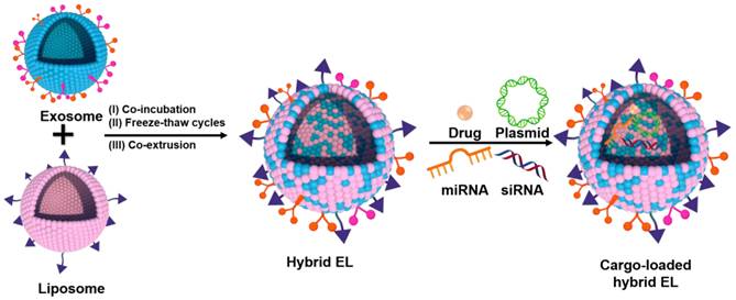
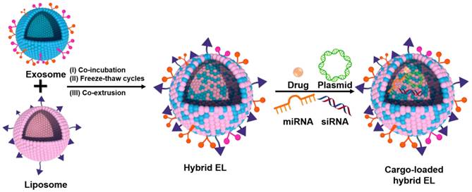
Although several reviews have discussed hybrid ELs, few have focused primarily on their therapeutic applications, and detailed elaboration on their use in drug delivery remains limited [36-40]. This review summarizes the advantages and disadvantages of liposomes and exosomes separately. As illustrated in Figure 1, the fabrication methods and diverse loading strategies for hybrid EL cargoes are also discussed. Moreover, the applications of these state-of-the-art nanovesicles for drug delivery are elaborated upon.
Schematic illustration of the fabrication and cargo loading of hybrid exosome-liposome (EL). There are three main strategies for the fabrication of hybrid EL: the co-incubation method, the freeze-thaw method, and the co-extrusion method. Diverse cargos can be loaded into the hybrid EL, including chemotherapeutic drugs, miRNAs, siRNAs, and plasmids.
2. Liposomes for drug delivery
Liposomes are a classic drug delivery system with various advantages, such as high loading efficiency, minimal adverse effects, and ease of fabrication and modification [17,41]. Both hydrophilic and lipophilic drugs can be loaded in liposomes. Hydrophilic drugs, such as mRNA [42], miRNA [43], and siRNA [44], can be entrapped in aqueous lumens of liposomes. In contrast, lipophilic drugs can be inserted into the lipid bilayer [45]. Various nanoparticles can also be entrapped within liposomes, improving nanoparticle biocompatibility and prolonging circulation time [46,47]. For instance, upconversion nanoparticles (UCNPs) were incorporated into membrane-integrated liposomes, enhancing luminescence, improving cellular imaging, and enabling targeted delivery [48]. Consequently, liposomes are widely used for co-loading diverse cargos as multifunctional carriers to achieve therapeutic synergy [49, 50]. Liposomes also offer convenient industrial-scale manufacturing, with several liposomal products, such as Doxil® (doxorubicin), Lipusu® (paclitaxel), Depocyt® (cytarabine), and Ambisome® (amphotericin B), already in clinical use.
There are two primary strategies to functionalize liposomes: surface modification and composition design. Surface modification involves conjugating functional molecules to the liposomal surface [16, 51]. PEGylation is a common strategy to prolong circulation time by forming a hydrophilic layer on the liposomal surface, which reduces plasma protein absorption and opsonization [52]. Additionally, targeting ligands, including peptides [53], proteins [54], and aptamers [15], can also be anchored to the liposomal surface to fabricate targeted liposomes. For instance, Luo et al. developed Tet1 peptide-modified liposomes (Lipo-T) for retinal ganglion cell (RGC)-targeting [55]. The Tet1 peptide exhibits high affinity for GT1b, which is enriched on RGCs but minimally expressed on glial cells, enabling precise targeting. Tet1 modification peptide enhanced RGC uptake, reduced glial cell activation, and minimized inflammatory responses and adverse side effects. Moreover, Tet1 peptide modification maintained stable drug encapsulation, controlled drug release, improved drug bioavailability, and enabled lower therapeutic doses, thereby reducing systemic toxicity.
During liposome fabrication, functional lipids, including pH-responsive [56], thermo-responsive [57], and ROS-responsive lipids [58], can be incorporated into the lipid bilayer of liposomes. For example, Gao and co-workers synthesized a ROS-responsive lipid, iDAPC, and used it to fabricate ROS-responsive liposomes loaded with the immunostimulatory CpG oligodeoxynucleotides (ODNs). Upon ROS exposure, DAPC underwent structural changes that destabilized ROS-responsive liposomes and selectively released CpG ODNs [58]. These ROS-responsive liposomes preferentially released CPG ODNs in malignant cells, promoting dendritic cell maturation and subsequent activation of CD8+ T cells. Thus, functionalized liposomes can achieve targeted delivery and prolonged circulation. Nevertheless, targeted liposomes still face challenges, such as non-specific biodistribution, limited circulation time, and potential immunogenicity [59].
3. Exosomes for drug delivery
Exosomes are rich in various biomolecules, including proteins, nucleic acids, and metabolites [60, 61]. As intercellular messengers, exosomes play a key role in delivering biomolecules and regulating physiological functions in recipient cells [62]. As listed in Table 1, exosomes originate from diverse sources that show cell specificity and innate homing capabilities [63]. Some exosomal biomarkers include tetraspanins (CD9, CD63, CD81), Alg2-interacting protein X (ALIX), and tumor susceptibility gene 101 (TSG101) [64]. Negative control biomarkers, including calnexin, cytochrome c oxidase subunit IV (COX IV), and histone H3, are associated with the endoplasmic reticulum, mitochondria, and cellular nucleus, separately [65]. Drug loading into exosomes generally follows two principal strategies: passive loading and active loading [66,67].
Comparisons of exosomes from different sources.
| Source | Advantages | Disadvantages | Examples |
|---|---|---|---|
| Body fluid | Easy collection | Batch differences | Plasma, semen, urine, saliva, ascites fluid, lavage fluid, cerebrospinal fluid, bile, lymph |
| Mammalian cell | Steady batch production | High large-scale production cost | MSC, macrophage, dendritic cell, B cell, T cell, platelet, epithelial cell, neuron |
| Edible plant | Inherent anti-inflammatory effect | Relative low yield | Ginger, garlic, grapefruit, carrot, broccoli |
| Edible food | Scalable and safe | Batch differences | Milk, strawberry, blueberry, grape, apple, lemon |
3.1. Passive loading
Passive loading relies on physicochemical interactions between exosomes and cargos [68]. Hydrophobic molecules predominantly partition into the lipid bilayer of exosomes, leading to moderate loading efficiencies [69,70]. A major merit is the preservation of membrane integrity, which keeps native surface proteins, such as tetraspanins and integrins [30,71]. Despite its simplicity, passive loading often requires substantial quantities of drugs, making purification and optimization challenging [30].
3.2. Active loading
Active loading overcomes diffusion limitations, including electroporation, sonication, and freeze-thaw cycles [72,73]. Electroporation transiently permeabilizes the exosomal membrane, facilitating nucleic acid encapsulation, although improper conditions may induce cargo aggregation and exosome clumping [28,74]. Sonication enhances drug loading by membrane permeabilization, but localized heat can increase the risk of protein denaturation [5,75]. Freeze-thaw cycles improve loading ratios of hydrophobic cargoes and preserve exosomal morphology [76]. Collectively, active loading facilitates precise loading of diverse cargoes, including small molecules, nucleic acids, and gene-editing systems, but may compromise membrane integrity, accelerate blood clearance, and damage surface proteins [35,77]. Thus, passive loading keeps biological function but limits loading efficiency, whereas active loading increases loading efficiency at the cost of exosomal performance [74,78].
4. Strategies to fabricate hybrid EL
Though exosomes possess innate biocompatibility and homing capabilities, they face some challenges, such as limited loading capacity and complex fabrication processes [13,14]. Their natural composition supports immune evasion and physiological barrier penetration, but leads to variability in production yield and functional consistency [79,80]. Conversely, liposomes offer efficient drug loading and scalable manufacturing, yet lack endogenous targeting ligands [5,17,18]. Hybrid ELs integrate the strength of both systems – combining exosomal targeting and prolonged circulation with liposomal flexibility and high loading capacity. Three primary strategies to fabricate hybrid EL: co-incubation, freeze-thaw, and co-extrusion, listed in Table 2.
Comparisons of characteristics between different fabrication strategies for hybrid EL.
| Category | Co-incubation method | Freeze-thaw method | Co-extrusion method |
|---|---|---|---|
| Mechanism of fusion | Passive intermolecular interactions | Exosomes and liposomes breakdown into smaller nanovesicles because of freeze-thaw cycles | Lipid bilayer rearrangement and reassembly to decrease the free energy |
| Advantages | Ease to prepare without any additional operational steps Negligible impact on cargos and exosomal membrane | High fusion efficiency | Controllable particle size and size distribution Short fabrication time |
| Disadvantages | Limits in fusion efficiency Heterogeneous size distribution and poor stability | Cargo leakage susceptibility Potential impairment of surface protein integrity and function | Low entrapment efficiency for hydrophilic drugs Potential impairment of surface protein integrity and function |
| Fusion efficiency | Low | High | Medium |
| Surface protein of exosome | Almost no impact | Potential impairment of surface protein bioactivity | Potential impairment of surface protein integrity and function |
| Stability | Low | Medium | High |
4.1. Co-incubation method
The co-incubation method enables membrane fusion between exosomes and liposomes by passive intermolecular interactions, allowing hybridization without compromising structural integrity or bioactivity [81]. This method requires no specialized conditions, keeps exosomal surface markers, and integrates liposomal loading capabilities under physiologically compatible environments [82]. Though fusion efficiency remains less than that achieved by mechanical or chemical methods, co-incubation minimizes disruptive forces, and is well-suited for keeping labile drugs [30]. However, hybrid ELs acquired by co-incubation may show increased particle size and size distributions [83]. Optimization of lipid composition and incubation parameters can enhance the stability and functionality of hybrid ELs.
4.2. Freeze-thaw method
Freeze-thaw cycles induce membrane fusion by repeated disruption and reformation of membranes, providing an efficient integration of exosome and liposome [84]. This method is based on the concept that large vesicles break into smaller ones during freeze-thaw cycles, which promotes fusion [85]. Reported fusion efficiencies reach up to 97.4% [86]. The process of fabricating freeze-thaw based hybrid ELs keeps the exosomal surface markers essential for targeting and uses the loading capacity of liposomes [87]. However, repeated freeze-thaw cycles may compromise membrane integrity or cause cargo leakage, necessitating a balance between fusion efficiency and vesicle stability [88].
4.3. Co-extrusion method
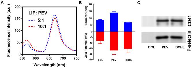
The co-extrusion method is widely used to prepare hybrid ELs by mechanically fusing exosomes and liposomes by repeated extrusion across porous filters, producing uniform nanovesicles [89]. Shear forces fragment nanovesicles into membrane sheets, which subsequently self-assemble into spherical nanovesicles, a process that reduces the free energy [90]. Membrane fusion structures of hybrid EL could be verified by a decline in the Forster resonance energy transfer (FRET) (Figure 2A), due to the membrane fusion of liposomes and exosomes [91]. The membrane fusion process was further confirmed by zeta drift (Figure 2B) and retention of platelet-specific markers, such as CD41/P-selectin (Figure 2C).
Verification of the membrane fusion structures of hybrid EL prepared by co-extrusion. (A) The membrane of platelet-derived exosomes labeled with fluorescent dyes and fused with a growing number of liposomes, indicating the FRET effect. (B) The hydrodynamic diameter and zeta potential of different formulations. All data are presented as the mean ± SD (n = 3). (C) The markers of platelet-derived exosomes, including CD41 and P-selectin, were detected using Western blotting. Adapted with permission from [91], copyright 2023 Cell Press.
Co-extrusion enhances drug-loading efficiency of exosomes by utilizing liposomal loading capacity [92]. Hybrid ELs fabricated by co-extrusion inherit environmental responsiveness from specialized liposomes and retain exosomal properties that improve targeted accumulation compared with conventional liposomes [89,93]. These hybrid ELs also demonstrate superior stability and prolonged circulation compared with co-incubation and freeze-thaw counterparts [94]. Optimal extrusion parameters, including pore size, pressure, and number, are essential to prevent excessive membrane disruption and preserve exosomal surface proteins [95,96]. For hydrophilic cargos, co-extrusion may yield lower encapsulation efficiency due to concentration constraints during extrusion [97].
5. Hybrid exosome-liposome for cargo delivery
5.1. Chemotherapeutics delivery
Hybrid ELs combine the biocompatibility and targeting properties of exosomes with the high loading capacity of liposomes, making them highly effective carriers for the delivery of chemotherapeutics. Importantly, these hybrid ELs can enhance therapeutic efficacy while reducing off-target toxicity. For example, doxorubicin (DOX), a widely used anti-cancer drug, suffers from non-specific biodistribution and dose-limiting toxicities, resulting in limited therapeutic efficacy [68,98,99]. Hybrid ELs offer a promising strategy to overcome these limitations.
Rayamajhi et al. developed a DOX-loaded hybrid EL by co-extruding macrophage-derived exosomes and pH-sensitive liposomes [83]. The DOX-loaded hybrid EL retained the innate targeting properties of macrophage-derived exosomes and the stability and high drug-loading capacity of liposomes. Compared with naive macrophage-derived exosomes, the DOX-loaded hybrid ELs improved homogeneity and colloidal stability, exhibited pH-sensitive drug release in acidic tumor environments, and facilitated efficient cellular uptake. Moreover, the DOX-loaded hybrid ELs exhibited potent cytotoxicity against cancer cells while sparing normal cells, demonstrating their potential for tumor-selective delivery and reduced off-target toxicity.
In another study, tumor-derived exosomes and liposomes were fused to generate DOX-loaded biomimetic nanovesicles (DOX@LINV) for cancer immunotherapy [27]. DOX@LINV achieved an 82% tumor suppression rate at a 4-fold reduced DOX dose required for conventional DOX-loaded liposomes (79% suppression), significantly reducing systemic toxicity. Furthermore, DOX@LINV preserved tumor antigens and danger signals, promoting dendritic cell maturation and immunogenic cell death (ICD). Of note, DOX-induced ICD and exosome-mediated immune activation together led to a potent anti-tumor response, including increased T-cell infiltration and modulation of the tumor immunosuppressive microenvironment. These findings highlight the versatility of hybrid ELs in improving chemotherapeutic outcomes and enabling synergistic chemo-immunotherapy.
Plant-derived exosomes have also been explored for drug delivery [5,69]. For instance, balloon flower root-derived exosome-like nanoparticles (BDEs), which were isolated by TFF, were co-extruded with silymarin (SM)-loaded liposomes to mitigate acetaminophen-induced hepatotoxicity [6]. The platycodin D-enriched BDEs were obtained at high yield and fused with SM-loaded liposomes, resulting in suppression of hepatocyte apoptosis and the MAPK pathway. The hybrid EL combined the intrinsic anti-inflammatory properties of BDEs with the high SM loading of liposomes. BDEs also facilitated intercellular communication through endogenous biomolecules, amplifying the detoxification effects of SM without inducing cytotoxicity. Thus, plant-derived hybrid ELs show great potential as both drug carriers and active therapeutic agents.
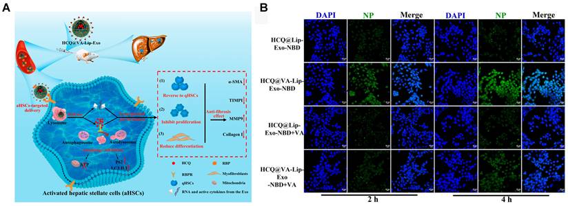
In addition to mitigating hepatotoxicity, hybrid ELs have also been applied to treat liver fibrosis. Zhang et al. developed hybrid ELs composed of BMSC-derived exosomes and hydroxychloroquine (HCQ)-loaded vitamin A (VA, retinol)-modified liposomes (HCQ@VA-Lip-Exo) [100] (Figure 3A). BMSC-derived exosomes provided innate tropism to activate aHSCs, while VA-modified liposomes enhanced targeting via retinol-binding protein, which is overexpressed on the surface of aHSCs. Accordingly, as shown in Figure 3B, HCQ@VA-Lip-Exo accumulated in aHSCs and inhibited autophagy, a key driver of fibrogenesis. Moreover, HCQ@VA-Lip-Exo minimized off-target effects. These findings highlight the potential of hybrid ELs to combine exosomal specificity with liposomal engineering to achieve efficient and safe chemotherapeutic delivery.
(A) Schematic illustration of HCQ@VA-Lip-Exo for aHSC-targeted autophagy inhibition and synergistic treatment of liver fibrosis. (B) Targeting uptake of HCQ@VA-Lip-Exo by aHSCs (Scale bar = 20 μm). Adapted with permission from [100], copyright 2023 American Chemical Society.
5.2. Peptide delivery
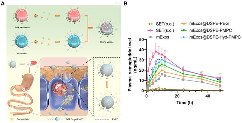
Oral therapeutic peptide delivery is hindered by rapid enzymatic degradation and poor gastrointestinal permeability [101,102]. Hybrid ELs offer innovative strategies to overcome these obstacles. Xiao et al. prepared semaglutide (SET)-loaded milk exosome -liposome nanovesicles (mExos@DSPE-Hyd-PMPC) designed to navigate sequential gastrointestinal barriers [103] (Figure 4A). Milk exosomes contributed membrane proteins that resisted enzymatic degradation, while liposomes improved SET loading efficiency. The hybrid EL mExos@DSPE-Hyd-PMPC incorporated pH-responsive zwitterionic polymers enabling adaptive surface properties: a hydrophilic, mucus-penetrating state in the jejunal lumen and a charge-reversed, cell-adhesive state at the mildly acidic epithelial surface. This design facilitated efficient traversal of the mucus barrier. Upon reaching jejunal epithelial cells, the membrane proteins and positively charged surfaces of mExos@DSPE-Hyd-PMPC enabled efficient transport across apical, intracellular, and basolateral barriers. Consequently, mExos@DSPE-Hyd-PMPC significantly improved SET oral bioavailability (Figure 4B). These findings demonstrated the ability of hybrid ELs to overcome limitations of oral peptide delivery, including protease degradation and poor mucosal permeability.
(A) Schematic illustration of mExos@DSPE-Hyd-PMPC for oral delivery of semaglutide. (B) Plasma semaglutide concentration-time profiles in rats. All data are presented as the mean ± SD (n = 5). SET (p.o.): oral semaglutide solution group; SET (s.c.): subcutaneous injection semaglutide solution group. *p < 0.05, and **p < 0.01 versus the mExos group. Adapted with permission from [103], copyright 2024 American Chemical Society.
5.3. RNA delivery
Both exosomes and liposomes have been used for RNA delivery, but each has limitations: exosomes exhibit low loading efficiency for anionic RNA, while liposomes lack targeting ability for RNA delivery [104,105]. Hybrid ELs have emerged as a promising strategy to overcome these limitations. Pre-encapsulation of RNA into liposomes enhances entrapment efficiency compared with direct exosomal electroporation, while preservation of exosomal surface proteins facilitates cell-/organ-selective targeting [106,107].
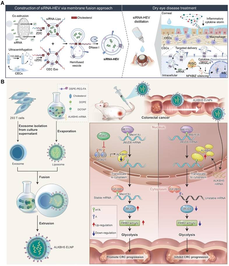
Xie et al. fabricated a hybrid EL (siRNA-HEV) using DNA zipper-mediated membrane fusion between corneal epithelium-derived exosomes and siRNA-loaded liposomes for treating dry eye disease (DED) [46] (Figure 5A). siRNA-HEV retained exosomal homing capabilities and improved siRNA loading efficiency of liposomes. By avoiding destructive methods, siRNA-HEV kept exosomal integrity. siRNA-HEV showed enhanced gene silencing and effectively alleviated inflammation in a DED mouse model. In another study using siRNA-loaded hybrid ELs, Jhan et al. prepared a hybrid EL by co-extruding tumor-derived exosomes with liposomes, and loading siRNA by electroporation [92]. The hybrid EL promoted effective gene silencing, compared with commercial reagents. In addition, the hybrid EL enabled scalable production and kept exosomal targeting. This example highlighted the potential of hybrid ELs for RNAi therapeutics in cancer treatment.
(A) Schematic illustration of the membrane fusion strategy to load anti-NFKBIZ siRNA into HEV construct for the treatment of dry eye disease [46]. Copyright 2024, John Wiley and Sons. (B) Schematic illustration of the ALKBH5-mediated regulatory network in colorectal cancer progression, and ALKBH5-ELNP mediated ALKBH5 mRNA delivery inhibits colorectal tumorigenesis. Adapted with permission from [110], copyright 2023 American Chemical Society.
In another study, Evers et al. developed siRNA-loaded hybrid ELs that activated endothelial signaling and migration, demonstrating cell-type-dependent uptake and gene-silencing efficacy [108]. Furthermore, hybrid ELs retained exosome-mediated biological functions, such as endothelial cell activation, and improved biocompatibility relative to conventional liposomes.
Hu et al. engineered C-X-C motif chemokine receptor 4 (CXCR4)-overexpressing NIH-3T3 exosomes, which were co-extruded with antagomir-188-loaded liposomes to treat age-related bone loss [109]. The hybrid ELs targeted bone marrow via stromal cell-derived factor 1 (SDF1) -CXCR4 interactions, facilitated osteogenic differentiation, suppressed adipogenesis in BMSCs, reversed bone loss, and reduced cortical bone porosity in an age-related osteoporosis mouse model. For colorectal cancer (CRC), folic acid (FA)-functionalized hybrid ELs (ELNPs) enhanced tumor-specific targeting via folate receptor-mediated uptake, and delivered ALKBH5 mRNA to modulate cancer-associated metabolic pathways [110] (Figure 5B). ELNPs restored tumor-suppressive gene expression, reshaped the tumor microenvironment by inhibiting glycolysis, and achieved potent inhibitory effects in preclinical colorectal tumor models. Furthermore, the FA-modified hybrid ELs enhanced RNA stability, facilitated precise targeting, and tackled intricate disease mechanisms through multimodal therapy. Collectively, hybrid ELs address critical challenges in RNA delivery, including stability, specificity, and functional integration.
5.4. DNA delivery
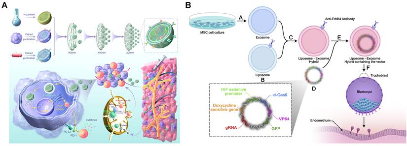
Hybrid ELs also enable targeted and safe delivery of DNA cargoes. Tong et al. developed Lipo@HEV, a novel tumor-derived antigen-loaded hybrid EL, combining immunostimulatory bacterial outer membrane vesicles (OMVs) with cationic liposomes [70] (Figure 6A). Lipo@HEV promoted dendritic cell maturation, activated cytotoxic T-cells, and exhibited efficient lymph node trafficking and deep tissue penetration, highlighting its ability to bypass physiological barriers.
(A) Schematic illustration of Lipo-PD-L1@HEV for synergistic cancer immunotherapy. Lipo-PD-L1@HEV was fabricated by fusing extracellular vesicles with a plasmid-loaded liposome. Upon peritumoral injection, Lipo-PD-L1@HEV can localize in tumor for PD-L1 blockade, and penetrate into lymph node to efficiently initiate dendritic cell, antigen presentation and cytotoxic T cell activation, synergistically inhibiting the tumor [70]. Reproduced under terms of the CC BY license. Copyright 2023, The Authors, published by Elsevier. (B) Schematic illustration of hypoxia-sensitive ErbB4-targeted hybrid EL for CRISPR/dCas9 delivery system. A: Isolation of MSC exosomes. B: Antibodies against ErbB4 conjugated on the surface of liposomes. C: The hybrid EL will be fabricated by co-incubating MSC-derived exosomes and ErbB4-targeted liposomes. D: Non-viral S/MAR episomal vector will be constructed. E: Electroporation will be used to encapsulate the designed episomal S/MAR vector into the hybrid EL. F: The hybrid EL will be added to the trophoblast cell media, and the episomal vector will specifically enter the trophoblast. Adapted with permission from [10], copyright 2023 Frontiers.
CRISPR/Cas9 gene editing requires cell-specific delivery to minimize systemic toxicity [111-114]. Hybrid ELs address this challenge by integrating exosomal tropism with liposomal plasmid-loading. As a paradigm, Liang and co-workers engineered CRISPR/Cas9 plasmid-loaded hybrid ELs modified with a CAP peptide and matrix metalloproteinase 13 (MMP13), enabling penetration of dense cartilage matrices in arthritic rats and selectively delivering plasmids to chondrocytes [115]. The hybrid ELs suppressed MMP-13 expression and attenuated cartilage degradation.
Lin et al. used membrane fusion to prepare hybrid ELs for CRISPR/Cas9 delivery to MSCs, which are typically resistant to liposomal transfection [116]. The hybrid ELs retained exosomal surface proteins, facilitating MSC uptake, while liposomal components protected CRISPR/Cas9 plasmid integrity. The system successfully eliminated β-catenin in human MSCs, demonstrating its ability to overcome cellular resistance mechanisms.
Yaghoobi et al. developed ErbB4-targeted hybrid ELs for hypoxia-sensitive CRISPR/dCas9 delivery to prevent placental insufficiency [10] (Figure 6B). Hybrid ELs encapsulated S/MAR episomal vectors with hypoxia-responsive promoters, upregulated microRNA-30d in trophoblasts, enhanced angiogenesis and implantation, and achieved trophoblast-specific uptake without affecting the inner cell mass. ErbB4-targeted hybrid ELs crossed the placental barrier and demonstrated potential for spatiotemporal gene editing control. These findings highlight the ability of hybrid ELs to combine exosomal biological advantages with liposomal engineering flexibility to facilitate precise CRISPR/Cas9 delivery across physiological barriers.
5.5. Co-delivery
Because both exosomes and liposomes possess lipid bilayers capable of encapsulating hydrophilic or lipophilic cargoes, hybrid ELs acquire this dual loading capability [91,117,118]. This co-loading capacity of hybrid ELs facilitates combination therapies. Hybrid ELs combine exosomal biocompatibility and innate targeting with liposomal tunability and high loading capacity, promoting targeted co-delivery to diseased tissues.
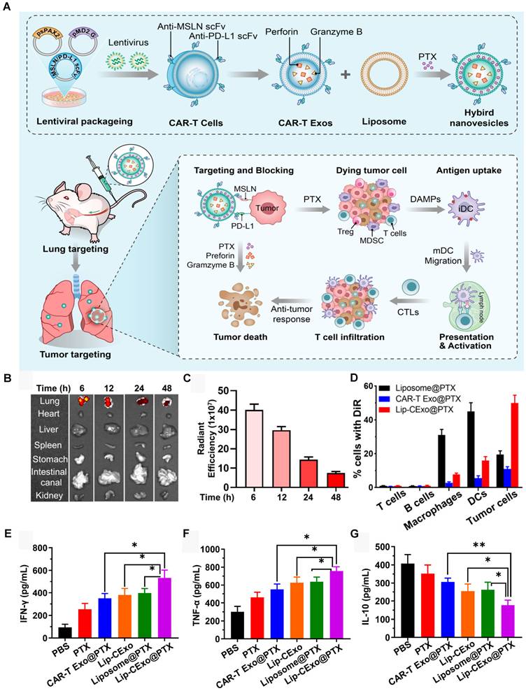
As a paradigm, Zhu et al. fabricated hybrid ELs (Lip-CExo@PTX) co-loaded with paclitaxel and cytotoxic granules, based on CAR-T-derived exosomes and lung-targeted liposomes to treat systemic malignancies [119] (Figure 7A). As displayed in Figure 7B-C, liposomes mediated primary lung accumulation through charge-directed tropism, while single-chain variable fragments (scFvs) from CAR-T-derived exosomes enabled secondary tumor-specific targeting (Figure 7D). Lip-CExo@PTX co-delivered paclitaxel and cytotoxic granules, and simultaneously engaged in PD-L1 blockade. This multi-modal activity caused chemotherapy-induced immunogenic cell death, T cell-mediated toxicity, and checkpoint inhibition (Figure 7E-G). Thus, Lip-CExo@PTX addressed pharmacokinetic limitations of free drugs and resistance mechanisms within the tumor microenvironment.
(A) Schematic illustration of Lip-CExo@PTX for lung cancer therapy. (B,C) Following injection of DiR-labeled Lip-CExo, the fluorescence of the major organs was analyzed by the IVIS system ex vivo at different time points. (D) The fluorescence of different cells in the tumor microenvironment 8 h post-injection of DiR-labeled CAR Exos, liposomes, or Lip-CExos was analyzed by flow cytometry. (E-G) The concentrations of IFN-γ, TNF-α, and IL-10 in tumor tissues were measured by ELISA after treatment with different drugs. All data are presented as the mean ± SD (n = 3). *p < 0.05, and **p < 0.01. Adapted with permission from [119], copyright 2023 American Chemical Society.
Hybrid ELs have also been applied to co-deliver chemotherapeutics for the targeted treatment of cancer. Zhou et al. developed IHEL, a hybrid EL co-loaded with IR780 and hemin, using exosomes derived from M1-polarized RAW264.7 cells and liposomes, for targeted melanoma therapy [120]. IHEL exploited exosomal tumor-homing properties, enabling selective accumulation in melanoma tissues. Under NIR laser irradiation, IHEL demonstrated superior tumor elimination due to the combined photodynamic/photothermal therapy and immunomodulation via ferroptosis activation, glycolytic suppression, and the repolarization of tumor-associated macrophages toward an M1 phenotype. IHEL thus represents a promising photo-immunotherapy strategy for melanoma.
For glioma treatment, Wang et al. developed pHybrid/SAB-CPT, a hybrid EL co-loaded with cryptotanshinone (CPT) and salvianolic acid B (SAB), using blood cell-derived exosomes and tLyp-1-modified liposomes. pHybrid/SAB-CPT leveraged exosomal transferrin receptors for BBB penetration and tLyp-1-modified targeting for enhanced tumor parenchyma penetration via neuropilin-1 interactions [121]. This dual-targeting strategy enabled efficient co-delivery of CPT and SAB to glioma. pHybrid/SAB-CPT maintained exosomal surface proteins for physiological barrier navigation and incorporated liposomal components for controlled release and tumor targeting. Blood cell-derived exosomes contributed to immune evasion, while liposomes provided tunable pharmacokinetics.
Hybrid ELs can also co-deliver RNA cargoes and chemotherapeutics for brain-targeted glioma therapy. Ma et al. fabricated a brain-targeted hybrid EL (PMRT) by co-incubating macrophage-derived exosomes with thermo-sensitive liposomes, to co-deliver temozolomide (TMZ) and siRNAs (si-mTOR and si-c-Myc) for glioma treatment [71]. PMRT utilized exosomal surface proteins, such as integrins, to enhance BBB penetration, while thermo-sensitive liposomes provided stable encapsulation and controlled release of TMZ and siRNAs. PMRT effectively reduced glioma stem cell (GSC) viability, inhibited tumorsphere formation, and synergistically suppressed GSCs by inhibiting the mTOR-c-Myc signaling pathway, thereby reversing TMZ-resistance in glioma. In orthotopic glioma xenograft mice, PMRT significantly suppressed tumor burden and prolonged survival, demonstrating strong therapeutic potential. In orthotopic glioma xenograft mice, PMRT significantly suppressed tumor growth and prolonged the lifespan of mice, highlighting the promising potential of PMRT for the efficient management of glioma.
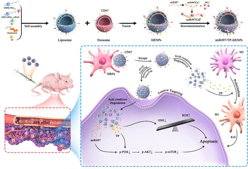
In another study, Li et al. developed miR497/TP-HENPs, a hybrid EL combining tumor-derived exosomes with cRGD-modified liposomes to overcome chemoresistance [122]. The hybrid EL co-delivered the small-molecule therapeutic drug triptolide (TP) and a tumor-suppressing microRNA (miR497) (Figure 8). Dual tumor-targeting was achieved through exosomal homologous tropism and cRGD-mediated integrin receptor recognition. miR497/TP-HENPs simultaneously blocked chemoresistance-associated signaling pathways and modulated the immunosuppressive tumor microenvironment. In addition, pH-responsive cargo release enabled spatiotemporal drug activation within an acidic tumor microenvironment. TP enhanced miR497 efficacy by depleting intracellular antioxidants, while miR497 amplified TP sensitivity to tumor cells by downregulating survival pathways. The hybrid EL exemplified multimodal therapies against complex resistance mechanisms. Hybrid ELs have also been explored for neurodegenerative diseases.
Schematic illustration of the preparation process and therapeutic mechanism of miR497/TP-HENPs. When miR497/TP-HENPs reach OC cells, miR497 and TP synergistically inhibited the PI3K/AKT/mTOR pathway. TP stimulated ROS production, and TP modulated polarization of M2 macrophages into M1 macrophages, synergistically overcoming OC resistance. Adapted with permission from [122], copyright 2022 BMC Press.
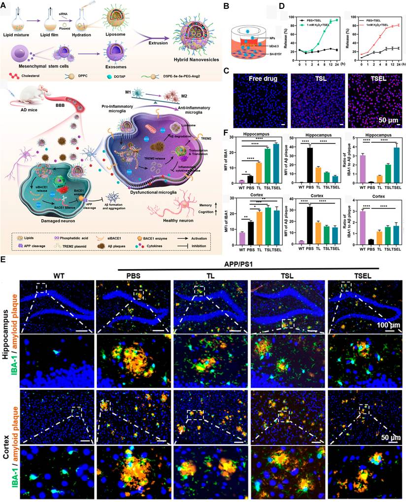
Jiang et al. developed BBB-penetrating hybrid ELs to co-deliver BACE1 siRNA (siBACE1) and TREM2 plasmid (pTREM2) for AD [123] (Figure 9A). Hybrid ELs combined MSC-derived exosomes with ROS-responsive liposomes. As displayed in Figure 9B-C, the exosomal membrane proteins facilitated neuronal targeting and lysosomal escape, while the liposomes enabled ROS-triggered release of cargoes (Figure 9D). This sequential delivery allowed initial siRNA-mediated inhibition of Aβ production followed by sustained plasmid-driven enhancement of microglial phagocytosis. The dual-gene therapeutic strategy could simultaneously target multiple pathological hallmarks, thereby establishing a self-perpetuating therapeutic cycle. As shown in Figure 9E-F, in vivo results indicated that hybrid ELs reduced Aβ burden, alleviated neuroinflammation, and improved microglial clearance, demonstrating strong therapeutic potential for AD.
(A) Schematic illustration of the preparation process and therapeutic mechanism of TSEL. (B) Schematic diagram of the in vitro BBB model. (C) CLSM images of Cy5-siRNA fluorescence signal to evaluate the BBB penetration capability of TSEL (Scale bar = 50 μm). (D) The cumulative release of pTREM2 and siRNA from the TSEL with or without 1 mM H2O2. All data are presented as the mean ± SD (n = 3). (E) Representative immunofluorescence images of amyloid plaque (orange) with microglia (green) colocalization in the hippocampus (Scale bar = 100 μm) and cortex (Scale bar = 50 μm) of AD mice treated with PBS, TL, TSL, or TSEL. The bottom row of each image displays an enlarged version of the white boxes in the upper image. (F) Quantification analysis of Aβ intensity and the number of IBA1-positive microglia engulfing Aβ in the hippocampus and cortex of AD mice. All data are presented as the mean ± SD (n = 3). *p < 0.05, **p < 0.01, and ***p < 0.001. Adapted with permission from [123], copyright 2024 American Chemical Society.
6. Limitations and challenges
Hybrid ELs offer promising potential to treat various diseases, yet some challenges hinder clinical translation.
6.1. Stability
Though hybrid ELs are generally more stable than exosomes, they remain susceptible to physiological fluctuations, like temperature, pH, and ROS, which can destroy structural integrity and destabilize cargo [124]. Dynamic in vivo physiological variables may reduce therapeutic effect, underscoring the need for long-term in vivo stability studies.
6.2. Immunogenicity
While the immunogenicity of exosomes is low due to their endogenous origin, the introduction of liposomes may alter immune responses [24]. For example, PEGylated liposomes can induce anti-PEG antibodies, triggering ABC [125]. Additionally, heterologous exosome sources may provoke immune responses [126]. Thus, rigorous immunogenicity is essential to ensure the efficacy of hybrid ELs.
6.3. Clearance and Biodegradation
Ensuring biodegradation of hybrid ELs and their cargoes is critical to avoid chronic toxicity. Clearance pathways must be thoroughly studied.
6.4. Interspecies gap
Most hybrid EL studies rely on murine models, limiting translational relevance. Patient-derived organoids and organ-on-a-chip offer physiologically relevant human surrogates for evaluating in vivo PK, PD, and immunogenicity.
6.5. Industrialization
Exosomal heterogeneity complicates batch-to-batch reproducibility and therapeutic consistency [127]. Exosomes vary with donor cell and extraction methods, affecting hybrid EL homogeneity. Scalability remains challenging due to limited yields from current isolation techniques and the difficulty of balancing regulatory frameworks for hybrid EL biosafety and characterization, which are underdeveloped, contributing to limited industrial expertise, which in turn could engender further potential delays in commercialization [24,128].
7. Conclusion and future directions
Future development of hybrid ELs depends on several actionable advances. Tangential flow filtration (TFF) enables large-scale exosome purification with high yield and low impurity and is already used for bulk production [129]. High-throughput microfluidic platforms allow single-step, continuous-flow isolation of exosomes, outperforming conventional exosome preparation methods such as ultracentrifugation [130,131]. Together, large-scale TFF and microfluidics production can reduce costs while enabling high-throughput, controlled fabrication of hybrid ELs.
Current optimization of hybrid ELs, including lipid compositions, exosome-to-liposome ratios, and fusion conditions, remains empirical. Artificial intelligence-driven high-throughput screening, including METiS technologies for lipid nanoparticle design optimization, could systematically map design-efficacy relationships, and thus accelerate the development of optimal hybrid ELs from years to months [132]. Also, regulatory agencies must develop parallel frameworks to evaluate in vivo biodistribution, long-term stability, and immunogenicity of hybrid ELs, ensuring therapeutic efficacy and biosafety. By integrating these advances, hybrid ELs have the promising potential to overcome the limitations of exosomes and liposomes and pioneer next-generation precision therapies for the treatment of various diseases.
Abbreviations
aHSC: activate hepatic stellate cell; ABC: accelerated blood clearance; AD: Alzheimer’s disease; ALIX: Alg2-interacting protein X; BBB: blood-brain barrier; BDEs: balloon flower root-derived exosome-like nanoparticles; BMSC: bone marrow mesenchymal stem cell; BRB: blood-retinal barrier; COX IV: cytochrome c oxidase subunit IV; CPT: cryptotanshinone; CRC: colorectal cancer; cRGD: cyclic arginyl-glycyl-aspartic acid; CXCR4: C-X-C motif chemokine receptor 4; DED: dry eye disease; DOX: doxorubicin; FA: folic acid; FRET: Forster resonance energy transfer; GSC: glioma stem cell; HCQ: hydroxychloroquine; ICD: immunogenic cell death; MAPK: mitogen-activated protein kinase; mExo: milk exosome; MISEV2018: minimal information for studies of extracellular vesicles 2018; MMP13: matrix metalloproteinase 13; MPS: mononuclear phagocyte system; MSC: mesenchymal stem cell; NPs: nanoparticles; OMVs: outer membrane vesicles; PD: pharmacodynamics; PEG: polyethylene glycol; PK: pharmacokinetics; RGC: retinal ganglion cell; ROS: reactive oxygen species; SAB: salvianolic acid B; SDF1: stromal cell-derived factor 1; SET: semaglutide; scFvs: single-chain variable fragments; SM: silymarin; TFF: tangential flow filtration; TMZ: temozolomide; TP: triptolide; TSG101: tumor susceptibility gene 101; UCNPs: upconversion nanoparticles; VA: vitamin A.
Author contributions
Jian Yu: Writing-Original draft preparation. Lujiao Yu: Writing-Reviewing and Editing. Fei Ren: Writing-Reviewing and Editing. Kai Cui: Supervision. Gongping Sun: Conceptualization. Hong Pan: Supervision. All authors have read and approved the final manuscript.
Competing interests
The authors have declared that no competing interest exists.
References
1. Ning B, Huang Z, Youngquist BM, Scott JW, Niu A, Bojanowski CM. et al. Liposome-mediated detection of SARS-CoV-2 RNA-positive extracellular vesicles in plasma. Nat Nanotechnol. 2021;16:1039-44
2. Zhang Q, Jeppesen DK, Higginbotham JN, Graves-Deal R, Trinh VQ, Ramirez MA. et al. Supermeres are functional extracellular nanoparticles replete with disease biomarkers and therapeutic targets. Nat Cell Biol. 2021;23:1240-54
3. Wang H, Alarcón CN, Liu B, Watson F, Searles S, Lee CK. et al. Genetically engineered and enucleated human mesenchymal stromal cells for the targeted delivery of therapeutics to diseased tissue. Nat Biomed Eng. 2022;6:882-97
4. Johnson J, Wu Y-W, Blyth C, Lichtfuss G, Goubran H, Burnouf T. Prospective therapeutic applications of platelet extracellular vesicles. Trends Biotechnol. 2021;39:598-612
5. Erana-Perez Z, Igartua M, Santos-Vizcaino E, Hernandez RM. Genetically engineered loaded extracellular vesicles for drug delivery. Trends Pharmacol Sci. 2024;45:350-65
6. Kim J, Gao C, Guo P, Sheng J, Wang J. A novel approach to alleviate acetaminophen-induced hepatotoxicity with hybrid balloon flower root-derived exosome-like nanoparticles (BDEs) with silymarin via inhibition of hepatocyte MAPK pathway and apoptosis. Cell Commun Signal CCS. 2024;22:334
7. Ran B, Ren X, Lin X, Teng Y, Xin F, Ma W. et al. Glycyrrhetinic acid loaded in milk-derived extracellular vesicles for inhalation therapy of idiopathic pulmonary fibrosis. J Control Release. 2024;370:811-20
8. Li B, Chen X, Qiu W, Zhao R, Duan J, Zhang S. et al. Synchronous disintegration of ferroptosis defense axis via engineered exosome-conjugated magnetic nanoparticles for glioblastoma therapy. Adv Sci. 2022;9:e2105451
9. Tian Y, Zhang F, Qiu Y, Wang S, Li F, Zhao J. et al. Reduction of choroidal neovascularization via cleavable VEGF antibodies conjugated to exosomes derived from regulatory T cells. Nat Biomed Eng. 2021;5:968-82
10. Yaghoobi A, Nazerian Y, Meymand AZ, Ansari A, Nazerian A, Niknejad H. Hypoxia-sensitive miRNA regulation via CRISPR/dCas9 loaded in hybrid exosomes: A novel strategy to improve embryo implantation and prevent placental insufficiency during pregnancy. Front Cell Dev Biol. 2023;10:1082657
11. Xu J, Liu H, Wang T, Wen Z, Chen H, Yang Z. et al. CCR7 mediated mimetic dendritic cell vaccine homing in lymph node for head and neck squamous cell carcinoma therapy. Adv Sci. 2023;10:2207017
12. Yang L, Huang X, Guo H, Wang L, Yang W, Wu W. et al. Exosomes as efficient nanocarriers in osteosarcoma: biological functions and potential clinical applications. Front Cell Dev Biol. 2021;9:737314
13. Bittel M, Reichert P, Sarfati I, Dressel A, Leikam S, Uderhardt S. et al. Visualizing transfer of microbial biomolecules by outer membrane vesicles in microbe-host-communication in vivo. J Extracell Vesicles. 2021;10:e12159
14. Shamshiripour P, Rahnama M, Nikoobakht M, Rad VF, Moradi A-R, Ahmadvand D. Extracellular vesicles derived from dendritic cells loaded with VEGF-A siRNA and doxorubicin reduce glioma angiogenesis in vitro. J Controlled Release. 2024;369:128-45
15. Wang T, Tang J, Yang H, Yin R, Zhang J, Zhou Q. et al. Effect of apatinib plus pegylated liposomal doxorubicin vs pegylated liposomal doxorubicin alone on platinum-resistant recurrent ovarian cancer: The APPROVE randomized clinical trial. JAMA Oncol. 2022;8:1169-76
16. Cao Z, Wang X, Pang Y, Cheng S, Liu J. Biointerfacial self-assembly generates lipid membrane coated bacteria for enhanced oral delivery and treatment. Nat Commun. 2019;10:5783
17. Kong L, Yu Y, Yang R, Guo R, Zhang L, Wang J. et al. Development and efficacy evaluation of nanoliposomes targeting CAFs-LCSCs communication for hepatocellular carcinoma treatment. Chem Eng J. 2024;496:154173
18. Wang W, Wu X, Kevin Tang KW, Pyatnitskiy I, Taniguchi R, Lin P. et al. Ultrasound-triggered in situ photon emission for noninvasive optogenetics. J Am Chem Soc. 2023;145:1097-107
19. Xia J, Ma S, Zhu X, Chen C, Zhang R, Cao Z. et al. Versatile ginsenoside Rg3 liposomes inhibit tumor metastasis by capturing circulating tumor cells and destroying metastatic niches. Sci Adv. 2022;8:eabj1262
20. Patel D, Solanki J, Kher MM, Azagury A. A review: Surface engineering of lipid-based drug delivery systems. Small. 2024;20:e2401990
21. Almeida B, Nag OK, Rogers KE, Delehanty JB. Recent progress in bioconjugation strategies for liposome-mediated drug delivery. Molecules. 2020;25:5672
22. Eroğlu İ, İbrahim M. Liposome-ligand conjugates: A review on the current state of art. J Drug Target. 2020;28:225-44
23. Yuan D, Zhao Y, Banks WA, Bullock KM, Haney M, Batrakova E. et al. Macrophage exosomes as natural nanocarriers for protein delivery to inflamed brain. Biomaterials. 2017;142:1-12
24. Wu K, Yu B, Li D, Tian Y, Liu Y, Jiang J. Recent advances in nanoplatforms for the treatment of osteosarcoma. Front Oncol. 2022;12:805978
25. Yuyama K, Sun H, Sakai S, Mitsutake S, Okada M, Tahara H. et al. Decreased amyloid-β pathologies by intracerebral loading of glycosphingolipid-enriched exosomes in Alzheimer model mice. J Biol Chem. 2014;289:24488-98
26. Wang X, Wan W, Lu J, Liu P. Inhalable FN-binding liposomes or liposome-exosome hybrid bionic vesicles encapsulated microparticles for enhanced pulmonary fibrosis therapy. Int J Pharm. 2024;656:124096
27. Hu M, Zhang J, Kong L, Yu Y, Hu Q, Yang T. et al. Immunogenic hybrid nanovesicles of liposomes and tumor-derived nanovesicles for cancer immunochemotherapy. ACS Nano. 2021;15:3123-38
28. Bao P, Gu H-Y, Ye J-J, He J-L, Zhong Z, Yu A-X. et al. Chimeric exosomes functionalized with STING activation for personalized glioblastoma immunotherapy. Adv Sci. 2024;11:e2306336
29. Yuan S, Mu W, Liu S, Liu M, Xia Z, Liang S. et al. Transforming cancer-associated fibroblast barrier into drug depots to boost chemo-Immunotherapy in “Shooting Fish in a Barrel” pattern. ACS Nano. 2023;17:13611-26
30. Xu X, Xu L, Wen C, Xia J, Zhang Y, Liang Y. Programming assembly of biomimetic exosomes: An emerging theranostic nanomedicine platform. Mater Today Bio. 2023;22:100760
31. Bao P, Gu H-Y, Jiang Y-C, Wang J-W, Wu M, Yu A. et al. In situ sprayed exosome-cross-linked gel as artificial lymph nodes for postoperative glioblastoma immunotherapy. ACS Nano. 2024;18:13266-76
32. Zhang J, Ji K, Ning Y, Sun L, Fan M, Shu C. et al. Biological hyperthermia-inducing nanoparticles for specific remodeling of the extracellular matrix microenvironment enhance pro-apoptotic therapy in fibrosis. ACS Nano. 2023;17:10113-28
33. Liu X, Zhang J, Zheng S, Li M, Xu W, Shi J. et al. Hybrid adipocyte-derived exosome nano platform for potent chemo-phototherapy in targeted hepatocellular carcinoma. J Controlled Release. 2024;370:168-81
34. Mondal J, Pillarisetti S, Junnuthula V, Saha M, Hwang SR, Park I. et al. Hybrid exosomes, exosome-like nanovesicles and engineered exosomes for therapeutic applications. J Controlled Release. 2023;353:1127-49
35. Pegtel DM, Gould SJ. Exosomes. Annu Rev Biochem. 2019;88:487-514
36. Mukherjee A, Bisht B, Dutta S, Paul MK. Current advances in the use of exosomes, liposomes, and bioengineered hybrid nanovesicles in cancer detection and therapy. Acta Pharmacol Sin. 2022;43:2759-76
37. Moholkar DN, Kandimalla R, Gupta RC, Aqil F. Advances in lipid-based carriers for cancer therapeutics: Liposomes, exosomes and hybrid exosomes. Cancer Lett. 2023;565:216220
38. Zheng L, Hu B, Zhao D, Liu W, Liu Q, Huang Y. et al. Recent progresses of exosome-liposome fusions in drug delivery. Chin Chem Lett. 2024;35:108647
39. Liu A, Yang G, Liu Y, Liu T. Research progress in membrane fusion-based hybrid exosomes for drug delivery systems. Front Bioeng Biotechnol. 2022;10:939441
40. Sundaram V, Aryal S. Emerging biomimetic drug delivery nanoparticles inspired by extracellular vesicles. WIREs Nanomed Nanobi. 2025;17:e70025
41. Hu S, Wang X, Li Z, Zhu D, Cores J, Wang Z. et al. Platelet membrane and stem cell exosome hybrid enhances cellular uptake and targeting to heart injury. Nano Today. 2021;39:101210
42. Pedersen GK, Alhakeem RS, Tami A, Christensen D, Shabanian Z, Jensen RF. et al. A stable two-component cationic liposome platform for mRNA delivery induces CD8+ T-cell responses and protection in a murine lymphoma model. J Nanobiotechnology. 2026;24:350
43. Tsugane M, Kato K, Shinohara K, Okita T, Sato R, Suzuki H. Triggering isothermal exponential amplification of microRNA via liposome fusion. Analyst. 2026;151:842-50
44. Zhao Y, Qin J, Yu D, Liu Y, Song D, Tian K. et al. Polymer-locking fusogenic liposomes for glioblastoma-targeted siRNA delivery and CRISPR-Cas gene editing. Nat Nanotechnol. 2024;19:1869-79
45. Fan L, Du P, Li Y, Chen X, Liu F, Liu Y. et al. Targeted liposomes sensitize plastic melanoma to ferroptosis via senescence induction and coenzyme depletion. ACS Nano. 2024;18:7011-23
46. Xie M, Wu Y, Zhang Y, Lu R, Zhai Z, Huang Y. et al. Membrane fusion-mediated loading of therapeutic siRNA into exosome for tissue-specific application. Adv Mater. 2024;36:e2403935
47. Yao C, Wang P, Li X, Hu X, Hou J, Wang L. et al. Near-infrared-triggered azobenzene-liposome/upconversion nanoparticle hybrid vesicles for remotely controlled drug delivery to overcome cancer multidrug resistance. Adv Mater. 2016;28:9341-8
48. Wang L-C, Chen H-K, Wang W-J, Hsu F-Y, Huang H-Z, Kuo R-T. et al. Boosting upconversion efficiency in optically inert shelled structures with electroactive membrane through electron donation. Adv Mater. 2024;36:e2404120
49. Huang K, Guo R, Luo H, Liu H, Chen D, Deng T. et al. Mucoadhesive liposomal delivery system synergizing anti-inflammation and anti-oxidation for enhanced treatment against dry eye disease. J Controlled Release. 2024;368:318-28
50. Yu Y, Huang Y, Feng W, Yang M, Shao B, Li J. et al. NIR-triggered upconversion nanoparticles@thermo-sensitive liposome hybrid theranostic nanoplatform for controlled drug delivery. RSC Adv. 2021;11:29065-72
51. Wang L, Chang Y, Feng Y, Li X, Cheng Y, Jian H. et al. Nitric oxide stimulated programmable drug release of nanosystem for multidrug resistance cancer therapy. Nano Lett. 2019;19:6800-11
52. Liu Y, Quan X, Li J, Huo J, Li X, Zhao Z. et al. Liposomes embedded with PEGylated iron oxide nanoparticles enable ferroptosis and combination therapy in cancer. Natl Sci Rev. 2023;10:nwac167
53. Meng N, Lu J, Zhou J, Yang S, Zhang C, Jia R. et al. Improved immunocompatibility of active targeting liposomes by attenuating nucleophilic attack of cyclic RGD peptides on complement 3. Biomaterials. 2025;321:123350
54. Qi N, Duan W, Gao D, Ma N, Zhang J, Feng J. et al. ‘Guide’ of muscone modification enhanced brain-targeting efficacy and anti-glioma effect of lactoferrin modified DTX liposomes. Bioeng Transl Med. 2023;8:e10393
55. Luo B, Cheng T, Xiang Y, Sun S, Liu Y, Yan J. et al. Promoting retinal ganglion cell regeneration with targeted liposome-based delivery of MHY1485 for optic nerve repair. J Controlled Release. 2025;383:113778
56. Su Y, Zhang Z, Lee LTO, Peng L, Lu L, He X. et al. Amphiphilic dendrimer doping enhanced pH-sensitivity of liposomal vesicle for effective co-delivery toward synergistic ferroptosis-apoptosis therapy of hepatocellular carcinoma. Adv Healthc Mater. 2023;12:e2202663
57. Sun S, Sun S, Sun Y, Wang P, Zhang J, Du W. et al. Bubble-manipulated local drug release from a smart thermosensitive cerasome for dual-mode imaging guided tumor chemo-photothermal therapy. Theranostics. 2019;9:8138-54
58. Gao Z, Zhang J, Hou Y, Lu J, Liang J, Gao Y. et al. Boosting the synergism between cancer ferroptosis and immunotherapy via targeted stimuli-responsive liposomes. Biomaterials. 2024;305:122442
59. Xia J, Chen C, Dong M, Zhu Y, Wang A, Li S. et al. Ginsenoside Rg3 endows liposomes with prolonged blood circulation and reduced accelerated blood clearance. J Control Release. 2023;364:23-36
60. Welsh JA, Goberdhan DCI, O’Driscoll L, Buzas EI, Blenkiron C, Bussolati B. et al. Minimal information for studies of extracellular vesicles (MISEV2023): From basic to advanced approaches. J Extracell Vesicles. 2024;13:e12404
61. Burns JM, Vankayala R, Mac JT, Anvari B. Erythrocyte-derived theranostic nanoplatforms for near infrared fluorescence imaging and photodestruction of tumors. ACS Appl Mater Interfaces. 2018;10:27621-30
62. Wu J-Y, Li Y-J, Hu X-B, Huang S, Luo S, Tang T. et al. Exosomes and biomimetic nanovesicles-mediated anti-glioblastoma therapy: A head-to-head comparison. J Controlled Release. 2021;336:510-21
63. Wang X, Zhang T, Zhang D, Cao Y, Wang X, Liu X. et al. Homologous tumor cell exosome-based drug delivery system co-delivering temozolomide and resveratrol for orthotopic glioblastoma therapy. Pharm Sci Adv. 2025;3:100075
64. Théry C, Witwer KW, Aikawa E, Alcaraz MJ, Anderson JD, Andriantsitohaina R. et al. Minimal information for studies of extracellular vesicles 2018 (MISEV2018): a position statement of the International Society for Extracellular Vesicles and update of the MISEV2014 guidelines. J Extracell Vesicles. 2018;7:1535750
65. Zhao J, Yin T, Deng Y, Liu H, Wei M, Chu C. et al. Harnessing semen-derived exosomes for noninvasive fundus drug delivery: A paradigm for exosome-based ocular fundus therapeutics. Sci Adv. 2026;12:eadw7275
66. Soltanmohammadi F, Gharehbaba AM, Zangi AR, Adibkia K, Javadzadeh Y. Current knowledge of hybrid nanoplatforms composed of exosomes and organic/inorganic nanoparticles for disease treatment and cell/tissue imaging. Biomed Pharmacother. 2024;178:117248
67. Cui J, Wang X, Li J, Zhu A, Du Y, Zeng W. et al. Immune exosomes loading self-assembled nanomicelles traverse the blood-brain barrier for chemo-immunotherapy against glioblastoma. ACS Nano. 2023;17:1464-84
68. Wang Y, Huo Y, Zhao C, Liu H, Shao Y, Zhu C. et al. Engineered exosomes with enhanced stability and delivery efficiency for glioblastoma therapy. J Controlled Release. 2024;368:170-83
69. Kim J, Li S, Zhang S, Wang J. Plant-derived exosome-like nanoparticles and their therapeutic activities. Asian J Pharm Sci. 2022;17:53-69
70. Tong Q, Li K, Huang F, Dai Y, Zhang T, Muaibati M. et al. Extracellular vesicles hybrid plasmid-loaded lipid nanovesicles for synergistic cancer immunotherapy. Mater Today Bio. 2023;23:100845
71. Ma Y, Zhang J, Rui Y, Rolle J, Xu T, Qian Z. et al. Depletion of glioma stem cells by synergistic inhibition of mTOR and c-Myc with a biological camouflaged cascade brain-targeting nanosystem. Biomaterials. 2021;268:120564
72. Luo H, Zhang H, Mao J, Cao H, Tao Y, Zhao G. et al. Exosome-based nanoimmunotherapy targeting TAMs, a promising strategy for glioma. Cell Death Dis. 2023;14:235
73. Rehman FU, Liu Y, Zheng M, Shi B. Exosomes based strategies for brain drug delivery. Biomaterials. 2023;293:121949
74. Feng T, Tang Z, Karges J, Shen J, Jin C, Chen Y. et al. Exosome camouflaged coordination-assembled Iridium(III) photosensitizers for apoptosis-autophagy-ferroptosis induced combination therapy against melanoma. Biomaterials. 2023;301:122212
75. Liu Y, Bai L, Guo K, Jia Y, Zhang K, Liu Q. et al. Focused ultrasound-augmented targeting delivery of nanosonosensitizers from homogenous exosomes for enhanced sonodynamic cancer therapy. Theranostics. 2019;9:5261-81
76. Manček-Keber M, Lainšček D, Benčina M, Chen JG, Romih R, Hunter ZR. et al. Extracellular vesicle-mediated transfer of constitutively active MyD88L265P engages MyD88wt and activates signaling. Blood. 2018;131:1720-9
77. Li C, Qin S, Wen Y, Zhao W, Huang Y, Liu J. Overcoming the blood-brain barrier: Exosomes as theranostic nanocarriers for precision neuroimaging. J Controlled Release. 2022;349:902-16
78. Khosravi N, Pishavar E, Baradaran B, Oroojalian F, Mokhtarzadeh A. Stem cell membrane, stem cell-derived exosomes and hybrid stem cell camouflaged nanoparticles: A promising biomimetic nanoplatforms for cancer theranostics. J Controlled Release. 2022;348:706-22
79. Ishwar D, Haldavnekar R, Das S, Tan B, Venkatakrishnan K. Glioblastoma associated natural killer cell EVs generating tumour-specific signatures: Noninvasive GBM liquid biopsy with self-functionalized quantum probes. ACS Nano. 2022;16:10859-77
80. Li Y, Chen L, Chen Y, Shi H, Yu S, Funmilayo A. et al. Exosome-decorated bio-heterojunctions reduce heat and ROS transfer distance for boosted antibacterial and tumor therapy. Biomaterials. 2025;315:122921
81. Piffoux M, Silva AKA, Wilhelm C, Gazeau F, Tareste D. Modification of extracellular vesicles by fusion with liposomes for the design of personalized biogenic drug delivery systems. ACS Nano. 2018;12:6830-42
82. Koçak P, Unsal N, Canikyan S, Kul Y, Cohen SR, Tiryaki T. et al. The effect of hybrosome (umbilical cord blood exosome-liposome hybrid vesicles) on human dermal cells in vitro. Aesthetic Surg J Open Forum. 2023;5:ojad039
83. Rayamajhi S, Nguyen TDT, Marasini R, Aryal S. Macrophage-derived exosome-mimetic hybrid vesicles for tumor targeted drug delivery. Acta Biomater. 2019;94:482-94
84. Zhou X, Zhuang Y, Liu X, Gu Y, Wang J, Shi Y. et al. Study on tumour cell-derived hybrid exosomes as dasatinib nanocarriers for pancreatic cancer therapy. Artif Cells Nanomedicine Biotechnol. 2023;51:532-46
85. Talsma H, Van Steenbergen MJ, Crommelin DJA. The cryopreservation of liposomes: 3. Almost complete retention of a water-soluble marker in small liposomes in a cryoprotectant containing dispersion after a freezing/thawing cycle. Int J Pharm. 1991;77:119-26
86. Kannavou M, Marazioti A, Stathopoulos GT, Antimisiaris SG. Engineered versus hybrid cellular vesicles as efficient drug delivery systems: a comparative study with brain targeted vesicles. Drug Deliv Transl Res. 2021;11:547-65
87. Aliakbari F, Marzookian K, Parsafar S, Hourfar H, Nayeri Z, Fattahi A. et al. The impact of hUC MSC-derived exosome-nanoliposome hybrids on α-synuclein fibrillation and neurotoxicity. Sci Adv. 2024;10:eadl3406
88. Wang X, Li D, Li G, Chen J, Yang Y, Bian L. et al. Enhanced therapeutic potential of hybrid exosomes loaded with paclitaxel for cancer therapy. Int J Mol Sci. 2024;25:3645
89. Kim B, Park H, Liu H, Kim S, Lee Y, Kim Y-C. Hybrid nanoparticles of extracellular vesicles and gemcitabine prodrug-loaded liposomes with enhanced targeting ability for effective PDAC treatment. ACS Appl Bio Mater. 2024;7:6025-33
90. Hernández-Zapata E, Martínez-Balbuena L, Santamaría-Holek I. Thermodynamics and dynamics of the formation of spherical lipid vesicles. J Biol Phys. 2009;35:297-308
91. Ning S, Zhang X, Suo M, Lyu M, Pan Y, Jiang Y. et al. Platelet-derived exosomes hybrid liposomes facilitate uninterrupted singlet oxygen generation to enhance breast cancer immunotherapy. Cell Rep Phys Sci. 2023;4:101505
92. Jhan Y-Y, Prasca-Chamorro D, Palou Zuniga G, Moore DM, Arun Kumar S, Gaharwar AK. et al. Engineered extracellular vesicles with synthetic lipids via membrane fusion to establish efficient gene delivery. Int J Pharm. 2020;573:118802
93. Gomes ER, Carvalho AT, Barbosa TC, Ferreira LL, Calado HDR, Sabino AP. et al. Fusion of tumor-derived exosomes with long-circulating and pH-sensitive liposomes loaded with doxorubicin for the treatment of breast cancer. AAPS PharmSciTech. 2022;23:255
94. Huang X, Wu W, Jing D, Yang L, Guo H, Wang L. et al. Engineered exosome as targeted lncRNA MEG3 delivery vehicles for osteosarcoma therapy. J Controlled Release. 2022;343:107-17
95. Sun L, Fan M, Huang D, Li B, Xu R, Gao F. et al. Clodronate-loaded liposomal and fibroblast-derived exosomal hybrid system for enhanced drug delivery to pulmonary fibrosis. Biomaterials. 2021;271:120761
96. Gomes ER, Souza FR, Cassali GD, Sabino A de P, Barros ALB de, Oliveira MC. Investigation of the antitumor activity and toxicity of tumor-derived exosomes fused with long-circulating and pH-sensitive liposomes containing doxorubicin. Pharmaceutics. 2022;14:2256
97. Ducrot C, Loiseau S, Wong C, Madec E, Volatron J, Piffoux M. Hybrid extracellular vesicles for drug delivery. Cancer Lett. 2023;558:216107
98. Zhu Z, Shang Y, Lin C, Zhang D, Ai L, Li Y. et al. Targeted covalent nanodrugs reinvigorate antitumor immunity and kill tumors via improving intratumoral accumulation and retention of doxorubicin. ACS Nano. 2025;19:2315-33
99. Wang S-Q, Wang Y, Yang X, Liu Y, Li H, Yang Z. et al. High-nuclearity luminescent lanthanide nanocages for tumor drug delivery. Angew Chem Int Ed Engl. 2024;63:e202317775
100. Zhang Y-W, Hou L-S, Xing J-H, Zhang T-R, Zhou S-Y, Zhang B-L. Two-membrane hybrid nanobiomimetic delivery system for targeted autophagy inhibition of activated hepatic stellate cells to synergistically treat liver fibrosis. ACS Appl Mater Interfaces. 2023;15:50863-77
101. Xiao P, Yuan H, Liu H, Guo C, Feng Y, Zhao W. et al. Modulating the elasticity of milk exosome-based hybrid vesicles to optimize transepithelial transport and enhance oral peptide delivery. J Control Release. 2025;380:36-51
102. Yang X, Li W, Li S, Chen S, Hu Z, He Z. et al. Fish oil-based microemulsion can efficiently deliver oral peptide blocking PD-1/PD-L1 and simultaneously induce ferroptosis for cancer immunotherapy. J Control Release. 2024;365:654-67
103. Xiao P, Wang H, Liu H, Yuan H, Guo C, Feng Y. et al. Milk exosome-liposome hybrid vesicles with self-adapting surface properties overcome the sequential absorption barriers for oral delivery of peptides. ACS Nano. 2024;18:21091-111
104. Yang Z, Shi J, Xie J, Wang Y, Sun J, Liu T. et al. Large-scale generation of functional mRNA-encapsulating exosomes via cellular nanoporation. Nat Biomed Eng. 2020;4:69-83
105. El-Andaloussi S, Lee Y, Lakhal-Littleton S, Li J, Seow Y, Gardiner C. et al. Exosome-mediated delivery of siRNA in vitro and in vivo. Nat Protoc. 2012;7:2112-26
106. Huang Y, Zhu X, Lu R, Jiang K, Zhu L, Zhou X. et al. Retina-targeted siRNA delivery via exosome-liposome hybrid vesicles for AMD treatment. Biomater Sci. 2026;14:3266-80
107. Wang M, Xue W, Liu J, Jiang Y, Luo J, Shi S. et al. Liposome/exosome hybrid loaded with FGF2 mRNA for diabetic wound healing. ACS Appl Mater Interfaces. 2026;18:18769-80
108. Evers MJW, van de Wakker SI, de Groot EM, de Jong OG, Gitz-François JJJ, Seinen CS. et al. Functional siRNA delivery by extracellular vesicle-liposome hybrid nanoparticles. Adv Healthc Mater. 2022;11:2101202
109. Hu Y, Li X, Zhang Q, Gu Z, Luo Y, Guo J. et al. Exosome-guided bone targeted delivery of Antagomir-188 as an anabolic therapy for bone loss. Bioact Mater. 2021;6:2905-13
110. Wu S, Yun J, Tang W, Familiari G, Relucenti M, Wu J. et al. Therapeutic m6A eraser ALKBH5 mRNA-loaded exosome-liposome hybrid nanoparticles inhibit progression of colorectal cancer in preclinical tumor models. ACS Nano. 2023;17:11838-54
111. Freitas FP, Alborzinia H, dos Santos AF, Nepachalovich P, Pedrera L, Zilka O. et al. 7-Dehydrocholesterol is an endogenous suppressor of ferroptosis. Nature. 2024;626:401-10
112. Tsvetkov P, Coy S, Petrova B, Dreishpoon M, Verma A, Abdusamad M. et al. Copper induces cell death by targeting lipoylated TCA cycle proteins. Science. 2022;375:1254-61
113. Leuzzi G, Vasciaveo A, Taglialatela A, Chen X, Firestone TM, Hickman AR. et al. SMARCAL1 is a dual regulator of innate immune signaling and PD-L1 expression that promotes tumor immune evasion. Cell. 2024;187:861-881.e32
114. Qin G, Liu Z, Yang J, Liao X, Zhao C, Ren J. et al. Targeting specific DNA G-quadruplexes with CRISPR-guided G-quadruplex-binding proteins and ligands. Nat Cell Biol. 2024;26:1212-24
115. Liang Y, Xu X, Xu L, Iqbal Z, Ouyang K, Zhang H. et al. Chondrocyte-specific genomic editing enabled by hybrid exosomes for osteoarthritis treatment. Theranostics. 2022;12:4866-78
116. Lin Y, Wu J, Gu W, Huang Y, Tong Z, Huang L. et al. Exosome-liposome hybrid nanoparticles deliver CRISPR/Cas9 system in MSCs. Adv Sci. 2018;5:1700611
117. Shan S, Chen J, Sun Y, Wang Y, Xia B, Tan H. et al. Functionalized macrophage exosomes with panobinostat and PPM1D-siRNA for diffuse intrinsic pontine gliomas therapy. Adv Sci. 2022;9:e2200353
118. Wu Q, Dong Q-Q, Wang S-H, Lu Y, Shi Y, Xu X-L. et al. Tumor cell-derived exosomal hybrid nanosystems loaded with rhubarbic acid and tanshinone IIA for sepsis treatment. J Inflamm Res. 2024;17:5093-112
119. Zhu T, Chen Z, Jiang G, Huang X. Sequential targeting hybrid nanovesicles composed of chimeric antigen receptor T-cell-derived exosomes and liposomes for enhanced cancer immunochemotherapy. ACS Nano. 2023;17:16770-86
120. Zhou H, Zhu C, Li Y, Zhao F, Feng Q, Liu S. et al. Exosome/liposome hybrid nanovesicles for enhanced phototherapy and boosted anti-tumor immunity against melanoma. Eur J Med Chem. 2025;289:117485
121. Wang R, Wang X, Zhao H, Li N, Li J, Zhang H. et al. Targeted delivery of hybrid nanovesicles for enhanced brain penetration to achieve synergistic therapy of glioma. J Controlled Release. 2024;365:331-47
122. Li L, He D, Guo Q, Zhang Z, Ru D, Wang L. et al. Exosome-liposome hybrid nanoparticle codelivery of TP and miR497 conspicuously overcomes chemoresistant ovarian cancer. J Nanobiotechnology. 2022;20:50
123. Jiang S, Cai G, Yang Z, Shi H, Zeng H, Ye Q. et al. Biomimetic nanovesicles as a dual gene delivery system for the synergistic gene therapy of Alzheimer’s disease. ACS Nano. 2024;18:11753-68
124. You Q, Liang F, Wu G, Cao F, Liu J, He Z. et al. The landscape of biomimetic nanovesicles in brain diseases. Adv Mater. 2024;36:e2306583
125. Guo C, Yuan H, Wang Y, Feng Y, Zhang Y, Yin T. et al. The interplay between PEGylated nanoparticles and blood immune system. Adv Drug Deliv Rev. 2023;200:115044
126. Ijaz M, Aslam B, Hasan I, Ullah Z, Roy S, Guo B. Cell membrane-coated biomimetic nanomedicines: productive cancer theranostic tools. Biomater Sci. 2024;12:863-95
127. Santos A, Domingues C, Jarak I, Veiga F, Figueiras A. Osteosarcoma from the unknown to the use of exosomes as a versatile and dynamic therapeutic approach. Eur J Pharm Biopharm. 2022;170:91-111
128. Tao B, Du R, Zhang X, Jia B, Gao Y, Zhao Y. et al. Engineering CAR-NK cell derived exosome disguised nano-bombs for enhanced HER2 positive breast cancer brain metastasis therapy. J Controlled Release. 2023;363:692-706
129. Hua X, Zhu Q, Liu Y, Zhou S, Huang P, Li Q. et al. A double tangential flow filtration-based microfluidic device for highly efficient separation and enrichment of exosomes. Anal Chim Acta. 2023;1258:341160
130. Cui K, Ren F, Yu J, Pan H. Bioinspired nanomedicines for the management of osteosarcoma: Recent progress and perspectives. Mater Today Bio. 2025;32:101607
131. Luo L, Zheng W, Li J, Chen T, Xue W, Lin T. et al. 3D-printed titanium trabecular scaffolds with sustained release of hypoxia-induced exosomes for dual-mimetic bone regeneration. Adv Sci. 2025 e2500599
132. Wang Y, Liu M, Chen X, Wang S, Li J, Sun Y. et al. The drug discovery and therapeutic nano-strategies targeting cellular senescence. Mater Today Bio. 2025;35:102480
Author contact
Corresponding authors: Hong Pan, Email: hpanedu.cn; Gongping Sun, Email: gpsunedu.cn; Kai Cui, Email: 20101402edu.cn;