Impact Factor




Theranostics 2027; 17(1):345-365. doi:10.7150/thno.141599 This issue Cite
Review
The biomechanics of the gut in mechanical regulation of microbial ecology and translational opportunities
Wenjing Yu1#, Kewei Hu1#, Zixian Feng1, Xiao Liu2  , Huazhe Yang1 , Jinyou Yang1
1. School of Intelligent Medicine, China Medical University, Shenyang, 110122, China.
2. Key Laboratory of Biomechanics and Mechanobiology (Beihang University), Ministry of Education, Beijing Advanced Innovation Center for Biomedical Engineering, School of Biological Science and Medical Engineering, Beihang University, Beijing, 100083, China.
# These authors contributed equally to this work.
Received 2026-7-27; Accepted 2026-9-29; Published 2027-1-1
Abstract
The intestine is a mechanically dynamic habitat in which motility, luminal flow, mucosal geometry, mucus rheology, and wall deformation jointly shape microbial transport, retention, washout, adhesion, and spatial organization. Emerging studies further show that bacteria can respond to mechanical cues through force-dependent adhesion, appendage-mediated surface sensing, rheotaxis, c-di-GMP signaling, and flow-regulated signal transport. Host mechanotransduction also remodels microbial habitats by influencing mucus integrity, epithelial function, intestinal motility, inflammation, and tissue mechanics. This review integrates current evidence linking intestinal biomechanics to bacterial behavior, community assembly, and reciprocal host-microbiota interactions. We distinguish direct mechanistic evidence from findings derived from model organisms, simplified flow systems, and animal models. We also evaluate organoids, gut-on-chip platforms, live imaging, and multiscale computational models, and discuss translational opportunities in motility modulation, mechanics-informed microbial delivery, and host mechanosensor targeting. Together, these findings position intestinal mechanics as one of several context-dependent factors that contribute to microbial ecology. Further studies combining regional mechanical measurements, anaerobic multispecies models, and longitudinal human data may help clarify the relevance of these mechanisms across experimental and clinical settings.
Keywords: intestinal biomechanics, mechanomedicine, gut motility, gut microbiota, microbial mechanosensation
1. Introduction
Irritable bowel syndrome (IBS) [1,2], inflammatory bowel disease (IBD) [3], chronic constipation [4], and small intestinal bacterial overgrowth (SIBO) are clinically important but heterogeneous intestinal disorders and syndromes [5]. Alterations in gut microbial composition and function are closely linked to intestinal barrier dysfunction and inflammation in IBD [3,6], whereas microbiota-associated changes in disorders of gut–brain interaction have been related to dysmotility, visceral sensory abnormalities, immune dysfunction, and impaired barrier function [7-9]. These observations have motivated the investigation of microbiota-targeted interventions, particularly probiotics and fecal microbiota transplantation (FMT) [10-13]. Their efficacy, however, varies across diseases, clinical studies, and patients [11-13]. One potential determinant of treatment outcome is whether administered microorganisms can reach, persist within, and remain functionally active in region-specific intestinal habitats [14-16]. This possibility is supported indirectly by studies showing that intestinal transit and luminal transport [15,17], mucus organization [14,16,18], and local ecological interactions regulate microbial residence and spatial distribution [15-17,19].
The intestine is a mechanically active habitat rather than a static biochemical compartment [20,21]. Peristaltic and segmental contractions generate pressure gradients, luminal flow and mixing [21-23], near-wall shear fields [24,25], and cyclic deformation of the intestinal wall [26,27]. Luminal geometry [25,28] and the rheological properties of chyme and mucus [16,20,23,29,30] further shape the spatial and temporal distribution of these mechanical conditions. Together, these processes influence microbial transport, retention and washout, near-wall contact, and the distribution of nutrients and metabolites [15,17,31-34]. Intestinal mucus forms a regionally organized [18,29,35], viscoelastic [16,29,30], and selectively permeable [14,16] interface that regulates microbial movement and access to the epithelial surface [16,18,35]. Intestinal mechanics should therefore be considered one component of a broader ecological system that also includes nutrient availability, oxygenation, bile acids, host secretions, immune activity, microbial metabolism, and interspecies interactions [9,19,34-37].
At the bacterial scale, defined experimental systems have identified several mechanisms by which mechanical conditions can modify bacterial behavior. These include force-dependent adhesion [38-42], type IV pilus-associated surface sensing and cyclic dimeric guanosine monophosphate (c-di-GMP) [43-46], flagellar motor-load responses [47], rheotaxis and mechanotaxis [48,49], and flow-dependent modulation of quorum-sensing signal accumulation [50,51]. Most of these mechanisms have been characterized in a limited number of pathogens and model organisms, particularly Pseudomonas aeruginosa (P. aeruginosa), Escherichia coli (E. coli), and Bacillus subtilis (B. subtilis) [52-54]. Their relevance to obligate anaerobic commensals, multispecies communities, and the intact mammalian intestine remains insufficiently defined. Host intestinal cells also respond to fluid and solid mechanical cues through mechanosensitive ion channels, including PIEZO1 (Piezo-type mechanosensitive ion channel component 1), adhesion complexes, and cytoskeletal signaling pathways [26,55,56]. In mice, goblet-cell-specific PIEZO1 deficiency weakened the colonic mucus barrier and was accompanied by changes in fecal and mucosa-associated microbiota [57], whereas enteric-neuronal PIEZO1 contributed to force-evoked intestinal motility and inflammatory homeostasis [58]. These studies define specific host and bacterial mechanosensitive pathways, but few investigations have connected a quantified mechanical input to a reproducible community-level ecological outcome in vivo [54,59,60].
Recent advances in intestinal organ chips [60-66], longitudinal live imaging, animal models [61,63,67], and computational modeling [68-71] allow selected mechanical variables to be imposed, observed, or reconstructed under controlled conditions [61-70]. Anaerobic intestine-on-chip systems can maintain human intestinal epithelium together with complex microbial communities [62], whereas tunable-flow chips [64] and zebrafish imaging models [63,67] enable analysis of microbial spatial dynamics. Computational models further connect intestinal motility and transport with microbial growth, taxis, washout, and spatial organization [69-71]. Each platform, however, reproduces only selected features of the intestinal environment [59,60,72,73]. Unlike previous reviews centered on intestinal motility [9], gut biogeography [19], bacterial mechanobiology [53,54], or microphysiological modeling [59,60,72,73] alone, this review connects mechanical inputs to microbial sensing, spatial ecological outcomes, reciprocal host remodeling, and translational decision-making within a single multiscale framework. In this review, we define the mechanical processes most relevant to microbial transport and habitat formation. We then evaluate bacterial mechanosensation, mechanics-dependent microbial spatial organization, and reciprocal remodeling of the intestinal environment by host and microbial signals. Finally, we assess current experimental and computational approaches and examine translational opportunities for mechanics-informed microbiota modulation. Throughout, direct mechanistic evidence is distinguished from model-dependent observations and translational hypotheses.
2. Fundamentals of intestinal mechanics relevant to microbial ecology
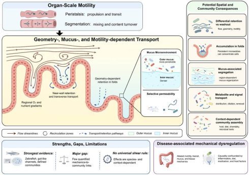
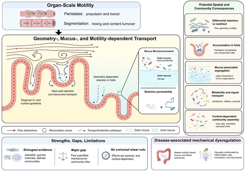
The intestinal mechanical environment arises from the coupling between luminal fluid mechanics and the solid mechanics of the intestinal wall. Intestinal motility generates pressure gradients, fluid flow, wall shear stress, and cyclic tissue deformation, whereas luminal geometry, the rheology of chyme and mucus, and extracellular matrix properties modulate their spatial and temporal distributions. As summarized in Figure 1, these mechanical processes generate heterogeneous physical conditions that influence microbial transport, retention, spatial organization, and host-mediated habitat remodeling, while interacting with chemical, biological, and host-derived factors. This section focuses on the mechanical processes most directly relevant to microbial transport, retention, spatial organization, and host-mediated habitat remodeling.
Context-dependent effects of intestinal mechanics and physicochemical heterogeneity on microbial spatial organization. Peristalsis and segmentation generate propulsion, mixing, wall deformation, and heterogeneous near-wall transport. These processes interact with mucosal geometry, luminal rheology, mucus organization, and regional physicochemical gradients to influence microbial retention, washout, spatial segregation, metabolite and signal transport, and community assembly. Geometry-dependent accumulation has been demonstrated in selected animal, microfluidic, and computational models, whereas direct evidence that human intestinal crypt geometry functions as a hydrodynamic microbial reservoir in vivo remains limited. The colonic inner–outer mucus organization shown is region specific. Mechanical effects are therefore context dependent and should not be interpreted as a universal upstream determinant of microbial organization. This figure was created using Figdraw (www.figdraw.com).
2.1 Intestinal motility and wall kinematics
Peristalsis and segmentation are major intestinal motor patterns [21,22]. Peristaltic contractions primarily generate propagating wall deformation and directional transport [21], whereas segmentation is more strongly associated with luminal mixing and limited net propulsion [22,23]. The enteric nervous system provides neural control of gastrointestinal motor activity [21], whereas interstitial cells of Cajal contribute to the generation and coordination of intestinal electrical and motor patterns [22,23]. These coordinated signals ultimately regulate smooth-muscle contraction and produce region- and state-dependent patterns of wall motion [21,22,74].
Peristalsis consists of propagating contractions that generate axial pressure gradients and directional luminal transport [21,75]. In larval zebrafish, anterograde peristaltic waves contributed predominantly to directional pumping, whereas retrograde waves enhanced luminal mixing [76]. Peristaltic wall motion also produces time-dependent changes in luminal cross-sectional area and provides moving-boundary conditions for the intraluminal flow field [76-78].
During fasting, phase III of the migrating motor complex consists of recurrent propagating contractions in the stomach and small intestine [75,79]. The timing and recurrence of these motor events show substantial intraindividual and interindividual variability [80]. Segmental contractions are characterized by spatially alternating contraction and relaxation [22,23]. Computational studies show that segmentation can generate oscillatory luminal motion, enhance radial mixing, and increase repeated interaction of luminal contents with the intestinal wall while producing relatively limited net axial propulsion [23]. Circular folds may also influence mixing by reshaping the flow field produced during intestinal contraction [28].
2.2 Intraluminal hydrodynamics and geometry-dependent flow heterogeneity
Intestinal transport occurs within a confined and deforming lumen. Small-intestinal flow is often treated as a low-Reynolds-number problem in theoretical and computational studies, but this approximation is not universally applicable. Its validity varies with lumen size, contraction pattern and frequency, as well as the rheological properties of the luminal contents [20,23,24]. Transport within the lumen is governed primarily by advection, molecular diffusion, wall-driven mixing, and interactions with luminal boundaries [20,23,25]. Wall deformation over time continuously reshapes the flow field within the tube lumen, and under specific geometric and kinematic conditions, flow reversal and localized recirculation may occur [23,24]. The situation becomes more complex when the luminal contents behave as non-Newtonian fluids, because changes in viscosity with deformation can alter both velocity gradients and local transport [20,23]. Thus, intestinal velocity and shear-rate distributions can differ substantially from those predicted by idealized rigid-tube models [23,24].
Mucosal geometry introduces additional spatial heterogeneity into the local flow field. In the small intestine, the mucosal surface contains villi and crypts, whereas the colonic epithelium lacks villi and is organized primarily around crypt openings [81]. The surface of the colon is further covered by a structured Muc2-dependent mucus layer [18,35]. Circular folds can redirect local streamlines and enhance mixing [28], while villus-scale geometry and wall motion modify near-surface velocity gradients and mass-transfer boundary layers [24,25,82]. Shear rate, defined as the local velocity gradient that quantifies fluid shear, is locally amplified or dampened by the combined effects of mucosal topography and wall motion, with its magnitude shaped by bulk flow direction, contraction kinematics, and geometric confinement [22,24]. Villi and intestinal folds should thus be regarded as geometric features that redistribute local flow and shear rather than as uniformly low-shear structures [24,28].
2.3 Rheological properties of chyme and mucus
As summarized in Table 1, chyme and intestinal mucus exhibit composition-, region-, and measurement-dependent rheological properties. Chyme is a heterogeneous multiphase material whose flow behavior changes with composition and digestive state [20,83], whereas intestinal mucus is a mucin-rich viscoelastic material whose mechanical response varies with anatomical origin, hydration, composition, and measurement conditions [16,20,29,30]. Depending on these conditions, both materials may exhibit non-Newtonian behavior [20,29,30], which can modify luminal flow, mixing, and near-wall mass transport [20,23,25].
Composition, rheological characteristics, and microbial relevance of intestinal chyme and mucus [14-18,20,29,30,35,36,83,85-88,90].
| Feature | Chyme | Intestinal mucus |
|---|---|---|
| Material composition | Heterogeneous multiphase digesta containing water, electrolytes, digestive products, food particles, enzymes, gases, and microorganisms | Highly hydrated, mucin 2-based gel containing electrolytes, lipids, immunoglobulins, trefoil factors, antimicrobial molecules, and other secreted components |
| Rheological behavior | Composition-dependent and often non-Newtonian; may exhibit shear-dependent viscosity, viscoelasticity, plasticity, and multiphase flow | Nonlinear viscoelastic behavior with shear thinning, apparent yielding, and partial recovery. Bulk rheology reflects volume-averaged properties, whereas microrheology is probe-size dependent; nanoscale particles, individual bacteria, and larger aggregates may experience markedly different local mechanical resistance. |
| Major determinants | Food composition, solids content, particle size, hydration, digestive stage, temperature, and applied shear conditions | Intestinal region, mucin and protein concentration, hydration, lipid and ionic composition, disease state, sample handling, and test protocol |
| Regional and temporal variation | Changes during gastrointestinal transit as digestion, secretion, absorption, and luminal composition evolve | Varies among intestinal regions. Rat small-intestinal mucus shows regional differences in viscosity and viscoelasticity; colonic mucus contains a dense inner layer and a more penetrable outer layer |
| Microbial relevance | Influences luminal mixing, nutrient and metabolite transport, microbial residence, growth, and washout | Influences bacterial movement through mucus, epithelial access, and the spatial separation of luminal bacteria from the colonic epithelium |
2.3.1 Chyme rheology
Chyme is a heterogeneous multiphase material composed of liquid, suspended and particulate matter, and, in some intestinal regions, gas [20]. Its rheological behavior depends on solids content, particle size and composition, hydration, and the stage of digestion [20,83]. Reported behaviors include shear-dependent viscosity, viscoelasticity, and yield-like responses, although their relative importance varies among digesta compositions and experimental conditions [20]. When shear-thinning behavior is present, increases in local shear rate reduce apparent viscosity and thereby modify the velocity field and mixing generated by intestinal contractions [20,23]. Computational studies further indicate that the interaction between contraction kinematics and luminal rheology can alter axial transport and solute transfer toward the mucosal surface [23,25,84]. These properties evolve continuously during gastrointestinal transit as digestion, secretion, absorption, and changes in particulate structure modify the composition of the luminal contents [20,83].
2.3.2 Mucus rheology
Intestinal mucus is a highly hydrated, mucin-rich hydrogel in which Muc2 forms the principal gel-forming scaffold in the intestine [16,35,85]. Its mechanical behavior is nonlinear and time dependent, with reported values varying according to intestinal region, hydration, mucin concentration, ionic composition, disease state, sample handling, and the rheological protocol used [16,29,30,86]. Native and model mucus preparations can exhibit shear-thinning behavior [29,86,87], apparent yielding [86], and partial recovery of viscoelastic properties after deformation [30,88]. These responses should not be interpreted as fixed material constants because they depend strongly on sample origin, preparation, and deformation history [29,86-88].
The mucus network derives its mechanical properties from mucin polymerization, chain entanglement, and reversible molecular interactions [29,85,88]. Regional differences in composition and organization further contribute to spatial variation in rheology [16,29]. In the colon, mucus secretion and expansion generate inner-to-outer differences in density and material organization [18,35,85]. Small-intestinal mucus also shows regional variation in viscosity and viscoelasticity [29], although direct measurements of layer-specific rheological gradients remain limited [16,29].
Bulk rheology and microrheology describe mucus mechanics at different spatial scales. Conventional bulk rheometry measures properties averaged over the sample volume, including viscosity and viscoelastic moduli, while microrheological approaches can resolve the local mechanical environment encountered by micro- and nanoscale entities [30,87]. This distinction is particularly important for mucus because its structure is highly heterogeneous. The resistance encountered by a probe depends not only on the material itself but also on probe size and its interactions with the mucin network [30]. For example, small non-adhesive particles may diffuse through relatively fluid regions and experience an apparent viscosity far below the bulk value. As particle size approaches the characteristic dimensions of the mucin mesh, steric constraints become increasingly important [30]. Individual bacteria and larger bacterial aggregates may therefore encounter different local mechanical environments, although direct quantitative measurements at these biological length scales remain limited. Bulk viscosity should not be interpreted directly as the effective mechanical resistance experienced by individual motile bacteria.
These observations emphasize that intestinal mucus should be treated as a spatially, temporally, and scale-dependent viscoelastic material rather than as a uniform hydrogel.
2.4 Solid mechanics of the intestinal wall and extracellular matrix
The intestinal wall is subjected to both fluid-derived and tissue-generated mechanical loads [26,55,56]. Intraluminal pressure and wall shear stress act at the luminal interface, whereas smooth-muscle contraction produces circumferential and longitudinal deformation, including local stretch and compression [26,56]. These deformations are distributed across the layered intestinal wall, whose extracellular-matrix properties, stiffness, and viscoelasticity influence local stress distribution and mechanical coupling [26,55,56]. More generally, dynamic fluid-solid interfaces couple solid-phase strain and stiffness with fluid shear stress, illustrating that these mechanical variables cannot always be considered independently [89]. The intestinal mechanical environment therefore emerges from the interaction among luminal loading, active wall deformation, tissue architecture, and extracellular-matrix properties [26,55,56,89].
3. Bacterial mechanosensation in the intestinal mechanical environment
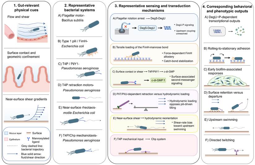
Representative examples of bacterial mechanosensing systems and their downstream responses are summarized in Figure 2. These examples illustrate diverse force-responsive strategies identified in selected bacterial models, but their conservation and ecological relevance within complex intestinal communities remain to be established. Within the intestine, bacteria experience diverse mechanical cues arising from flow, surface interactions, mucus confinement, and spatial constraints. Mechanosensation enables bacteria to detect these physical stimuli and convert them into intracellular signaling responses through force-responsive structures, adhesion systems, and regulatory networks. Current knowledge of bacterial mechanosensing has been derived mainly from selected model organisms and pathogens, including P. aeruginosa, E. coli, and B. subtilis. These studies have revealed representative mechanisms involving flagellar load sensing, force-dependent adhesion, type IV pilus-associated signaling, and mechanically regulated second-messenger pathways. However, whether these mechanisms are conserved in complex intestinal microbial communities, particularly anaerobic commensals, remains largely unknown. This section focuses on the molecular processes underlying bacterial mechanosensing and signal transduction, whereas their consequences for bacterial behavior and spatial organization are discussed in Section 4. Representative mechanical cues and bacterial responses in experimental models are summarized in Table 2.
Representative bacterial responses to gut-relevant physical cues and transport processes. Fluid shear, surface contact, geometric confinement, near-surface flow, and signal transport regulate bacterial adhesion, motility, surface residence, and biofilm-associated responses through distinct mechanisms. Representative examples include DegS–DegU signaling following flagellar arrest in B. subtilis, FimH catch-bond adhesion in E. coli, type IV pili/PilY1-mediated c-di-GMP signaling and PilT/PilU-dependent surface retention in P. aeruginosa, near-surface rheotaxis, Chp-dependent mechanotaxis, and flow-modulated quorum sensing. Rows A–F represent matched examples across columns 2–4 and do not constitute a universal sequential pathway. Solid arrows indicate experimentally supported mechanism–output relationships in the indicated models. This figure was created using Figdraw (www.figdraw.com).
Representative mechanical cues and bacterial responses in experimental models.
| Mechanical parameter | Model bacterium | Experimental system | Observed response | Supported mechanism | Reference |
|---|---|---|---|---|---|
| Cell-scale hydrodynamic force, 0.1–10 pN | P. aeruginosa | Microfluidic flow over an abiotic surface | Increasing force opposed cell tilting, promoted a surface-parallel orientation, and prolonged surface residence | Hydrodynamic loading counteracted PilT/PilU-dependent type IV pilus retraction and pilus-driven surface departure | [38] |
| Wall shear stress 0.25–4 Pa | Type 1-fimbriated E. coli | Mannose-coated flow chamber | Higher shear reduced inhibitor-mediated detachment and stabilized FimH-dependent adhesion | Force loading increased the lifetime of the FimH–mannose catch bond | [40] |
| Wall shear stress, approximately 5–100 mPa | Crohn’s disease-associated adherent-invasive E. coli and other E. coli pathovars | Surface-plasmon-resonance flow assay on mannosylated surfaces | AIEC isolates generally showed weaker shear-dependent changes in adhesion; LF82 was nearly shear-insensitive under the tested conditions | Natural FimH sequence variation, including interdomain substitutions, was associated with altered shear responsiveness | [99] |
| Tensile loading of FimH | Type 1-fimbriated E. coli | Structural, kinetic, and molecular studies | Stabilized the high-affinity ligand-bound state of FimH | Force-induced separation and rearrangement of the lectin and pilin domains regulate catch-bond formation | [41,91-93] |
| Flagellar rotation arrest | B. subtilis | Genetic inhibition, clutch-mediated arrest, and physical restriction of flagellar rotation | Increased degU transcription and DegU~P-dependent exoprotease and poly-γ-glutamate production | Flagellar motor arrest activated the DegS–DegU regulatory pathway; the upstream coupling mechanism remains unresolved | [47,101] |
| Substrate stiffness, 0.5–12 kPa; controlled series 1.5–5 kPa | Myxococcus xanthus | Uncoated polyacrylamide hydrogels and motility mutants | Colony expansion increased with stiffness; adventurous motility was more stiffness-sensitive than type IV pilus-dependent social motility | A-motility-associated surface adhesion machinery contributed to stiffness-dependent expansion; surface chemistry also affected the response | [100] |
3.1 Mechanical sensors and force-responsive structures
3.1.1 Flagellar motor: a mechanical sensor
In B. subtilis, the flagellar motor functions not only as a motility apparatus but also as a force-responsive sensory structure [47]. Genetic, clutch-mediated, or physical inhibition of flagellar rotation increased degU transcription and DegU-dependent outputs, including exoprotease production and poly-γ-glutamate biosynthesis, implicating the DegS–DegU two-component system in signaling downstream of altered motor activity [47]. However, the molecular mechanism linking rotational arrest to DegS activation remains unresolved, and whether this pathway is conserved among gut-associated bacteria remains unknown [52,53].
3.1.2 Type I pili (FimH): force-dependent adhesion through catch bonds
Flow-chamber studies using mannosylated surfaces demonstrated that tensile loading enhances FimH-mediated adhesion under shear conditions [39,40]. Structural and single-molecule studies further showed that mechanical force promotes allosteric rearrangement between the FimH lectin and pilin domains, stabilizing a high-affinity ligand-bound state [41,91,92]. The magnitude and force dependence of this catch-bond response vary with FimH sequence, ligand presentation, and mechanical loading conditions [39-41,91-93]. These studies establish FimH-mediated catch-bond adhesion as a representative force-dependent adhesion mechanism in selected E. coli strains. A distinct catch-bond mechanism has also been identified in the cellulosome-anchoring complex of Ruminococcus champanellensis, where force stabilizes attachment to cellulose fibers rather than host epithelial receptors [42].
3.1.3 Type IV pili: PilY1-mediated force sensing
Type IV pili (T4P) are dynamic surface filaments involved in adhesion and twitching motility in several bacterial species [45]. Their mechanosensory functions and downstream signaling pathways are species- and strain-dependent [45,46]. In P. aeruginosa, experiments have shown that both T4P and the pilus-associated protein PilY1 are necessary for the increase in intracellular c-di-GMP triggered by shear [43]. Force-induced conformational changes of PilY1 contribute to surface sensing and downstream c-di-GMP signaling [46], while single-molecule studies further demonstrate the force-dependent mechanical behavior of PilY1 [44]. These studies identify a T4P/PilY1-dependent mechanotransduction pathway in defined P. aeruginosa strains [43,46].
3.2 Second messenger signaling under mechanical stimulation
Cyclic dimeric guanosine monophosphate (c-di-GMP) is a widespread bacterial second messenger that controls the balance between motile and surface-associated lifestyles and regulates a broad range of adaptive behaviors [52,53]. Mechanical stimuli, including fluid shear [43] and surface contact or surface mechanics [94], can modulate c-di-GMP signaling through force-responsive regulatory pathways in selected bacterial models.
One such pathway has been characterized in P. aeruginosa. Here, mechanical input sensed through T4P and PilY1 can be transmitted to the c-di-GMP network [43]. Genetic studies identified the diguanylate cyclase SadC as a downstream component of PilY1-dependent mechanosensing, although the molecular events linking mechanical force perception to SadC activation remain incompletely resolved [95]. Biophysical and structural evidence is consistent with this model, showing that PilY1 changes conformation under mechanical loading [44,46]. Once c-di-GMP levels rise, the response extends beyond force sensing itself, promoting transcriptional programs involved in surface adaptation and extracellular matrix production [52,53,94].
Mechanical signals can also be integrated through phosphorylation-based regulatory systems. In B. subtilis, inhibition of flagellar rotation activates the DegS–DegU two-component signaling pathway, leading to increased degU transcription and DegU~P-dependent regulation of downstream targets, including exoprotease production and poly-γ-glutamate biosynthesis [47]. These examples illustrate that bacteria can convert mechanical perturbations into intracellular regulatory states through both second messenger systems and two-component signaling pathways [52,53]. However, whether these mechanisms are conserved among intestinal commensals and contribute substantially to host-associated microbial communities remains unclear [52-54].
3.3 Molecular effectors downstream of mechanical signaling
Mechanical signaling pathways ultimately influence bacterial phenotypes through regulation of specific molecular effectors [52,53]. In P. aeruginosa, activation of the PilY1–SadC–c-di-GMP pathway induces transcriptional programs associated with surface-associated growth and extracellular matrix production [43]. The c-di-GMP-regulated transcriptional regulator FleQ has been implicated in controlling biofilm-associated gene expression, including pathways involved in extracellular matrix synthesis [96].
In B. subtilis, activation of the DegS–DegU pathway following flagellar motor perturbation regulates downstream transcriptional responses, including genes involved in extracellular enzyme production and poly-γ-glutamate biosynthesis [47]. This provides a clear example of how a mechanical disturbance can be coupled to a specific transcriptional response through an established bacterial regulatory system [47,52,53].
Mechanical forces can also directly regulate adhesion-associated proteins. In type 1-fimbriated E. coli, tensile loading induces structural rearrangement of the FimH adhesin, stabilizing a high-affinity ligand-binding conformation through a catch-bond mechanism [41,91,92]. Thus, mechanically regulated bacterial responses can occur through multiple molecular mechanisms, including transcriptional regulation, second messenger-dependent pathways, and force-dependent modulation of adhesion proteins [41,47,91,92].
3.4 Mechanical regulation of extracellular signaling environments
Mechanical conditions can influence bacterial communication by regulating the transport and accumulation of extracellular signaling molecules. Quorum-sensing activation depends on signal production, degradation, molecular diffusion, advective transport, and spatial confinement [50]. Flow therefore affects local signal concentrations by modifying molecular accumulation, dilution, and removal, thereby influencing the threshold required for signal activation [50].
Computational analyses have shown that flow geometry and bacterial spatial organization can alter quorum-sensing dynamics by changing signal transport and retention [50]. Experimental studies further indicate that shear conditions influence quorum-sensing-associated responses in a context-dependent manner. In P. aeruginosa, different shear environments altered acyl-homoserine-lactone production, although the magnitude and direction of these changes depended on strain background and environmental conditions [97].
Extracellular signaling may also interact with intracellular regulatory networks. In several bacterial systems, quorum sensing and c-di-GMP signaling regulate overlapping surface-associated programs, although the molecular relationship between these pathways varies among species [98]. In a gut-inspired porous microfluidic system, flow altered the spatial distribution of autoinducer-2 (AI-2), generating chemical gradients that affected bacterial accumulation through changes in chemotactic behavior [51]. It is not yet clear whether comparable transport–signaling interactions occur in the native intestinal lumen.
3.5 Context dependence of bacterial mechanosensing
Bacterial responses to mechanical cues vary with genetic background, surface characteristics, and experimental setting [40,93,99]. FimH-mediated adhesion under shear provides a representative example of such variability. Comparative analyses of Crohn’s disease-associated adherent-invasive E. coli (AIEC) and other pathogenic isolates demonstrated strain-dependent differences in shear-responsive adhesion, with the AIEC strain LF82 showing limited shear-dependent changes under the tested conditions [99]. Thus, species identity alone is not sufficient to predict how a bacterium will respond mechanically. Several factors may contribute to this variability, including differences in adhesin sequence [99], ligand presentation, mechanical loading conditions [40,93], and the surrounding physicochemical environment. Each of these factors can alter force-dependent responses. Mechanical conditions in the intestine may therefore contribute to bacterial fitness, but evidence that they directly select for, or drive the emergence of, specific bacterial traits is still limited [52-54,99].
3.6 Non-intestinal models revealing bacterial mechanical responses
Non-intestinal bacterial systems have provided additional insights into how microorganisms respond to mechanical properties of their surrounding environments [52,53]. In the soil bacterium Myxococcus xanthus (M. xanthus), substrate stiffness influences collective colony expansion [100]. Experiments using motility mutants demonstrated that this response was primarily associated with adventurous motility, whereas type IV pilus-dependent social motility showed weaker stiffness dependence [100]. The response of M. xanthus was also influenced by substrate surface chemistry, indicating that bacterial adaptation can integrate both mechanical and chemical properties of the extracellular environment [100]. Although substrate stiffness sensing has not been directly demonstrated in intestinal microbial communities, these studies broaden the known range of bacterial mechanobiological responses beyond fluid-derived forces. Further investigations are required to determine whether similar mechanisms contribute to bacterial adaptation within mechanically complex host-associated environments.
4. Intestinal mechanics and microbial spatial organization
Intestinal mechanics contributes to microbial spatial organization by modifying physical constraints on microbial transport and habitat accessibility [15,19,31-33]. Motility-driven flow and intestinal deformation [15,63], mucosal architecture [19,31,32], and mucus organization [18,35] collectively generate heterogeneous physical environments that influence microbial retention and community assembly [15,19,31-33,63]. Mechanical influences do not act in isolation. Their effects are shaped by nutrient conditions, host-derived signals, and interactions among microbial populations [19,33,34]. Microbial distribution in the gut is therefore more appropriately understood as the outcome of interacting physical and biological processes, with mechanics representing only one part of this system [19,33,34].
4.1 Geometry- and motility-dependent microbial retention
Intestinal geometry can generate spatial heterogeneity in microbial distribution by modifying local transport pathways and bacterial escape dynamics [15,31-33]. In larval zebrafish, motile E. coli preferentially accumulated within intestinal folds, and biomimetic microfluidic systems further demonstrated that fold-like geometries could promote bacterial retention by restricting escape from confined regions [31]. Computational work reached a similar conclusion, identifying both hydrodynamic confinement and steric interactions as contributors to bacterial persistence in groove-like geometries [32]. These studies provide direct evidence that geometric features can influence bacterial localization in defined systems, although the resulting spatial patterns also depend on bacterial motility, chemotaxis, and adhesion properties.
Mucosal structures do not simply generate uniformly protected microbial niches. Villi, folds, and wall motion continually alter the local flow field, so the mechanical environment near the mucosal surface is spatially and temporally variable [24,25,28,82]. The effect of mucosal geometry depends on contraction kinematics, fluid rheology, and anatomical scale [20,23-25,28]. Although intestinal crypts have been proposed as potential microbial habitats [19], direct evidence that human crypt geometry functions as a hydrodynamic reservoir remains limited. In the healthy colon, the dense inner mucus layer also restricts bacterial access to the epithelial surface [18,35] and may counteract geometry-driven accumulation.
Bacterial motility further determines the balance between retention and washout. In gut-like microfluidic systems, flow velocity and peristaltic-like deformation altered bacterial distribution and growth patterns [15]. In larval zebrafish, intestinal motility affected competition between Aeromonas and Vibrio; when motility was reduced, coexistence between the two became more likely [63]. These findings suggest that host-generated motion can modify microbial community structure in simplified or tractable model systems. Whether the same type of mechanical control operates in complex mammalian microbiomes remains uncertain.
4.2 Mucus-associated spatial segregation
Mucus influences how readily intestinal bacteria can reach the epithelial surface through both its mechanical properties and chemical composition [14,16,35]. In the healthy colon, the dense inner Muc2-dependent mucus layer largely excludes bacteria from direct contact with epithelial cells, while the outer layer is more accessible to microbial colonization [18,35]. This organization differs across intestinal regions, as mucus thickness, attachment, composition, and rheological properties vary between the small intestine and colon [16,29,35].
Microbial transport within mucus cannot be explained solely by physical exclusion based on pore size [14,16]. Bacterial movement depends on cell size and shape, motility, adhesin-mediated interactions, mucus composition, and local viscoelastic properties [14,16,30]. This complexity is important when interpreting in vitro studies. Purified mucins and artificial mucus formulations reproduce selected features of the native material, but they do not necessarily capture its full structural heterogeneity or its continual remodeling in vivo [16,87,88].
Access to mucus-associated habitats depends on both physical transport and the ability of bacteria to interact with and adapt to their local environment [14,16,35]. Flagella, pili, adhesins, mucus-degrading enzymes, and resistance mechanisms can contribute to mucus-associated colonization in selected species [16,35], but no single appendage-mediated mechanism explains microbial positioning across the intestinal microbiota. Mucus is better understood as a dynamic and selective habitat that continually shapes microbial localization, rather than as an inert barrier separating bacteria from the epithelium [14,16,35].
4.3 Flow-mediated metabolic exchange and community assembly
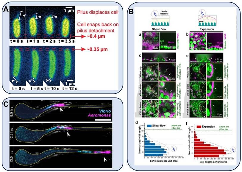
Intestinal flow affects more than bacterial transport and retention. By changing the local distribution of nutrients, metabolites, and extracellular signaling molecules, it can also alter the conditions under which microbial populations interact [15,33,34]. Representative experimental systems illustrating how mechanical conditions influence microbial surface residence, spatial distribution, and community interactions are summarized in Figure 3. Flow may wash out weakly retained cells and dilute extracellular products, while also facilitating metabolite exchange between spatially separated populations [15,33].
Representative experimental evidence linking intestinal mechanical conditions to microbial spatial organization. (A) Flow-dependent surface residence of Pseudomonas aeruginosa mediated by type IV pili. Microfluidic flow experiments showed that hydrodynamic loading opposed pilus-driven cell tilting and influenced surface retention versus departure behavior. Adapted with permission from [103], copyright 2024 American Physical Society. (B) Spatial redistribution of motile Escherichia coli Nissle in a mechanically dynamic three-dimensional gut-on-chip model. Cyclic shear flow and epithelial expansion altered bacterial localization relative to epithelial structures and mucus-like regions. Adapted with permission from [64], copyright 2025 Wiley-VCH GmbH. (C) Motility-dependent microbial competition in the larval zebrafish intestine. Host intestinal motility influenced the spatial interaction and competitive dynamics between Aeromonas and Vibrio populations. Adapted with permission from [63], copyright 2016 Public Library of Science. Together, these studies provide representative evidence that mechanical conditions can influence microbial residence, spatial distribution, and ecological interactions in defined experimental systems. However, they do not establish a universal mechanical rule governing intestinal microbial organization across species or communities.
In defined gut microbial communities, flow altered the spatial organization of metabolic interactions and influenced community composition by redistributing extracellular metabolites and shared resources [33]. Under specific experimental conditions, intermediate transport rates have been reported to facilitate metabolic interactions between neighboring populations, whereas stronger removal can reduce local metabolite availability [33]. Flow can therefore reorganize metabolic interactions rather than simply wash microorganisms out of the system.
The relationship between flow and microbial attachment is similarly context dependent. As described in Section 3, force-responsive adhesion and mechanically regulated surface-associated responses vary among bacterial species, strains, and molecular systems [38-40,43]. A change in shear rate therefore does not predict attachment or dispersal in a uniform way. Surface structures, adhesion mechanisms, environmental chemistry, physiological state, and the surrounding microbial community can all influence the outcome. In surface-colonization models, flow has also been shown to affect growth and population heterogeneity within attached communities [102].
Regional patterns in the intestinal microbiota are likewise shaped by more than local mechanics. Transit and luminal flow operate alongside nutrient availability and local chemical conditions [34,36], oxygen gradients [37], mucus organization and host-derived factors [19,35], and microbial interactions [19,33]. The microbial patterns observed along the gastrointestinal tract emerge from these processes acting together, with mechanical conditions representing only one part of the local ecological environment.
4.4 Evidence gaps in mechanically mediated microbial organization
Altered motility and transit dynamics [4,9], mucus organization [35], and tissue mechanics [104] are associated with inflammatory and functional gastrointestinal disorders. These changes may influence microbial residence and washout [15,17,31,32] and mucosal accessibility [16,18,35]. However, their specific contributions are difficult to separate from concurrent alterations in diet, medication exposure, inflammation, epithelial permeability, and immune activity [19,105,106]. Mechanical dysregulation should therefore be considered as a potential modulating factor in microbial imbalance rather than a general initiating mechanism [106].
Crohn’s disease-associated adherent-invasive E. coli isolates exhibit strain-dependent FimH-mediated adhesion responses under flow conditions. The LF82 strain, a widely used AIEC reference isolate, showed limited shear-dependent changes in adhesion across the tested shear conditions [99]. Force-responsive adhesion clearly varies among E. coli strains, but the available data do not show that these phenotypes arose directly from inflammatory mechanical conditions [99]. Differences in host receptor presentation, inflammation, pre-existing bacterial variation [99], and other selective pressures may also contribute to pathobiont expansion [105].
Tissue stiffening adds another possible mechanical connection. Increased intestinal stiffness associated with fibrosis has been reported in inflammatory bowel disease [104]. Luminal bacteria, however, are physically separated from the extracellular matrix by the mucus layer, epithelium, and basement membrane [18,35,36]. Changes in tissue mechanics are therefore more likely to affect microbial communities indirectly, for example by altering epithelial deformation, mucus organization, host signaling, inflammation, or intestinal motility, rather than through direct sensing of matrix stiffness by luminal bacteria [26,104].
A major limitation of the current literature is that most experimental systems reproduce only part of the mechanical and microbial complexity of the intestine [59,60,72,73]. Models that more closely resemble the native environment will need to account for segment-specific flow, native mucus properties, anaerobic conditions, host-derived signals, and interactions among multiple microbial species [59,60,62,64,65,72]. Combining these models with bacterial-scale force measurements and genetic perturbation could help distinguish direct responses to mechanical cues from indirect effects associated with host pathology.
Intestinal mechanics can affect microbial retention, washout, mucus-associated segregation, and metabolite exchange, but the magnitude and direction of these effects vary with bacterial traits, community composition, and local physicochemical conditions [19,106]. A simple distinction between low- and high-shear environments is therefore not sufficient to explain microbial organization in the gut. Section 5 examines the opposite direction of this relationship by considering how microbial activity can modify intestinal mechanical properties.
5. Reciprocal host–microbiota influences on the intestinal mechanical environment
Host-generated mechanical conditions regulate microbial transport, retention, and spatial organization, whereas microbial activity and inflammatory responses can modify the physical properties of the intestinal habitat. These reciprocal interactions involve changes in mucus organization, epithelial barrier function, intestinal motility, and tissue remodeling. However, current evidence remains heterogeneous across microbial taxa, host cell types, and experimental models. This section discusses how host responses to mechanical stimuli influence microbial habitats and how microbial and inflammatory signals reshape intestinal mechanical properties.
5.1 Host mechanosensation modulates microbial habitats
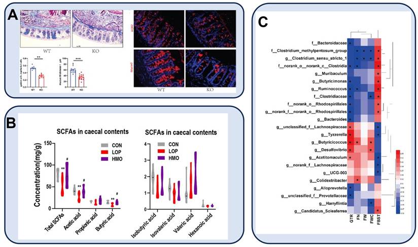
Host mechanosensation can influence microbial habitats by regulating mucus integrity [57] and intestinal motility and inflammatory homeostasis [58]. Among identified mechanosensitive pathways, PIEZO1 provides direct evidence linking mechanical sensing with habitat-relevant intestinal functions [57,58]. In mice, goblet-cell-specific PIEZO1 deficiency reduced goblet-cell abundance, Muc2 expression, and inner mucus-layer integrity, accompanied by alterations in fecal and mucosa-associated microbiota [57]. Enteric-neuronal PIEZO1 also contributes to force-dependent regulation of intestinal motility and inflammatory homeostasis [58]. These findings indicate that host mechanosensing can modify microbial habitats through cell-type-specific regulation of mucus organization and intestinal physiology. However, microbiota alterations following PIEZO1 perturbation may result from multiple downstream changes, including altered mucus properties, antimicrobial defense, epithelial function, and inflammation, rather than from mechanical sensing alone [57]. Direct evidence connecting individual host mechanotransduction pathways with microbial spatial organization remains limited [59,60]. Representative evidence linking host mechanosensation with mucus-barrier remodeling and microbiota-associated changes is summarized in Figure 4A.
Representative evidence linking host mechanosensation and intestinal mechanical alterations with microbiota-associated phenotypes. (A) Direct evidence from goblet-cell-specific PIEZO1-deficient mice. Loss of goblet-cell PIEZO1 altered goblet-cell abundance, mucin 2 expression, and colonic mucus barrier integrity, accompanied by changes in fecal and mucosa-associated microbiota. Adapted with permission from [57], copyright 2022 Frontiers Media SA. (B-C) Disease-model-associated changes in intestinal metabolites and microbiota. In a loperamide-induced constipation mouse model, altered intestinal transit was associated with changes in cecal short-chain fatty acid profiles (B) and correlations between bacterial taxa, constipation-related phenotypes, gastrointestinal transit, and enteric nervous system indices (C). These findings represent associations between altered intestinal function and microbial/metabolic profiles rather than direct evidence of mechanical causality. Adapted with permission from [115], copyright 2023 Frontiers Media SA. Together, these examples illustrate different levels of evidence linking intestinal mechanical regulation with microbiota-associated changes, ranging from mechanistic host genetic perturbation to disease-associated correlations.
5.2 Microbial and inflammatory regulation of intestinal mechanical properties
Microbial activity and inflammatory signaling can modify host properties that help shape the intestinal mechanical environment, including mucus organization, epithelial integrity, smooth-muscle activity and tissue stiffness [35,104,107-109]. Inflammatory disorders are frequently accompanied by altered mucus production and organization [35], which can change the material properties [16,30,86] and protective function of the mucus layer [35,57]. In epithelial models and experimental colitis, tumor necrosis factor-like cytokine 1A (TL1A) promotes bacterial internalization and transcytosis through myosin light chain kinase 2 (MLCK2), the shorter splice variant of long MLCK, contributing to epithelial barrier dysfunction [110]. Although this pathway does not directly represent bacterial sensing of mechanical cues, it illustrates how inflammatory signaling can remodel host structures that regulate microbial accessibility. Inflammation-associated tissue remodeling can also modify the solid mechanical properties of the intestine. Fibrotic remodeling in inflammatory bowel disease is associated with increased intestinal stiffness [104]. Luminal microorganisms are separated from the extracellular matrix by mucus, epithelium and basement membrane [18,35,36]. Changes in tissue mechanics are therefore more likely to affect the microbiota indirectly through epithelial deformation, mucus organization, immune signaling, or altered motility than through direct sensing of matrix stiffness by luminal bacteria.
Microbial metabolites can also regulate intestinal motility through interactions with the enteric nervous system [107-109]. Short-chain fatty acids and other microbially derived signals influence enteric neural activity and smooth-muscle function, although these effects depend on metabolite concentration, receptor distribution, intestinal region and host state [107-109]. What is still poorly defined is how individual microbial signals translate into measurable changes in luminal flow, mucus rheology, or intestinal wall mechanics [59,60].
5.3 Causal uncertainty of mechanical–microbial alterations in gastrointestinal disorders
Altered intestinal motility and microbial composition frequently coexist in gastrointestinal disorders, including irritable bowel syndrome, chronic constipation, and small intestinal bacterial overgrowth [4,5,9,111,112]. Irritable bowel syndrome is also associated with barrier dysfunction and altered sensory signaling [111]. The temporal relationship among these abnormalities is difficult to resolve. Mechanical dysfunction may precede changes in the microbiota, arise as a consequence of them, or develop alongside microbial alterations through shared inflammatory and metabolic processes [9,111].
In irritable bowel syndrome, altered bowel habits, microbial composition, immune activity, and sensory signaling may occur simultaneously [111]. In mice, enteric-neuronal PIEZO1 regulates gastrointestinal motility and inflammatory homeostasis [58], but a comparable mechanistic link has not yet been established in human irritable bowel syndrome. In a post-infectious irritable bowel syndrome mouse model, reduced interstitial cells of Cajal and increased intraepithelial lymphocytes were associated with small intestinal bacterial overgrowth [113]. The model connects inflammation, impaired motor regulation, and microbial expansion, but it does not clarify which event occurs first or which process drives the others.
A similar problem applies to chronic constipation. Slower transit prolongs luminal residence time and is associated with differences in microbial composition [4,9]. Changes in metabolites and microbiota-associated features have also been reported in constipation models (Figure 4B–C), but these observations do not establish the direction of causality. Fecal microbiota transplantation has improved bowel symptoms and modified microbial profiles in selected patients with functional constipation [114]. However, whether these effects arise from restoration of intestinal mechanics, microbial metabolites, immune modulation, or other mechanisms remains unclear.
Small intestinal bacterial overgrowth is associated with impaired intestinal clearance, but also with anatomical abnormalities, altered antimicrobial defenses, medication exposure, and underlying host diseases [5,112]. Impaired motility alone is therefore insufficient to explain microbial accumulation in the small intestine. Across these disorders, the available evidence points to close interaction between microbial and mechanical alterations, while the direction of causality is often difficult to establish [4,5,9,112].
5.4 Causal boundaries and research priorities
Establishing causal relationships between intestinal mechanics and microbiota requires evidence that defined mechanical perturbations produce specific host or microbial responses [43,46,57,58] and lead to reproducible changes in microbial organization or function. Few studies currently integrate these processes within the same experimental system [59,60,72,73].
Future studies should integrate mechanical, microbial, and molecular measurements, including intestinal motility, regional transit, mucus rheology [88], tissue mechanics, microbial spatial organization [64,65], and metabolite profiles. Cell-type-specific host perturbations [57,58], microbial genetic approaches [43,46], anaerobic multispecies models [62], and longitudinal human studies will be required to distinguish direct mechanical effects from those mediated by inflammation, diet, medication, and host-derived factors [59,60,62]. Such integrated approaches will clarify when host–microbiota mechanical interactions represent causal regulatory mechanisms rather than secondary consequences of intestinal dysfunction.
6. Frontier technologies and models for intestinal mechanical–microbial research
6.1 Reductionist epithelial and mucus models
Enteroids and colonoids derived from adult intestinal stem cells, and human intestinal organoids derived from pluripotent stem cells, retain distinct epithelial, regional and donor-associated properties and should not be treated as interchangeable model systems [81,116,117]. Air–liquid interface and mucus-producing epithelial cultures enable investigation of mucus secretion and in situ rheology [88,118]. These reductionist systems are well suited to epithelial and mucus mechanisms but generally lack physiological luminal flow, vascular and neural components, immune complexity and stable multispecies microbial communities [81,116,117]. Native mucus, collected mucus and mucus surrogates should also be distinguished because sample origin, collection and preparation substantially affect measured rheological properties [16,29,30,87].
6.2 Coupled microphysiological systems
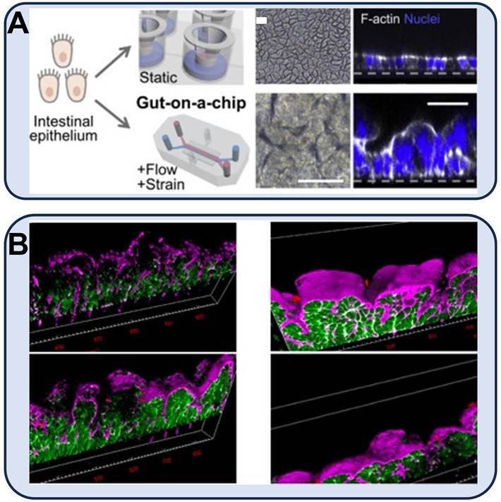
Microphysiological systems permit controlled manipulation of luminal flow, cyclic deformation, oxygen availability, epithelial geometry and microbial exposure [61,62,64,89,119]. Representative examples of mechanodynamic gut-on-chip applications are shown in Figure 5, illustrating how controlled mechanical inputs can regulate epithelial organization and host–microbe interactions. Anaerobic intestine-on-chip systems can maintain human intestinal epithelium together with complex aerobic and anaerobic microbial communities under controlled oxygen gradients [62]. Mechanodynamic chips have been used to examine epithelial differentiation [119], single-cell transcriptional responses [66] and pathogen invasion under peristalsis-like deformation [61,66,119,120]. Tunable-flow microbiome chips and bioprinted intestinal models extend these approaches to microbial spatial organization and geometry-dependent transport [64,65,121]. Model relevance nevertheless depends on epithelial source, mucus production, oxygen control, microbial complexity, surface material and the imposed flow or deformation waveform [60,72,73].
Representative mechanodynamic gut-on-chip models for studying host mechanical responses and microbial interactions. (A) Flow- and strain-dependent epithelial remodeling in a gut-on-chip model. Caco-2 intestinal epithelial cells cultured under dynamic flow and cyclic strain exhibited distinct morphological organization compared with static Transwell cultures, demonstrating that mechanical cues influence epithelial architecture. Adapted with permission from [66], copyright 2022 Elsevier Inc. (B) Peristalsis-like mechanical regulation of Entamoeba histolytica invasion in a three-dimensional intestinal model. Three-dimensional reconstructions demonstrate altered parasite invasion patterns under different mechanical deformation conditions and protease inhibition treatments, indicating that host mechanical states can influence pathogen–epithelium interactions. Adapted with permission from [61], copyright 2022 American Association for the Advancement of Science. Together, these examples illustrate the capability of mechanodynamic gut-on-chip systems to dissect how controlled mechanical perturbations influence host epithelial states and microbial interactions. These models nevertheless reproduce only selected features of the native intestinal environment.
6.3 In vivo models and longitudinal imaging
Larval zebrafish provide optical access to intestinal motility, bacterial localization, aggregation and competition at high spatial and temporal resolution [31,63,67,122]. These studies have directly demonstrated motility-dependent redistribution and competitive exclusion [63], but quantitative translation to the mammalian intestine remains limited by anatomical and physiological differences [122]. Mouse models enable cell-type-specific genetic perturbation and whole-organism analysis. Goblet-cell and enteric-neuronal PIEZO1 models have linked host mechanosensation to mucus-barrier properties, motility, inflammation and microbiota-associated changes [57,58]. Laboratory mouse microbiota and immune phenotypes are also influenced by housing and microbial history, and microbiota-depleted or transplantation models should therefore be treated as perturbation systems rather than neutral physiological baselines [108].
Live and four-dimensional imaging provide longitudinal information on tissue deformation, bacterial localization, aggregation and pathogen invasion [61,63,67]. However, local forces, stresses and pressure fields are commonly inferred from observed deformation or computational reconstruction rather than measured directly.
6.4 Computational and data-integrated models
6.4.1 Flow and transport models
Flow and transport models quantify how wall motion, mucosal geometry, luminal rheology and absorption influence velocity fields, mixing, shear and solute transport [17,23-25,76,78,82,123,124]. These approaches range from idealized active-wall and peristaltic-tube models to villi-resolved and multiscale transport simulations [24,25,76,82]. Their predictions are sensitive to constitutive equations, boundary conditions, contraction waveforms and parameter values, many of which are obtained from simplified, animal or non-physiological systems [59,124]. Geometry-resolved microfluidic and computational models provide evidence for bacterial retention within idealized fold- or groove-like structures [31,32], but these findings do not establish that human intestinal crypts function as hydrodynamic microbial reservoirs in vivo.
6.4.2 Microbial population and community models
Microbial population models couple hydrodynamic transport with bacterial growth, nutrient utilization, taxis, diffusion and washout [17,68,69,71]. In a zebrafish intestinal model, biological taxis generated bacterial aggregation, whereas intestinal peristalsis reduced aggregation through its mixing effect [71]. This result illustrates that microbial activity and hydrodynamics must be modeled jointly rather than treating flow as the sole determinant of spatial organization. Colon-scale models further show that dietary substrate input, microbial growth and luminal transport interact to generate longitudinal community structure [69].
6.4.3 Hybrid multiscale and data-driven models
Hybrid models combine continuum transport equations, agent-based representations and genome-scale metabolic networks to examine microbial spatial organization and metabolic interactions. MetaBiome integrates agent-based modeling, finite-volume transport and genome-scale metabolic models to simulate mucosal microbial communities [70]. Machine-learning methods may assist parameter inference, model reduction and integration of multi-omics data, but prospective validation, uncertainty quantification and direct incorporation of patient-specific mechanical measurements remain limited [125,126].
Medical digital-twin concepts provide a prospective framework for integrating imaging, motility and pressure measurements, microbial profiles, molecular data and clinical observations [127-130]. A patient-specific intestinal mechanical–microbial twin would require multimodal inputs spanning organ-scale to microscale physiology, potentially including high-resolution intestinal manometry for pressure and contractile patterns [131], dynamic magnetic resonance imaging for intestinal wall kinematics and regional transit [132], ex vivo microrheological characterization of mucus or tissue samples [30,87,88], and spatially resolved metagenomic profiling of microbial communities [133]. A major computational challenge is the coupling of continuum-scale descriptions of luminal flow and intestinal wall mechanics with discrete agent-based representations of individual microbial cells and communities [134]. The wide separation in spatial and temporal scales, together with the large number of microbial agents required for physiologically relevant simulations, creates substantial demands for parameterization, computation, and cross-scale validation [134]. To our knowledge, no prospectively validated patient-specific intestinal mechanical–microbial twin currently predicts microbiota responses to a predefined mechanical intervention. Digital twins should therefore be presented as a long-term research objective rather than an established intestinal research or clinical platform [127-130]. The applications and major limitations of current experimental and computational models are summarized in Table 3.
Comparison of models for intestinal mechanical–microbial research.
| Method | Applications | Main Limitations | References |
|---|---|---|---|
| Intestinal epithelial and organoid models | Analysis of epithelial and mucus mechanisms and defined host–microbe interactions | Conventional organoids lack physiological flow, neural and vascular components, systemic immunity, and stable multispecies communities; collected mucus and mucus surrogates may differ from native mucus | [81,87,116,117,135] |
| Native and model mucus systems | Characterization of mucus rheology and interactions among mucus, bacteria, and delivery systems | Collection and purification can disrupt native mucus structure; mucus substitutes do not fully reproduce native intestinal mucus | [16,29,87,88] |
| Intestinal organ chips and related microphysiological systems | Controlled study of mechanical effects on epithelial differentiation, pathogen invasion, anaerobic microbiota culture, and microbial spatial organization | Limited systemic regulation; results depend on epithelial source, mucus production, oxygen control, device material, microbial complexity, and applied mechanical waveform | [60-62,64-66,136] |
| Live and four-dimensional imaging | Longitudinal visualization of dynamic host–microbial interactions in organ chips and transparent animal models | Local force and stress are usually inferred rather than directly measured; imaging depth, lower phototoxicity, higher resolution, and longer observation times are difficult to achieve simultaneously. | [61,63,67] |
| Larval zebrafish models | Direct visualization and perturbation of motility-dependent microbial spatial dynamics | Major anatomical, physiological, immune, and microbial differences limit quantitative extrapolation to mammalian and human intestines. | [31,63,67] |
| Mouse models, including genetically modified and microbiota-manipulated mice | Cell-specific mechanotransduction studies and causal analysis of host–microbiota interactions in vivo | Anatomy, diet, motility, mucus properties, and microbiota differ substantially across species. Antibiotic treatment also alters host physiology, so antibiotic-depleted animals should not be regarded as a neutral “pseudo-germ-free” model. | [18,57,58,104,137] |
| Mechanistic flow, transport, and microbial population models | Quantification of coupled wall motion, flow, mixing, solute transport, bacterial growth, taxis, and washout; testing mechanistic hypotheses about microbial residence and spatial organization. | Model outputs are sensitive to assumptions about geometry, material behavior, boundary conditions, and parameter values. Many of these inputs are derived from simplified systems, animal studies, or in vitro experiments. | [17,24,32,59,68,82,84] |
| Hybrid multiscale and data-integrated models | Integration of agent-based dynamics, microbial metabolic networks, multi-omics data, and data-driven parameter estimation; conceptual development of future patient-specific modeling frameworks. | Prospective validation, uncertainty quantification, external testing, and incorporation of patient-specific intestinal mechanical measurements remain limited. | [70,126-128,138,139] |
7. Translational opportunities for mechanics-informed microbiota modulation
Direct clinical evidence for mechanics-guided microbiota modulation remains limited; the following sections therefore distinguish established associations, preclinical evidence, and design hypotheses.
7.1 Motility and transit as modifiable physical variables
Motility and transit influence microbial residence, washout, nutrient access and competitive dynamics in theoretical, gut-like channel and animal models [9,15,17,63]. Slower transit is associated with constipation-related microbial alterations [4], whereas impaired migrating motor activity and small-intestinal clearance are recognized contributors to SIBO risk [5,75,79,112,140]. These findings identify motility and transit as plausible intervention variables, but clinical studies rarely quantify regional transit, luminal mechanics, microbial engraftment and ecological outcomes within the same intervention. FMT in functional constipation has been associated with improvements in bowel symptoms and changes in microbial profiles [114], whereas Saccharomyces boulardii reduced breath-test-defined SIBO in patients with decompensated cirrhosis [141]. Neither study established that the observed benefit resulted from restoration of intestinal mechanical conditions.
7.2 Mechanics-informed delivery of microbiota-based therapies
The survival, regional exposure and engraftment of probiotics, engineered microorganisms and transplanted communities may be influenced by transit, luminal mixing, mucus interactions and hydrodynamic washout [16,20,77,87,88,142]. Mechanics-informed delivery should therefore be regarded as a design hypothesis that accounts for segment-specific transit and mucus properties. Candidate variables include formulation viscosity, release kinetics, mucus adhesion or penetration, carrier disintegration and resistance to advective removal [16,77,87,88]. However, evidence from drug-carrier and mucus studies cannot be directly extrapolated to microbial engraftment, and clinical evidence that matching formulation properties to regional intestinal mechanics improves colonization remains limited [142]. Strategies that enhance mucus penetration must also preserve the native barrier and avoid increasing epithelial exposure to microorganisms [14,16,18,35].
7.3 Host mechanosensors as experimental and preclinical targets
The cell-specific evidence summarized in Section 5.1 identifies host mechanosensors as potential experimental targets [57,58]. Therapeutic modulation would nevertheless require compartment-specific target engagement and safety evaluation because PIEZO1 signaling is strongly dependent on cell type and tissue context [56-58]. Available PIEZO1-modulating compounds and mechanosensitive-channel inhibitors remain primarily experimental tools, and their selectivity, cell-type specificity, systemic effects, and gastrointestinal safety require careful evaluation [56]. Yoda1 and Dooku1 remain pharmacological probes rather than clinically validated therapeutics. Yoda1 has limited aqueous solubility, and its apparent potency can vary with formulation and experimental conditions [143]. Possible off-target and context-dependent effects also remain a concern [143]. Dooku1 antagonizes Yoda1-evoked PIEZO1 activation [144], but its poor solubility in body fluids and incompletely characterized pharmacological profile limit its translational application [145]. More importantly, neither compound provides cell-type-specific modulation of PIEZO1, which is broadly expressed across multiple tissues and cell populations [56,143]. Systemic administration may therefore alter PIEZO1 activity outside the intended intestinal compartment. Localized or cell-targeted delivery strategies may be required to improve tissue specificity and reduce systemic effects before host mechanosensors can be considered viable therapeutic targets.
7.4 Requirements for clinical translation
Translational studies should measure mechanical, microbial and clinical endpoints within the same prospective design. Mechanical endpoints may include regional transit, contractile activity, pressure patterns and, where feasible, mucus or tissue properties; microbial endpoints should include engraftment, spatial localization, community composition and metabolite profiles. Studies targeting host mechanosensors should additionally demonstrate cell- or tissue-specific target engagement, dose–response relationships, reversibility and safety. Mechanistic claims will require evidence that modification of a predefined mechanical variable precedes and mediates a reproducible microbial outcome after major confounders, including diet, medication, inflammation and host secretion, have been measured and controlled. Intestinal organ chips, anaerobic multispecies systems and tunable-flow models provide platforms for preclinical testing, but prospective human validation remains necessary [59,60,62,64,65,72,73]. Future digital-twin approaches will additionally require prospective integration and validation of such multimodal mechanical, microbial and clinical inputs at the individual-patient level [127-130].
8. Conclusions and Outlook
Intestinal mechanics constitutes an important but incompletely characterized regulatory layer of microbial ecology. Motility, luminal flow, mucosal geometry, mucus rheology and tissue deformation can influence microbial transport, residence, adhesion, metabolite exchange and host responses. These physical processes operate together with nutrient availability, oxygenation, bile acids, immune activity, microbial metabolism and interspecies interactions rather than acting independently or hierarchically [9,15,59]. Most mechanistic evidence currently comes from controlled flow systems, selected bacterial or animal models, and engineered intestinal platforms. Much less is known about the mechanical conditions experienced locally within the human intestine, and direct evidence linking these conditions to changes in microbial behavior is still scarce.
Future studies will need measurements of intestinal mechanics at defined anatomical sites together with experiments in anaerobic, multispecies microbial communities. Collecting mechanical and microbial data from the same subjects over time may also help determine whether relationships identified in experimental models are reproduced in humans. This would provide a clearer basis for assessing whether mechanical measurements add useful information to microbiome-based diagnosis or treatment.
A more realistic translational approach may be to examine mechanical and microbial features together rather than treating intestinal mechanics as an independent therapeutic target. Regional measurements of transit, mixing, mucus properties, or tissue remodeling could be combined with spatial and functional microbiome profiling to identify conditions that favor or restrict microbial persistence. This type of information may eventually help refine patient stratification, treatment delivery, and monitoring of therapeutic responses.
Abbreviations
IBS: irritable bowel syndrome; IBD: inflammatory bowel disease; SIBO: small intestinal bacterial overgrowth; FMT: fecal microbiota transplantation; Muc2: mucin 2; T4P: type IV pili; c-di-GMP: cyclic dimeric guanosine monophosphate; AI-2: autoinducer-2; AIEC: adherent-invasive Escherichia coli; MLCK2: myosin light chain kinase 2; PIEZO1: Piezo-type mechanosensitive ion channel component 1; SCFAs: short-chain fatty acids.
Acknowledgements
The authors used artificial intelligence (AI)-assisted tools (ChatGPT, OpenAI) for language refinement during manuscript preparation. The authors take full responsibility for the content, interpretation, and conclusions presented in this manuscript.
Funding
This study was supported by the Liaoning Provincial Natural Science Foundation of China (Grant No. 2024-MS-066).
Contributions
Wenjing Yu, Kewei Hu and Jinyou Yang: Writing – original draft; Writing – review & editing. Wenjing Yu and Zixian Feng: Visualization. Xiao Liu, Huazhe Yang and Jinyou Yang: Conceptualization; Writing – review & editing; Supervision.
Competing Interests
The authors have declared that no competing interest exists.
References
1. Oka P, Parr H, Barberio B, Black CJ, Savarino EV, Ford AC. Global prevalence of irritable bowel syndrome according to Rome III or IV criteria: a systematic review and meta-analysis. Lancet Gastroenterol Hepatol. 2020;5:908-17
2. Sperber AD, Bangdiwala SI, Drossman DA, Ghoshal UC, Simren M, Tack J. et al. Worldwide prevalence and burden of functional gastrointestinal disorders, results of Rome Foundation global study. Gastroenterology. 2021;160:99-114.e3
3. Xie H, Yu S, Tang M, Xun Y, Shen Q, Wu G. Gut microbiota dysbiosis in inflammatory bowel disease: interaction with intestinal barriers and microbiota-targeted treatment options. Front Cell Infect Microbiol. 2025;15:1608025
4. Yang L, Wang Y, Zhang Y, Li W, Jiang S, Qian D. et al. Gut microbiota: a new avenue to reveal pathological mechanisms of constipation. Appl Microbiol Biotechnol. 2022;106:6899-913
5. Bures J, Cyrany J, Kohoutova D, Förstl M, Rejchrt S, Kvetina J. et al. Small intestinal bacterial overgrowth syndrome. World J Gastroenterol. 2010;16:2978-90
6. Nagao-Kitamoto H, Kitamoto S, Kuffa P, Kamada N. Pathogenic role of the gut microbiota in gastrointestinal diseases. Intest Res. 2016;14:127-38
7. Wei L, Singh R, Ro S, Ghoshal UC. Gut microbiota dysbiosis in functional gastrointestinal disorders: underpinning the symptoms and pathophysiology. JGH Open. 2021;5:976-87
8. Aggeletopoulou I, Triantos C. Microbiome shifts and their impact on gut physiology in irritable bowel syndrome. Int J Mol Sci. 2024;25:12395
9. Waclawiková B, Codutti A, Alim K, El Aidy S. Gut microbiota-motility interregulation: insights from in vivo, ex vivo and in silico studies. Gut Microbes. 2022;14:1997296
10. Knox NC, Forbes JD, Van Domselaar G, Bernstein CN. The gut microbiome as a target for IBD treatment: are we there yet? Curr Treat Options Gastroenterol. 2019;17:115-26
11. Su GL, Ko CW, Bercik P, Falck-Ytter Y, Sultan S, Weizman AV. et al. AGA clinical practice guidelines on the role of probiotics in the management of gastrointestinal disorders. Gastroenterology. 2020;159:697-705
12. Paramsothy S, Kamm MA, Kaakoush NO, Walsh AJ, Van Den Bogaerde J, Samuel D. et al. Multidonor intensive faecal microbiota transplantation for active ulcerative colitis: a randomised placebo-controlled trial. Lancet. 2017;389:1218-28
13. Halkjær SI, Lo B, Cold F, Højer Christensen A, Holster S, König J. et al. Fecal microbiota transplantation for the treatment of irritable bowel syndrome: a systematic review and meta-analysis. World J Gastroenterol. 2023;29:3185-202
14. Witten J, Samad T, Ribbeck K. Selective permeability of mucus barriers. Curr Opin Biotechnol. 2018;52:124-33
15. Cremer J, Segota I, Yang C, Arnoldini M, Sauls JT, Zhang Z. et al. Effect of flow and peristaltic mixing on bacterial growth in a gut-like channel. Proc Natl Acad Sci U S A. 2016;113:11414-9
16. Wang C-M, Fernez MT, Woolston BM, Carrier RL. Native gastrointestinal mucus: critical features and techniques for studying interactions with drugs, drug carriers, and bacteria. Adv Drug Deliv Rev. 2023;200:114966
17. Codutti A, Cremer J, Alim K. Changing flows balance nutrient absorption and bacterial growth along the gut. Phys Rev Lett. 2022;129:138101
18. Johansson MEV, Phillipson M, Petersson J, Velcich A, Holm L, Hansson GC. The inner of the two Muc2 mucin-dependent mucus layers in colon is devoid of bacteria. Proc Natl Acad Sci U S A. 2008;105:15064-9
19. Donaldson GP, Lee SM, Mazmanian SK. Gut biogeography of the bacterial microbiota. Nat Rev Microbiol. 2016;14:20-32
20. Lentle RG, Janssen PWM. Physical characteristics of digesta and their influence on flow and mixing in the mammalian intestine: a review. J Comp Physiol B. 2008;178:673-90
21. Spencer NJ, Hu H. Enteric nervous system: sensory transduction, neural circuits and gastrointestinal motility. Nat Rev Gastroenterol Hepatol. 2020;17:338-51
22. Huizinga JD, Chen J-H, Fang Zhu Y, Pawelka A, McGinn RJ, Bardakjian BL. et al. The origin of segmentation motor activity in the intestine. Nat Commun. 2014;5:3326
23. Avvari RK. Role of segmental contraction in the small intestinal digestion: a computational approach to study the physics behind the luminal mixing and transport. J Theor Biol. 2023;561:111418
24. Vernekar R, Ahmad F, Garic M, Yánez Martín DI, Loverdo C, Tanguy S. et al. Hydrodynamics in a villi-patterned channel due to pendular-wave activity. J Fluid Mech. 2025;1024:A9
25. Zhang Y, Wu P, Jeantet R, Dupont D, Delaplace G, Chen XD. et al. How motility can enhance mass transfer and absorption in the duodenum: taking the structure of the villi into account. Chem Eng Sci. 2020;213:115406
26. Mercado-Perez A, Beyder A. Gut feelings: mechanosensing in the gastrointestinal tract. Nat Rev Gastroenterol Hepatol. 2022;19:283-96
27. Wang Z, Xie N, Liang X, Shu Q, Hong Y, Shi H. et al. Gut mechanoimmunology: shaping immune response through physical cues. Phys Life Rev. 2024;50:13-26
28. Zha J, Zou S, Hao J, Liu X, Delaplace G, Jeantet R. et al. The role of circular folds in mixing intensification in the small intestine: a numerical study. Chem Eng Sci. 2021;229:116079
29. Klitgaard M, Jacobsen J, Kristensen MN, Berthelsen R, Müllertz A. Characterizing interregional differences in the rheological properties and composition of rat small intestinal mucus. Drug Deliv Transl Res. 2024;14:3309-20
30. Lai SK, Wang Y-Y, Wirtz D, Hanes J. Micro- and macrorheology of mucus. Adv Drug Deliv Rev. 2009;61:86-100
31. Yang J, Isaka T, Kikuchi K, Numayama-Tsuruta K, Ishikawa T. Bacterial accumulation in intestinal folds induced by physical and biological factors. BMC Biol. 2024;22:76
32. Yang J, Omori T, Kikuchi K, Ishikawa T. Hydrodynamic confinement of bacteria within intestinal folds. Proc Biol Sci. 2025;292:20243068
33. Wong JPH, Fischer-Stettler M, Zeeman SC, Battin TJ, Persat A. Fluid flow structures gut microbiota biofilm communities by distributing public goods. Proc Natl Acad Sci U S A. 2023;120:e2217577120
34. Cremer J, Arnoldini M, Hwa T. Effect of water flow and chemical environment on microbiota growth and composition in the human colon. Proc Natl Acad Sci U S A. 2017;114:6438-43
35. Gustafsson JK, Johansson MEV. The role of goblet cells and mucus in intestinal homeostasis. Nat Rev Gastroenterol Hepatol. 2022;19:785-803
36. Arnoldini M, Cremer J, Hwa T. Bacterial growth, flow, and mixing shape human gut microbiota density and composition. Gut Microbes. 2018;9:1-8
37. Konjar Š, Pavšič M, Veldhoen M. Regulation of oxygen homeostasis at the intestinal epithelial barrier site. Int J Mol Sci. 2021;22:9170
38. Palalay J-JS, Simsek AN, Reed JL, Koch MD, Sabass B, Sanfilippo JE. Shear force enhances adhesion of Pseudomonas aeruginosa by counteracting pilus-driven surface departure. Proc Natl Acad Sci U S A. 2023;120:e2307718120
39. Thomas WE, Nilsson LM, Forero M, Sokurenko EV, Vogel V. Shear-dependent ‘stick-and-roll’ adhesion of type 1 fimbriated Escherichia coli. Mol Microbiol. 2004;53:1545-57
40. Nilsson LM, Thomas WE, Sokurenko EV, Vogel V. Elevated shear stress protects Escherichia coli cells adhering to surfaces via catch bonds from detachment by soluble inhibitors. Appl Environ Microbiol. 2006;72:3005-10
41. Sauer MM, Jakob RP, Eras J, Baday S, Eriş D, Navarra G. et al. Catch-bond mechanism of the bacterial adhesin FimH. Nat Commun. 2016;7:10738
42. Liu Z, Liu H, Vera AM, Bernardi RC, Tinnefeld P, Nash MA. High force catch bond mechanism of bacterial adhesion in the human gut. Nat Commun. 2020;11:4321
43. Rodesney CA, Roman B, Dhamani N, Cooley BJ, Katira P, Touhami A. et al. Mechanosensing of shear by Pseudomonas aeruginosa leads to increased levels of the cyclic-di-GMP signal initiating biofilm development. Proc Natl Acad Sci U S A. 2017;114:5906-11
44. Cao-Garcia FJ, Walker JE, Board S, Alonso-Caballero A. Mechanical forces and ligand binding modulate Pseudomonas aeruginosa PilY1 mechanosensitive protein. Life Sci Alliance. 2025;8:e202403111
45. Craig L, Forest KT, Maier B. Type IV pili: dynamics, biophysics and functional consequences. Nat Rev Microbiol. 2019;17:429-40
46. Webster SS, Mathelié-Guinlet M, Verissimo AF, Schultz D, Viljoen A, Lee CK. et al. Force-induced changes of PilY1 drive surface sensing by Pseudomonas aeruginosa. mBio. 2022;13:e03754-21
47. Cairns LS, Marlow VL, Bissett E, Ostrowski A, Stanley-Wall NR. A mechanical signal transmitted by the flagellum controls signalling in Bacillus subtilis. Mol Microbiol. 2013;90:6-21
48. Kaya T, Koser H. Direct upstream motility in Escherichia coli. Biophys J. 2012;102:1514-23
49. Kühn MJ, Talà L, Inclan YF, Patino R, Pierrat X, Vos I. et al. Mechanotaxis directs Pseudomonas aeruginosa twitching motility. Proc Natl Acad Sci U S A. 2021;118:e2101759118
50. Jung H, Meile CD. Numerical investigation of microbial quorum sensing under various flow conditions. PeerJ. 2020;8:e9942
51. Scheidweiler D, Bordoloi AD, Jiao W, Sentchilo V, Bollani M, Chhun A. et al. Spatial structure, chemotaxis and quorum sensing shape bacterial biomass accumulation in complex porous media. Nat Commun. 2024;15:191
52. Persat A, Nadell CD, Kim MK, Ingremeau F, Siryaporn A, Drescher K. et al. The mechanical world of bacteria. Cell. 2015;161:988-97
53. Dufrêne YF, Persat A. Mechanomicrobiology: how bacteria sense and respond to forces. Nat Rev Microbiol. 2020;18:227-40
54. Ishikawa T, Omori T, Kikuchi K. Bacterial biomechanics-from individual behaviors to biofilm and the gut flora. APL Bioeng. 2020;4:041504
55. Xie N, Xiao C, Shu Q, Cheng B, Wang Z, Xue R. et al. Cell response to mechanical microenvironment cues via Rho signaling: from mechanobiology to mechanomedicine. Acta Biomater. 2023;159:1-20
56. Xiao B. Mechanisms of mechanotransduction and physiological roles of PIEZO channels. Nat Rev Mol Cell Biol. 2024;25:886-903
57. Liu Y, Fang F, Xiong Y, Wu J, Li X, Li G. et al. Reprogrammed fecal and mucosa-associated intestinal microbiota and weakened mucus layer in intestinal goblet cell-specific Piezo1-deficient mice. Front Cell Infect Microbiol. 2022;12:1035386
58. Xie Z, Rose L, Feng J, Zhao Y, Lu Y, Kane H. et al. Enteric neuronal Piezo1 maintains mechanical and immunological homeostasis by sensing force. Cell. 2025;188:2417-32.e19
59. Cheng HS, Tey YH, Hu SY, Yeo AYN, Ngo ZH, Kim JHS. et al. Advancements and challenges in modeling mechanobiology in intestinal host-microbiota interaction. ACS Appl Mater Interfaces. 2025;17:31698-713
60. Shin YC, Than N, Min S, Shin W, Kim HJ. Modelling host-microbiome interactions in organ-on-a-chip platforms. Nat Rev Bioeng. 2023;2:175-91
61. Boquet-Pujadas A, Feaugas T, Petracchini A, Grassart A, Mary H, Manich M. et al. 4D live imaging and computational modeling of a functional gut-on-a-chip evaluate how peristalsis facilitates enteric pathogen invasion. Sci Adv. 2022;8:eabo5767
62. Jalili-Firoozinezhad S, Gazzaniga FS, Calamari EL, Camacho DM, Fadel CW, Bein A. et al. A complex human gut microbiome cultured in an anaerobic intestine-on-a-chip. Nat Biomed Eng. 2019;3:520-31
63. Wiles TJ, Jemielita M, Baker RP, Schlomann BH, Logan SL, Ganz J. et al. Host gut motility promotes competitive exclusion within a model intestinal microbiota. PLoS Biol. 2016;14:e1002517
64. Lee J, Menon NV, Truong HD, Lim CT. Dynamics of spatial organization of bacterial communities in a tunable flow gut microbiome-on-a-chip. Small. 2025;21:2410258
65. Gao Z, Du H, Yu S, Li Q, Shi R, Jiang Z. et al. 3D bioprinted human-scale intestine models for physiological and microbial insights through fluid-driven heterogeneity. Sci Adv. 2025;11:eady6562
66. Shin W, Su Z, Yi SS, Kim HJ. Single-cell transcriptomic mapping of intestinal epithelium that undergoes 3D morphogenesis and mechanodynamic stimulation in a gut-on-a-chip. iScience. 2022;25:105521
67. Jemielita M, Taormina MJ, Burns AR, Hampton JS, Rolig AS, Guillemin K. et al. Spatial and temporal features of the growth of a bacterial species colonizing the zebrafish gut. mBio. 2014;5:e01751-14
68. Ishikawa T, Sato T, Mohit G, Imai Y, Yamaguchi T. Transport phenomena of microbial flora in the small intestine with peristalsis. J Theor Biol. 2011;279:63-73
69. Labarthe S, Polizzi B, Phan T, Goudon T, Ribot M, Laroche B. A mathematical model to investigate the key drivers of the biogeography of the colon microbiota. J Theor Biol. 2019;462:552-81
70. Aminian-Dehkordi J, Dickson A, Valiei A, Mofrad MRK. MetaBiome: a multiscale model integrating agent-based and metabolic networks to reveal spatial regulation in gut mucosal microbial communities. mSystems. 2025;10:e01652-24
71. Yang J, Shimogonya Y, Ishikawa T. What causes the spatial heterogeneity of bacterial flora in the intestine of zebrafish larvae? J Theor Biol. 2018;446:101-9
72. Valiei A, Aminian-Dehkordi J, Mofrad MRK. Gut-on-a-chip models for dissecting the gut microbiology and physiology. APL Bioeng. 2023;7:011502
73. Özkan A, LoGrande NT, Feitor JF, Goyal G, Ingber DE. Intestinal organ chips for disease modelling and personalized medicine. Nat Rev Gastroenterol Hepatol. 2024;21:751-73
74. Huizinga JD, Hussain A, Chen J-H. Interstitial cells of Cajal and human colon motility in health and disease. Am J Physiol Gastrointest Liver Physiol. 2021;321:G552-75
75. Deloose E, Tack J. Redefining the functional roles of the gastrointestinal migrating motor complex and motilin in small bacterial overgrowth and hunger signaling. Am J Physiol Gastrointest Liver Physiol. 2016;310:G228-33
76. Yang J, Shimogonya Y, Ishikawa T. Mixing and pumping functions of the intestine of zebrafish larvae. J Theor Biol. 2017;419:152-8
77. Naranjani B, Sinko PD, Bergström CAS, Gogoll A, Hossain S, Larsson P. Numerical simulation of peristalsis to study co-localization and intestinal distribution of a macromolecular drug and permeation enhancer. Int J Biol Macromol. 2023;240:124388
78. Carew EO, Pedley TJ. An active membrane model for peristaltic pumping: part I-periodic activation waves in an infinite tube. J Biomech Eng. 1997;119:66-76
79. Vantrappen G, Janssens J, Hellemans J, Ghoos Y. The interdigestive motor complex of normal subjects and patients with bacterial overgrowth of the small intestine. J Clin Invest. 1977;59:1158-66
80. Husebye E, Skar V, Aalen OO, Osnes M. Digital ambulatory manometry of the small intestine in healthy adults: estimates of variation within and between individuals and statistical management of incomplete MMC periods. Dig Dis Sci. 1990;35:1057-65
81. Dutton JS, Hinman SS, Kim R, Wang Y, Allbritton NL. Primary cell-derived intestinal models: recapitulating physiology. Trends Biotechnol. 2019;37:744-60
82. Wang Y, Brasseur JG, Banco GG, Webb AG, Ailiani AC, Neuberger T. A multiscale lattice Boltzmann model of macro- to micro-scale transport, with applications to gut function. Philos Trans A Math Phys Eng Sci. 2010;368:2863-80
83. Iqbal S, Wu P, Kirk TV, Chen XD. Amylose content modulates maize starch hydrolysis, rheology, and microstructure during simulated gastrointestinal digestion. Food Hydrocoll. 2021;110:106171
84. Fullard L, Lammers W, Wake GC, Ferrua MJ. Propagating longitudinal contractions in the ileum of the rabbit - efficiency of advective mixing. Food Funct. 2014;5:2731-42
85. Song C, Chai Z, Chen S, Zhang H, Zhang X, Zhou Y. Intestinal mucus components and secretion mechanisms: what we do and do not know. Exp Mol Med. 2023;55:681-91
86. Kavishvar D, Ramachandran A. The yielding behaviour of human mucus. Adv Colloid Interface Sci. 2023;322:103049
87. Huck BC, Hartwig O, Biehl A, Schwarzkopf K, Wagner C, Loretz B. et al. Macro- and microrheological properties of mucus surrogates in comparison to native intestinal and pulmonary mucus. Biomacromolecules. 2019;20:3504-12
88. Cai PC, Braunreuther M, Shih A, Spakowitz AJ, Fuller GG, Heilshorn SC. Air-liquid intestinal cell culture allows in situ rheological characterization of intestinal mucus. APL Bioeng. 2024;8:026112
89. Su H, Ma T, Liu X, Wang L, Shu F, Liang Z. et al. Microfluidic organ chip of fluid-solid dynamic curved interface. Appl Phys Rev. 2024;11:011404
90. Atuma C, Strugala V, Allen A, Holm L. The adherent gastrointestinal mucus gel layer: thickness and physical state in vivo. Am J Physiol Gastrointest Liver Physiol. 2001;280:G922-9
91. Yakovenko O, Sharma S, Forero M, Tchesnokova V, Aprikian P, Kidd B. et al. FimH forms catch bonds that are enhanced by mechanical force due to allosteric regulation. J Biol Chem. 2008;283:11596-605
92. Le Trong I, Aprikian P, Kidd BA, Forero-Shelton M, Tchesnokova V, Rajagopal P. et al. Structural basis for mechanical force regulation of the adhesin FimH via finger trap-like β sheet twisting. Cell. 2010;141:645-55
93. Languin-Cattoën O, Sterpone F, Stirnemann G. Binding site plasticity regulation of the FimH catch-bond mechanism. Biophys J. 2023;122:2744-56
94. Wang L, Wong Y-C, Correira JM, Wancura M, Geiger CJ, Webster SS. et al. The accumulation and growth of Pseudomonas aeruginosa on surfaces is modulated by surface mechanics via cyclic-di-GMP signaling. NPJ Biofilms Microbiomes. 2023;9:78
95. Kuchma SL, Ballok AE, Merritt JH, Hammond JH, Lu W, Rabinowitz JD. et al. Cyclic-di-GMP-mediated repression of swarming motility by Pseudomonas aeruginosa: the pilY1 gene and its impact on surface-associated behaviors. J Bacteriol. 2010;192:2950-64
96. Hickman JW, Harwood CS. Identification of FleQ from Pseudomonas aeruginosa as a c-di-GMP-responsive transcription factor. Mol Microbiol. 2008;69:376-89
97. Dingemans J, Monsieurs P, Yu S-H, Crabbé A, Förstner KU, Malfroot A. et al. Effect of shear stress on Pseudomonas aeruginosa isolated from the cystic fibrosis lung. mBio. 2016;7:e00813-16
98. Prentice JA, Bridges AA, Bassler BL. Synergy between c-di-GMP and quorum-sensing signaling in Vibrio cholerae biofilm morphogenesis. J Bacteriol. 2022;204:e00249-22
99. Szunerits S, Zagorodko O, Cogez V, Dumych T, Chalopin T, Alvarez Dorta D. et al. Differentiation of Crohn’s disease-associated isolates from other pathogenic Escherichia coli by fimbrial adhesion under shear force. Biology (Basel). 2016;5:14
100. Faiza N, Welch R, Patteson A. Substrate stiffness modulates collective colony expansion of the social bacterium Myxococcus xanthus. APL Bioeng. 2025;9:016104
101. Verhamme DT, Murray EJ, Stanley-Wall NR. DegU and Spo0A jointly control transcription of two loci required for complex colony development by Bacillus subtilis. J Bacteriol. 2009;191:100-8
102. Hubert A, Tabuteau H, Farasin J, Loncar A, Dufresne A, Méheust Y. et al. Fluid flow drives phenotypic heterogeneity in bacterial growth and adhesion on surfaces. Nat Commun. 2024;15:6161
103. Simsek AN, Koch MD, Sanfilippo JE, Gitai Z, Gompper G, Sabass B. Bacteria tune a trade-off between adhesion and migration to colonize surfaces under flow. PRX Life. 2024;2:023003
104. Stewart DC, Berrie D, Li J, Liu X, Rickerson C, Mkoji D. et al. Quantitative assessment of intestinal stiffness and associations with fibrosis in human inflammatory bowel disease. PLoS One. 2018;13:e0200377
105. Caruso R, Lo BC, Chen GY, Núñez G. Host-pathobiont interactions in Crohn’s disease. Nat Rev Gastroenterol Hepatol. 2025;22:395-414
106. Nguyen J, Pepin DM, Tropini C. Cause or effect? The spatial organization of pathogens and the gut microbiota in disease. Microbes Infect. 2021;23:104815
107. Margolis KG, Cryan JF, Mayer EA. The microbiota-gut-brain axis: from motility to mood. Gastroenterology. 2021;160:1486-501
108. Vicentini FA, Keenan CM, Wallace LE, Woods C, Cavin J-B, Flockton AR. et al. Intestinal microbiota shapes gut physiology and regulates enteric neurons and glia. Microbiome. 2021;9:210
109. Kaji I, Akiba Y, Furuyama T, Adelson DW, Iwamoto K, Watanabe M. et al. Free fatty acid receptor 3 activation suppresses neurogenic motility in rat proximal colon. Neurogastroenterol Motil. 2018;30:e13157
110. Pai Y-C, Weng L-T, Wei S-C, Wu L-L, Shih DQ, Targan SR. et al. Gut microbial transcytosis induced by tumour necrosis factor-like 1A-dependent activation of a myosin light chain kinase splice variant contributes to inflammatory bowel disease. J Crohns Colitis. 2021;15:258-72
111. Shaikh SD, Sun N, Canakis A, Park WY, Weber HC. Irritable bowel syndrome and the gut microbiome: a comprehensive review. J Clin Med. 2023;12:2558
112. Knez E, Kadac-Czapska K, Grembecka M. The importance of food quality, gut motility, and microbiome in SIBO development and treatment. Nutrition. 2024;124:112464
113. Chen B, Zhu S, Du L, He H, Kim JJ, Dai N. Reduced interstitial cells of Cajal and increased intraepithelial lymphocytes are associated with development of small intestinal bacterial overgrowth in post-infectious IBS mouse model. Scand J Gastroenterol. 2017;52:1065-71
114. Zhang X, Li N, Chen Q, Qin H. Fecal microbiota transplantation modulates the gut flora favoring patients with functional constipation. Front Microbiol. 2021;12:700718
115. Gao X, Yang W, Li S, Liu S, Yang W, Song S. et al. Moringa oleifera leaf alleviates functional constipation via regulating the gut microbiota and the enteric nervous system in mice. Front Microbiol. 2023;14:1315402
116. Xiang T, Wang J, Li H. Current applications of intestinal organoids: a review. Stem Cell Res Ther. 2024;15:155
117. Adeniyi-Ipadeola G, Nwanosike H, Ramani S. Human intestinal organoids as models to study enteric bacteria and viruses. Curr Opin Microbiol. 2023;75:102362
118. Sardelli L, Vangosa FB, Merli M, Ziccarelli A, Visentin S, Visai L. et al. Bioinspired in vitro intestinal mucus model for 3D-dynamic culture of bacteria. Biomater Adv. 2022;139:213022
119. Yin J, Sunuwar L, Kasendra M, Yu H, Tse C-M, Talbot CC. et al. Fluid shear stress enhances differentiation of jejunal human enteroids in Intestine-Chip. Am J Physiol Gastrointest Liver Physiol. 2021;320:G258-71
120. Bernheim-Dennery M, Gérémie L, Brun J, Chassatte L, Gropplero G, Bouras R. et al. A new 3D colon on a chip to decipher the influence of mechanical forces on the physiological cellular ecosystem. Adv Healthc Mater. 2026;15:e05483
121. Ugolini GS, Wang M, Secchi E, Pioli R, Ackermann M, Stocker R. Microfluidic approaches in microbial ecology. Lab Chip. 2024;24:1394-418
122. Cornuault JK, Byatt G, Paquet M-E, De Koninck P, Moineau S. Zebrafish: a big fish in the study of the gut microbiota. Curr Opin Biotechnol. 2022;73:308-13
123. Zhang B, Leishangthem P, Ding Y, Xu X. An effective and efficient model of the near-field hydrodynamic interactions for active suspensions of bacteria. Proc Natl Acad Sci U S A. 2021;118:e2100145118
124. Le Feunteun S, Mackie AR, Dupont D. In silico trials of food digestion and absorption: how far are we? Curr Opin Food Sci. 2020;31:121-5
125. Xie N, Tian J, Li Z, Shi N, Li B, Cheng B. et al. Invited review for 20th anniversary special issue of PLRev “AI for mechanomedicine”. Phys Life Rev. 2024;51:328-42
126. Kumar A, Xu C, Dakal TC. Microbiome based precision medicine through integrated multiomics and machine learning. Microbiol Res. 2026;303:128384
127. De Domenico M, Allegri L, Caldarelli G, d’Andrea V, Di Camillo B, Rocha LM. et al. Challenges and opportunities for digital twins in precision medicine from a complex systems perspective. NPJ Digit Med. 2025;8:37
128. Sadée C, Testa S, Barba T, Hartmann K, Schuessler M, Thieme A. et al. Medical digital twins: enabling precision medicine and medical artificial intelligence. Lancet Digit Health. 2025;7:100864
129. Emmert-Streib F, Parkkila S, Laubenbacher R, Mannermaa A, Hood L, Yli-Harja O. The role of digital twins in P4 medicine: a paradigm for modern healthcare. NPJ Digit Med. 2025;8:735
130. Johnson B, Curtius K. Digital twins are integral to personalizing medicine and improving public health. Nat Rev Gastroenterol Hepatol. 2024;21:740-1
131. Alcala-Gonzalez LG, Nieto A, Accarino A, Azpiroz F, Malagelada C. High-resolution versus conventional manometry for the diagnosis of small bowel motor dysfunction. Neurogastroenterol Motil. 2024;36:e14907
132. De Jonge CS, Smout AJPM, Nederveen AJ, Stoker J. Evaluation of gastrointestinal motility with MRI: advances, challenges and opportunities. Neurogastroenterol Motil. 2018;30:e13257
133. Sheth RU, Li M, Jiang W, Sims PA, Leong KW, Wang HH. Spatial metagenomic characterization of microbial biogeography in the gut. Nat Biotechnol. 2019;37:877-83
134. Nagarajan K, Ni C, Lu T. Agent-based modeling of microbial communities. ACS Synth Biol. 2022;11:3564-74
135. Yao Y, Kim G, Shafer S, Chen Z, Kubo S, Ji Y. et al. Mucus sialylation determines intestinal host-commensal homeostasis. Cell. 2022;185:1172-88.e28
136. Grassart A, Malardé V, Gobaa S, Sartori-Rupp A, Kerns J, Karalis K. et al. Bioengineered human organ-on-chip reveals intestinal microenvironment and mechanical forces impacting Shigella infection. Cell Host Microbe. 2019;26:435-44.e4
137. Rosshart SP, Herz J, Vassallo BG, Hunter A, Wall MK, Badger JH. et al. Laboratory mice born to wild mice have natural microbiota and model human immune responses. Science. 2019;365:eaaw4361
138. Roupar D, Berni P, Martins JT, Caetano AC, Teixeira JA, Nobre C. Bioengineering approaches to simulate human colon microbiome ecosystem. Trends Food Sci Technol. 2021;112:808-22
139. Tao F, Xiao B, Qi Q, Cheng J, Ji P. Digital twin modeling. J Manuf Syst. 2022;64:372-89
140. Soliman N, Kruithoff C, San Valentin EM, Gamal A, McCormick TS, Ghannoum M. Small intestinal bacterial and fungal overgrowth: health implications and management perspectives. Nutrients. 2025;17:1365
141. Efremova I, Maslennikov R, Zharkova M, Poluektova E, Benuni N, Kotusov A. et al. Efficacy and safety of a probiotic containing Saccharomyces boulardii CNCM I-745 in the treatment of small intestinal bacterial overgrowth in decompensated cirrhosis: randomized, placebo-controlled study. J Clin Med. 2024;13:919
142. Mingaila J, Atzeni A, Burokas A. A comparison of methods of gut microbiota transplantation for preclinical studies. Int J Mol Sci. 2023;24:12005
143. Kinsella JA, Debant M, Parsonage G, Morley LC, Bajarwan M, Revill C. et al. Pharmacology of PIEZO1 channels. Br J Pharmacol. 2024;181:4714-32
144. Evans EL, Cuthbertson K, Endesh N, Rode B, Blythe NM, Hyman AJ. et al. Yoda1 analogue (Dooku1) which antagonizes Yoda1-evoked activation of Piezo1 and aortic relaxation. Br J Pharmacol. 2018;175:1744-59
145. Nguyen DT, Nguyen HN, Duong TA, Baecker D. Piezo1 and its inhibitors: overview and perspectives. Eur J Med Chem. 2024;273:116502
Author contact
Corresponding authors: Xiao Liu (Email: liuxiaoedu.cn), Huazhe Yang (Email: hzyangedu.cn) and Jinyou Yang (Email: jyyangedu.cn).