Impact Factor




Theranostics 2027; 17(2):755-786. doi:10.7150/thno.142668 This issue Cite
Review
Engineering nanocarriers and nanomaterial-microbe hybrids for gut microbiome modulation: From targeted delivery to gut-X axis therapy
Na Qu1, Ju Guo1, Meng Zhang1, Mingxia Liu1, Huizhong Ma1, Xin Li2,3  , Chenguo Zheng2 , Qiao Li4
1. School of Pharmaceutical Science, Liaoning University, Shenyang, China.
2. Department of Coloproctology, The Second Affiliated Hospital and Yuying Children’s Hospital of Wenzhou Medical University, Wenzhou, China.
3. Department of Biomedical Engineering, City University of Hong Kong, Hong Kong, China.
4. Department of Ultrasonography, The First Affiliated Hospital of Wenzhou Medical University, Wenzhou, China.
Received 2026-8-17; Accepted 2026-10-24; Published 2027-1-1
Abstract
Dysbiosis of the gut microbiota is linked to many diseases. Traditional interventions (probiotics, antibiotics, FMT) suffer from gastric degradation, poor colonisation, non-specific toxicity and variability, limiting targeted regulation. Nanocarriers with tunable size, surface properties and environmental responsiveness enable GI stability, mucus penetration, targeting and on-demand release. Moreover, nanoscale materials can integrate with microbial cells to form nanomaterial-microbe hybrids. This review focuses on engineered nanocarriers and nanomaterial-microbe hybrids for gut microbiota regulation, from targeted delivery to gut-X axis therapy. It examines their direct and indirect effects on microbiota, intestinal microenvironment and host immunity, as well as bidirectional interactions with the microbiota. Key challenges in preclinical models, safety, manufacturing, personalised design and regulation are analysed, together with future directions toward axis-based regulation.
Keywords: gut microbiota, nanocarriers, nanomaterial-microbe hybrids, targeted delivery, gut-X axis
1. Introduction
Healthy intestinal flora maintains host health through species synergy and metabolic division of labour. Beneficial bacteria form stable communities to inhibit pathogens, and their genetic abundance positively correlates with metabolic health, influencing blood lipids, glucose, and the risk of obesity and type 2 diabetes [1,2]. Concurrently, microbial metabolites (e.g., short-chain fatty acids, SCFAs) modulate immunity, suppress pro-inflammatory factors like IL-6, and eliminate pathogenic bacteria [2].
Dysbiosis of the gut microbiota is associated with the onset and progression of various diseases. In inflammatory bowel disease (IBD), a reduction in the number of anti-inflammatory commensal bacteria and decreased levels of SCFAs can compromise the intestinal barrier, leading to a state of a “leaky gut” in which bacterial byproducts can activate immune cells and trigger chronic inflammation [3-5]. In metabolic syndrome, dysbiosis leads to increased levels of lipopolysaccharide (LPS) and trimethylamine N-oxide (TMAO), accompanied by a deficiency in SCFAs, which in turn contributes to insulin resistance, hypertension, and lipid metabolism abnormalities [6,7]. Dysbiosis also promotes neuroinflammation in neurodegenerative diseases by triggering abnormal protein aggregation and microglial activation, while influencing tumor growth, microenvironment, and treatment response in cancer [8,9].
Current strategies for modulating gut flora include probiotics, prebiotics, antibiotics, and FMT [10,11]. However, each approach faces significant limitations. Probiotics and prebiotics aim to introduce or stimulate beneficial bacteria. Yet traditional probiotics suffer from low viability due to gastric acid (pH<3.0) and bile salts, poor long-term colonisation due to resident microbial resistance (niche competition, inhibitory substances), and unpredictable effects due to inter-individual variability in gut flora and immune status [12]. Antibiotics reduce gut microbiota diversity, disrupt the balance between bacterial species, and stimulate the development of antibiotic resistance [13]. FMT has demonstrated clear efficacy and well-established mechanisms in recurrent Clostridioides difficile infection [11]. However, when applied to systemic diseases beyond Clostridioides difficile infection, FMT still faces challenges in standardisation, potential safety risks, and unclear mechanisms of action [14].
In the face of numerous limitations associated with traditional approaches to regulating the microbiome, nanotechnology offers a novel solution for the targeted modulation of the gut microbiota. Unlike the “broad-spectrum” mode of action of traditional probiotics and antibiotics, nanoparticles can be engineered to actively or passively target specific bacterial groups through surface modification, size control and material selection [15-17]. Nano-carriers can encapsulate probiotics and antimicrobial peptides, effectively protecting them from degradation by stomach acid, digestive enzymes and bile, and ensuring that the active ingredients reach their target site in the gut safely [15,18,19]. At the same time, nanoparticles with specific surface properties can efficiently penetrate the intestinal mucus layer to reach the intestinal epithelium or the deeper niches of the gut microbiota [20]. Furthermore, by functionalizing nanoparticles with antibodies, lectins, phage receptor binding domains, or specific polysaccharides, molecular-level recognition of bacterial surface markers can be achieved. This technology has been applied to pathogen-specific detection and targeted delivery of antibacterial drugs [21-24]. However, to achieve true microbial community regulation, that is, to selectively modify the composition and function of the microbial community without disrupting the overall ecological balance, more advanced strategies are required, such as engineered phage delivery systems with CRISPR-Cas systems to specifically eliminate target strains in complex gut microbiota [25]. Synthetic biology-designed probiotics sense disease biomarkers and secrete therapeutic molecules depending on environmental conditions [26]. Recombinant systems derived from phages achieve in-situ genome editing of specific taxonomic groups in the native microbial community [27]. Although ligand-functionalized nanoparticles have high binding specificity at the molecular level, their ecological-specific applications in multi-species ecological systems are still in the stage of research and exploration. The intelligent nanosystem can respond to the changes in pH value and enzyme activity in the intestinal microenvironment, and release the drugs to the lesion site or the vicinity of the target microbial community as needed [28,29]. Ultimately, a nanoscale platform can simultaneously carry multiple therapeutic drugs, thereby enabling synergistic treatment.
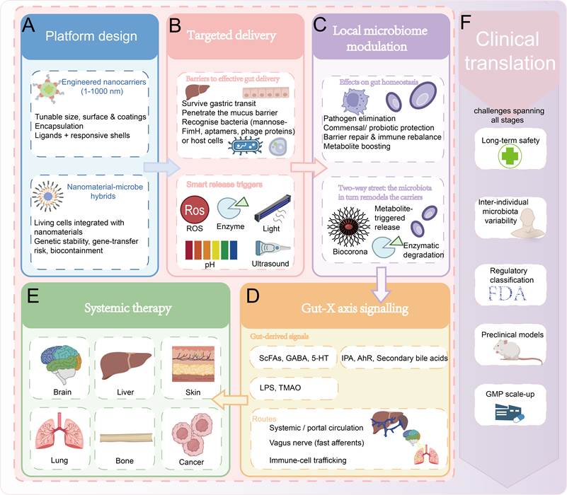
This review focuses on engineered nanocarriers and nanomaterial-microbe hybrids for targeted gut microbiome modulation, spanning rational material design, intestinal targeted delivery, and local gut microecology regulation. By reshaping the intestinal microenvironment and related gut-derived signalling, local intestinal modulation further triggers crosstalk along gut-organ axes, thereby exerting systemic regulatory effects on multiple peripheral organs and tumor microenvironments. On this basis, we further generalise the pervasive translational bottlenecks throughout the entire research pipeline, including preclinical evaluation, biosafety, manufacturability, personalised design and regulatory constraints, and systematically compare the distinct translational challenges faced by conventional nanocarriers and nanomaterial-microbe hybrids (Figure 1). In this review, engineered nanocarriers are defined as nanoscale delivery platforms (approximately 1-1000 nm); nanotherapeutics refer to therapeutic or theranostic formulations whose delivery or action primarily relies on such materials. Nanomaterial-microbe hybrids, in which living microbial cells are physically integrated with nanomaterials, are treated as a distinct category rather than conventional nanocarriers and are explicitly identified where discussed.
Engineered nanocarriers and nanomaterial-microbe hybrids for gut microbiome modulation. (A) Platform design: tunable engineered nanocarriers (1-1000 nm) and living nanomaterial-microbe hybrids; (B) Targeted delivery: surviving gastric transit, penetrating the mucus barrier and recognising bacteria or host cells, with smart release triggered by ROS, enzymes, light, pH or ultrasound; (C) Local microbiome modulation: pathogen elimination, commensal/probiotic protection, barrier repair, immune rebalance and metabolite boosting, while the microbiota in turn remodels the carriers (biocorona formation, metabolite-triggered release, enzymatic degradation); (D) Gut-X axis signalling: gut-derived signals (SCFAs, GABA, 5-HT, IPA/AhR ligands, secondary bile acids, LPS, TMAO) reach distant organs via systemic/portal circulation, vagal afferents and immune-cell trafficking; (E) Systemic therapy: gut-derived signals are translated into distal-organ therapy along gut-X axes; (F) Clinical translation: a foundational constraint spanning all stages of this framework, including preclinical model validity, long-term safety of materials and degradation products, GMP scale-up and batch consistency, inter-individual microbiota variability, and regulatory classification. Abbreviations: 5-HT, 5-hydroxytryptamine; AhR, aryl hydrocarbon receptor; GABA, γ-aminobutyric acid; IPA, indole-3-propionic acid; LPS, lipopolysaccharide; ROS, reactive oxygen species; SCFA, short-chain fatty acid; TMAO, trimethylamine N-oxide.
2. Rational design of theranostic systems: an “armamentarium” for gut microbiota modulation
Oral administration is the most convenient and patient-friendly way of drug administration. However, the gastrointestinal environment is harsh, characterized by extreme pH fluctuations, the presence of digestive enzymes and mucosal barriers, which pose significant challenges to the stability and absorption of drugs [30,31]. Delivery systems can provide a protective platform to enhance drug stability, prolong retention time in the gastrointestinal tract, and enable controlled release, thereby improving bioavailability and therapeutic efficacy [32-34]. Despite these advantages, these systems must still overcome three consecutive hurdles to effectively modulate the gut microbiota: penetrating the gastrointestinal mucosal and epithelial barriers, precisely recognizing and targeting specific microorganisms or host cells, and achieving on-demand drug release triggered by local environmental signals.
2.1. The first hurdle: penetrate barrier - self armed
The GI tract is the site where most nanoparticles with therapeutic properties and biological characteristics are absorbed. One of the core challenges for delivery systems in the GI environment is disruption of GI fluids and obstruction of the mucus barrier [35]. The varying pH (from extreme acidity in the stomach to mild alkalinity in the intestine) and high ionic strength in the GI tract can chemically disrupt the carrier structure or surface modification layer of nanoparticles, leading to particle agglomeration and drug leakage [36,37].
To overcome these chemical barriers, enteric coating materials that resist gastrointestinal digestion are based on enteric polymers, of which the Eudragit® series (e.g., Eudragit L100, L100-55, S100) is the most widely used. Eudragit L series (pH>5.5 solubility) can be targeted to the small intestine, and Eudragit S series (pH>7.0 solubility) can be targeted to the colon through the formation of dense hydrophobic membranes in the stomach to block gastric acid and digestive enzymes, and deprotonation dissolution of carboxyl groups in the intestine to achieve drug release [38]. Materials such as chitosan, citrus pectin, alginate, albumin, PLGA, etc., are often used in combination with Eudragit®, and these composite systems both improve resistance to gastric digestion and enhance targeting through targeting ligands or microbiota-responsive release [38]. This targeting logic relies on a more nuanced pH landscape than a simple stomach-to-colon gradient. While the terminal ileum reaches pH ~7.4–7.5, microbial fermentation of carbohydrates into SCFAs acidifies the cecum and ascending colon to approximately 5.7–6.5, with pH gradually rising toward 6.8–7.2 in the descending colon and rectum [37]. Consequently, Eudragit S100 remains intact through the acidic proximal colon and releases its payload primarily in the distal colonic segments, thereby avoiding premature dissolution in the ileum while surmounting the acidic challenge posed by SCFA-mediated luminal acidification [38].
Beyond chemical barriers, as a physical barrier on the inner surface of the GI tract, the mucus layer itself prevents nanoparticles from penetrating, and in scenarios such as active IBD, the increased thickness of the mucus layer and the rise in adherence can make this blockage even more pronounced. Mucus alterations in IBD vary with disease subtype and stage. Mucus hypersecretion and increased adhesiveness can trap nanoparticles, whereas the mucus depletion and goblet-cell loss typical of active UC compromise barrier integrity and expose carriers to epithelial and immune cells [39-41]. The mucus layer traps the nanoparticles, preventing them from reaching their target site; the immune response also perceives them as foreign, leading to premature clearance [32]. Therefore, it is necessary to modify the nanoparticles to enhance their mucus penetration ability.
To enhance mucus penetration, PEG modification is a classic strategy for overcoming this obstacle. Its hydrophilicity and steric effects reduce mucin adsorption; among these, low-molecular-weight (1 - 2 kDa) PEG forms a denser coating layer and thus exhibits superior penetration efficiency compared to high-molecular-weight PEG [36,42]. However, PEGylation may reduce cellular uptake, and long-term repeated administration may induce anti-PEG antibodies [42].
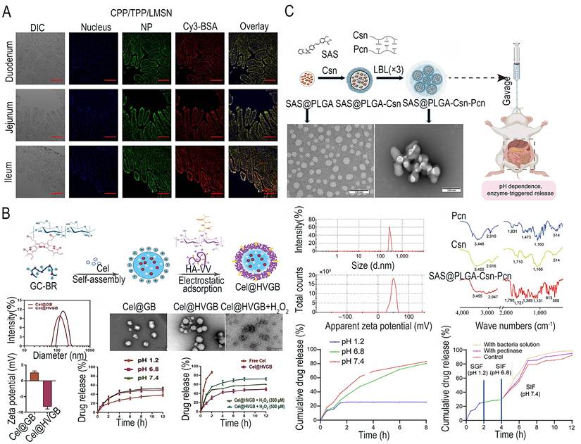
Building on these strategies, Tan et al. [42] further designed mesoporous silica nanoparticles co-modified with PEG and cell-penetrating peptides (CPPs), and by leveraging the active endocytosis of CPPs, achieved highly efficient transmembrane delivery of therapeutic proteins across multiple intestinal segments. (Figure 2A) The same self-arming logic protects the living component of nanomaterial-microbe hybrids. Zhu et al. [43] coated E. coli Nissle 1917 with a polyphenol-nanozyme armour (ECA@EcN), in which chitosan-modified epigallocatechin gallate coordinates gold nanozymes onto the bacterial surface. The armoured probiotic retained viability in simulated gastric fluid, intestinal fluid and bile salts, showed enhanced intestinal retention and ROS scavenging, and thereby ameliorated DSS-induced colitis. Having overcome the physical and chemical barriers of the GI environment, the carriers must then confront the second challenge: how to precisely identify their intended targets among the complex microbial and cellular community.
(A) LSCM images of duodenum, jejunum and ileum after administration with CPP/TPP/LMSN (blue, DAPI-stained nucleus; green, FITC-labeled silica NPs; red, Cy3-BSA; yellow, co-localization of silica NPs and Cy3-BSA) (scale bar: 200 μm). Adapted with permission from Ref. [42]. Copyright 2019. Royal Soc Chemistry (B) Synthesis diagram of the Cel@HVGB nanosystem, including its preparation process, particle size/zeta potential/morphology characterization, and pH- and H2O2-responsive drug release behaviour. Adapted with permission from Ref. [84]. Copyright 2023. Elsevier (C) The preparation and characterization of SAS@PLGA-Csn-Pcn NPs. Preparation scheme of NPs. Adapted with permission from Ref. [53]. Copyright 2024. AAAS. Abbreviations: BSA, bovine serum albumin; CPP, cell-penetrating peptide; DIC, differential interference contrast; DLS, dynamic light scattering; FT-IR, Fourier-transform infrared; HA-VV, hyaluronic acid-VV peptide conjugate; LSCM, laser scanning confocal microscopy; NP, nanoparticle; PLGA, poly(lactic-co-glycolic acid); SAS, sulfasalazine; SEM, scanning electron microscopy; SGF, simulated gastric fluid; SIF, simulated intestinal fluid; TEM, transmission electron microscopy; TPP, triphenylphosphonium.
2.2. The second challenge: selective identification - construction of the system
2.2.1 Bacteria-targeted delivery
The diverse functions exhibited by the gut microbiota during disease progression and treatment intervention provide engineers with insights into the multi-dimensional and targeted regulation of the microbiome. Designing and optimizing targeted ligands for specific bacteria is crucial for overcoming core bottlenecks such as “imbalanced microbiome caused by non-specific intervention” and “low efficiency of drug delivery” [44,45]. These ligands-including sugar-based antibody fragments, phage-derived proteins and aptamers-utilize the unique structures on the surface of bacteria (such as flagella, lipopolysaccharides and teichoic acids) or their metabolic characteristics to achieve selective identification of the target microbial community [46-50]. Based on these ligands, a delivery system can be designed for delivering antibacterial agents, gene editing tools and metabolic regulatory molecules. The drug release can be triggered by bacterial metabolites or specific pH values of the microbial microenvironment [51-53].
The bacteria-targeting systems directly interfere with pathogenic microorganisms by delivering antibacterial agents, narrow-spectrum antibiotics or photodynamic therapy drugs, thereby reducing the bacterial population and increasing the local drug concentration [54,55]. The delivery systems can also transport nutrients to the beneficial bacteria, thereby indirectly increasing their proportion in the intestinal tract [56]. For example, α-D-mannose binds to FimH on bacterial type I pili, and the Colicin E9 receptor-binding domain recognises the BtuB receptor on enteropathogenic Escherichia coli [57,58]. Phage-derived proteins, including endolysin, as well as engineered phages, modulate microbial community structure and function [59-61]. Aptamers offer specific recognition, easy modification via SELEX screening [62]. Aptamer-based ligands have also been screened against specific gut commensals for biosensing applications [63].
Nanomaterial-microbe hybrids represent a distinct approach to bacterial targeting. Ma et al. [47] developed a dual-targeted platform (Zn-MOF@Man/LRB) for the treatment of E. coli intestinal infections, in which a mannosylated Zn-MOF recognises the FimH fimbriae of E. coli and is co-assembled with a metabolically active Limosilactobacillus reuteri biofilm (9.68 × 10⁹ CFU g-1, viable cells densely packed in the EPS matrix) that provides gastric protection and facilitates intestinal delivery. CLSM and upregulated luxS expression confirmed the biofilm’s cellular viability and quorum-sensing activity. This system therefore exemplifies a nanomaterial-microbe hybrid rather than a conventional nanocarrier.
Beyond therapy, nanomaterials also enable diagnostic imaging of the gut microbiota. Bacteria-targeted fluorescent or magnetic nanoprobes permit real-time visualization of pathogen colonization and spatial distribution within the intestine [17,64,65]. Such nano-diagnostics provide critical feedback to guide the therapeutic interventions described above, bridging detection and treatment.
2.2.2 Host cell-targeted delivery
Beyond bacterial targeting, selective delivery to host cells-particularly intestinal epithelial cells and immune cells-constitutes another critical dimension of this second challenge [66]. The host cell targeting strategy delivers therapeutic drugs to intestinal epithelial cells and immune cells by recognizing surface receptors such as integrins, Toll-like receptors, and Fc receptors, using ligands such as antibody fragments, RGD peptides, and transferrin. These carriers carry anti-inflammatory drugs, immunomodulators, or barrier repair molecules, and drug release is triggered by changes in reactive oxygen species (ROS)/pH value or enzyme activity in the inflammatory microenvironment [67-71]. Under specific pathological conditions, the highly expressed receptors are exclusively enriched in intestinal epithelial cells, enabling specific recognition of target cells by the carrier and mediating endocytosis, thereby significantly increasing the local drug concentration [72-75]. Previous studies have shown that by utilizing receptors such as CD44, drugs or nanocarriers can be precisely delivered to intestinal immune cells [76]. This helps to promote macrophage polarization, T cell activation, and dendritic cell (DC) enhancement of antigen presentation, thereby reshaping the intestinal immune microenvironment. This approach avoids the off-target effects and risk of intestinal flora imbalance associated with systemic administration, thereby improving the efficacy and safety of treatment for intestinal-related diseases [69,74,77-80].
Representative studies on host-cell targeting demonstrate the efficacy of this strategy. The interaction between intestinal epithelial cells and immune cells can also lead to dysfunction of the intestinal epithelium and exacerbate the surrounding chronic inflammation [81]. Researchers have developed a kind of lipid particles that are modified with butyric acid and specifically target intestinal epithelial cells [82,83]. Chen et al. [84] developed the PepT1/CD44 dual-targeted nanocarrier Cel@HVGB for delivering celecoxib to inflammatory intestinal epithelial cells and M1 macrophages in UC (Figure 2B). This ROS-responsive carrier protects epithelial cells, restores barrier function, and regulates macrophage polarization, thereby alleviating UC symptoms and offering a new strategy for targeted UC treatment. Yang et al. [85] developed an oral laminarin-based nanogene delivery system (LA-G4@RNA) that protects miRNA-223 from GI degradation and enables targeted delivery to colonic macrophages via dectin-1 receptor recognition.
For nanomaterial-microbe hybrids, host targeting operates at a different granularity; rather than relying on receptor-level recognition of specific host cell types, the living component provides autonomous, tissue-level tropism toward pathological host sites. Quan et al. [86] coupled 5-ASA-loaded hairy microporous nanospheres to E. coli Nissle 1917 (E-xPAM@ASA), in which the bacterium’s intrinsic inflammation-homing ability drove selective accumulation and prolonged retention at inflamed colonic sites, while the porous nanospheres scavenged ROS and released 5-ASA sustainedly, achieving mucosal repair in a UC model. These distinct strategies are further compared in Table 1.
Comparison of delivery systems targeting strategies: bacteria vs. host cells
| Targeting bacteria | Targeting host cells | |
|---|---|---|
| Typical targets | Bacterial surface receptors (fimbriae, LPS, teichoic acids) [46] | Host cell surface receptors (integrins, Toll-like receptors, Fc receptors) [69,87,88] |
| Common ligands | Mannose, aptamers, phage-derived proteins, lectins [47-49,89] | Antibody fragments, targeting peptides, transferrin [67-69] |
| Design Objectives | Delivery of antimicrobial agents, gene-editing tools and metabolic regulatory molecules [51,90] | Delivery of anti-inflammatory drugs, immunomodulators and barrier-repairing molecules [91-93] |
| Requirements for the drug release environment | Bacterial metabolites trigger (such as azoreductase) or the pH of the microenvironment [52,53] | ROS/pH responses in the inflammatory microenvironment, enzyme-sensitive release [70,71] |
2.3.1 Microbiota-responsive release
The enzymes secreted by specific microbial communities in the colon (such as xanthanase and cellulase) or their metabolites (such as butyric acid) can directly cause the degradation of the carrier structure [94,95]. For instance, by using natural polysaccharides as the carrier shell and utilizing the polysaccharide-degrading enzymes secreted by the intestinal microbial community as the “switch”, drug delivery to the colon can be achieved. Li et al. [53] developed the prebiotic nanocarrier Sulfasalazine@Poly(lactic-co-glycolic acid)-Chitosan-Pectin (SAS@PLGA-Csn-Pcn), capable of delivering sulfasalazine to the colon (Figure 2C). This carrier exhibits dual responsiveness to both gut microbiota pectinase activity and pH, enabling on-demand drug release through synergistic triggering by microbial hydrolysis and intestinal pH gradients. This mechanism can regulate the balance of intestinal flora and alleviate colonic inflammation. This responsive-release logic is equally applicable beyond intestinal inflammation. For example, Tai et al. [96] developed niclosamide-loaded, hyaluronic acid-modified PLGA nanospheres (NIC@PLGA-HA) that exhibit pH-responsive controlled release at colonic pH for the treatment of Clostridioides difficile infection. The formulation killed Clostridioides difficile vegetative cells, inhibited spore germination and biofilm formation, and outperformed free niclosamide in a gerbil CDI model.
The azo reductase and esterase secreted by the intestinal microbiota can also break the azo bonds or ester bonds in the prodrug structure, thereby achieving colonic-specific drug activation [97]. However, because the abundance and enzymatic activity of these microbial populations, as well as the luminal metabolite levels they generate, vary substantially among individuals, the reliability of microbiota-responsive triggers can differ considerably from patient to patient [98,99].
2.3.2 Disease-microenvironment-responsive release
Beyond microbial enzymes, endogenous inflammatory signals serve as another powerful trigger for smart release. The inflamed area will release unique physiological signals (such as increased levels of reactive oxygen species or changes in pH value), which act as endogenous switches to activate carrier-mediated drug release, thereby achieving intelligent and targeted therapeutic responses within the lesion area [100]. Specifically, ROS-responsive nanocarriers typically utilize thioether bonds or other unstable bonds that break down when the level of reactive oxygen species increases at the inflammatory site, thereby enabling on-demand drug delivery [101-103]. Hypoxia is a hallmark feature of intestinal tissue inflammation and may also act as an inducer; hypoxia-responsive nanocarriers containing nitroimidazole or azo groups can achieve selective drug release under hypoxic conditions [103].
Additionally, the longitudinal pH gradient of the GI tract can also be engineered as a spatial trigger for release. Whereas the enteric coatings described in Section 2.1 exploit this gradient (stomach pH 1.2-3.0, terminal ileum pH ~7.4-7.5, colon pH ~5.7-6.5 proximally, rising to 6.8-7.2 distally) to survive gastric transit, pH-sensitive carriers can use the same gradient to time payload release, achieving gastric protection – colon delivery [37,104-107]. For instance, pH-responsive nanospheres loaded with prebiotics (such as xylooligosaccharides) or beneficial metabolic precursors (such as tryptophan) can penetrate the gastric acid barrier and achieve the release of nutrients for the colon, thereby selectively promoting the proliferation of beneficial bacteria including Bifidobacterium and Akkermansia [108].
Nanomaterial-microbe hybrids can likewise be programmed with such endogenous triggers. Shi et al. [109] sequentially coated probiotics with a ROS-scavenging MnO2 nanozyme and a colon-responsive degradation material. The dual coating protected the bacteria through the gastrointestinal tract and then underwent pH- and ROS-responsive degradation in the inflamed colon, “awakening” the probiotics in situ to relieve intestinal inflammation, repair the epithelial barrier and rebalance the gut microbiota.
The intensity of pathological signals such as reactive oxygen species and pro-inflammatory enzymes fluctuates considerably across disease subtypes and progression stages, so a trigger threshold tuned to one disease state may release prematurely during severe flares or insufficiently during milder stages, making such thresholds difficult to standardize [110].
2.3.3 Externally triggered release
Externally triggered systems represent a parallel strategy for on-demand release. The application of light, magnetic and other external stimulus response technologies in the gastrointestinal tract holds great potential, especially in areas such as targeted drug delivery, regulation of the intestinal flora, and targeted therapy. By using an external magnetic field, magnetic nanoparticles can be guided to the target intestinal segment, thereby increasing the local drug concentration or regulating gastrointestinal peristalsis [111,112]. Photothermal conversion materials, which generate local heat upon near-infrared (NIR) irradiation to trigger drug release or enable photothermal ablation, represent another light-responsive strategy [113].
Representative nanocarrier and hybrid platforms illustrate the potential of external triggers. Wang et al. [114] developed a PDA-BNN6/MSN Janus nanomotor that combines NIR-induced photothermal energy with NO propulsion to penetrate the mucus barrier and target the colon via Lacticaseibacillus rhamnosus GG cell wall ligands. This system increased cisplatin delivery efficiency by 3.5-fold and achieved a 98.6% tumor suppression rate in vivo. Nanomaterial-microbe hybrids can go one step further, making the microbial component an active actuator of the trigger rather than a passive cargo. Lu et al. [115] conjugated optogenetically engineered Shewanella oneidensis MR-1 with drug-loaded azo-bonded covalent organic frameworks (TA-COFs); under 660-nm red-light irradiation, bacterial extracellular electron transfer cleaved the azo bonds, disassembling the framework and triggering a 2.8-fold increase in on-demand drug release, which alleviated DSS-induced colitis.
However, externally triggered delivery systems face practical limitations. The complex microenvironment of the gastrointestinal tract (high acidity, enzymatic activity, mucus barrier, peristaltic shear force) may degrade stimulus-responsive carriers or impair their functionality [116,117]. Optical stimulation presents limitations in tissue penetration depth, hinders application in deeper intestinal segments, and carries potential phototoxicity risks [118]. The long-term biocompatibility of magnetic nanoparticles remains to be verified [119]. These are the fundamental parameters for designing intelligent delivery systems. Therefore, when designing environment-responsive “switches”, it is necessary to simultaneously optimize the particle size and surface charge in order to achieve efficient colonic targeting delivery [120,121].
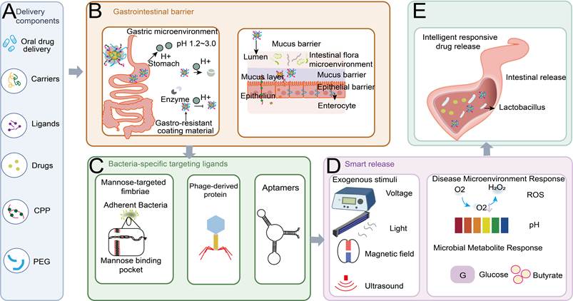
Table 2 summarises the environmental response mechanisms (including microbial enzymes, inflammatory signals such as ROS and hypoxia, pH gradients, and azo/ester bond cleavage) and the corresponding design strategies for intelligent drug delivery systems in the gut. Figure 3 provides a visual representation of the technical pathway through which nanomedicines exert their effects on the gut microbiota. After explaining how delivery systems are designed to survive in the gastrointestinal tract, reach their target sites, and release their drug components on demand, the next section will discuss the mechanisms by which they modulate the microbial community and reshape the intestinal microenvironment.
Environmental response mechanisms and design strategies for intelligent drug delivery systems in the gut
| Response type | Trigger | Design strategy | Refs |
|---|---|---|---|
| Microbial metabolite response | Azo-reductase, esterase | Azo-bond-linked prodrugs | [97,122] |
| Response of the disease microenvironment | pH, ROS, hypoxia | pH-sensitive bonds, ROS-sensitive bonds (thioketone bonds) | [101-103] |
| Response to external stimuli | Photo (UV/NIR), Magnetic | Photothermal conversion materials, magnetic nanoparticles | [113,123] |
Schematic illustration of smart oral drug delivery system targeting intestinal microbiota: from gastrointestinal barrier penetration to release. (A) Building blocks of the delivery platform: oral delivery platform loaded with therapeutic drugs and functionalised with targeting ligands, cell-penetrating peptides (CPP) and PEG coatings. (B) Gastrointestinal barriers encountered en route-the gastric microenvironment (pH 1.2-3.0 and digestive enzymes), overcome with gastro-resistant coating materials, and the intestinal mucus and epithelial barriers separating the luminal flora from enterocytes. (C) Bacteria-specific targeting ligands: mannosylated ligands targeting the mannose-binding pocket (FimH) of type 1 fimbriae on adherent bacteria, phage-derived proteins, and aptamers. (D) Smart release triggers: exogenous stimuli (voltage, light, magnetic field, ultrasound), disease-microenvironment cues and microbial metabolites. (E) Intelligent responsive drug release at the intestinal site, illustrated among colonising Lactobacillus. Abbreviations: CPP, cell-penetrating peptide; PEG, polyethylene glycol; ROS, reactive oxygen species.
3. Local remodeling of the intestinal microecology: direct intervention and indirect regulation by nanodevices
The preceding section described how rationally designed delivery systems overcome the gastrointestinal barrier by resisting gastric acid hydrolysis, penetrating the mucus layer, recognising their targets, and releasing payloads on demand. However, successful delivery does not by itself guarantee therapeutic benefit: conventional antimicrobial strategies, even when efficiently delivered, kill indiscriminately, damaging commensal bacteria and destabilising the microbial community. This section therefore examines how these systems remodel the intestinal microecology after reaching the target site through two complementary routes. On the one hand, they directly eliminate pathogens via membrane disruption, metal ion or ROS release, and ligand-mediated recognition; on the other hand, they indirectly restore the survival advantage of commensals by neutralising bacterial toxins and modulating host immune cell function [124-131]. Such a combination of targeted clearance and microenvironmental remediation circumvents the non-specific toxicity of broad-spectrum antimicrobials and promotes the restoration of microbial homeostasis. Yet even this picture remains incomplete, as current carrier design largely treats delivery as a one-way process. The gut microbiota can in turn reshape the in vivo fate of these systems through enzymatic degradation of carriers, microbial metabolism of payloads, and community-driven changes in the local microenvironment, and this reciprocal interaction is examined at the end of this section.
3.1. Direct intervention: Manipulation of the bacteria themselves
3.1.1 Direct antibacterial intervention
Direct intervention in the gut microbiota acts directly against pathogenic bacteria through physical or chemical mechanisms. Nanoantibiotics (e.g., silver and zinc oxide nanoparticles) disrupt bacterial membranes and induce oxidative stress by releasing metal ions such as Ag+ and Zn2+ [132-134]. Their apparent selectivity for pathogens over commensals is observed mainly under short-term in vitro conditions, and may not persist during chronic exposure, when sustained ion release within intestinal biofilms can progressively inhibit beneficial taxa such as bifidobacteria and lactobacilli [135-139].
To enhance the specificity of this direct antibacterial action, surface modification with targeted ligands such as DAPT and chitosan further enhances targeted binding to pathogenic bacteria [124,125]. Moreover, photodynamic and photothermal therapies can achieve targeted elimination of intestinal pathogens through energy conversion or reactive oxygen species generation, collectively forming a targeted antibacterial system whereby nanomedicines directly intervene with bacteria [140].
Representative engineering nanocarrier systems illustrate the therapeutic potential of these combined mechanisms. Li et al. [140] designed an IR/CP@BNP@HA (the hyaluronic acid (HA) shell and ROS-responsive chitosan biguanide-functionalized biopolymer core (IR/CP@BNP), which encapsulates chemotherapy drug camptothecin (CPT) and photothermal agent IR1048) nanosystem whose HA shell targets colorectal lesions while evading immune clearance. Upon arrival, the shell is shed via high-concentration intra-tumor hyaluronidase. Particle size reduction and potential reversal enable penetration through mucus and deep tissues. Excitation of IR1048 by 1064 nm laser generates localized photothermal heating up to ≈ 54 °C. Combined with guanidinated chitosan and lysozyme, this precisely eradicates intracellular Escherichia coli, disrupts biofilms, and blocks bacterial metabolic inactivation of camptothecin. This photothermal-chemotherapy-immunotherapy trimodal synergistic strategy achieves targeted clearance of intestinal pathogens while reversing immunosuppressive microenvironments and enhancing colorectal cancer (CRC) treatment efficacy. Chen et al. [141] developed vancomycin-loaded iron oxide nanoparticles that selectively bind Clostridioides difficile spores, delaying germination and sparing commensal flora in a murine CDI model. This case indicates that nanocarriers can balance the killing of pathogens and the protection of the original ecological niche, thereby resolving the fundamental dilemma of antibacterial treatment.
Nanomaterial-microbe hybrids also achieve this balance through a living mechanism. Chen et al. [142] doubly engineered E. coli Nissle 1917 to secrete TGF-β and coated it with a pH-responsive polymer (EcNT@L). In a nano-PET/Salmonella co-infection model, this hybrid eradicated Salmonella from the gastrointestinal tract and major organs with efficacy comparable to chloramphenicol, while-unlike the antibiotic-simultaneously upregulating tight-junction proteins, rebalancing Treg/Th17 responses and restoring microbial diversity, illustrating how hybrids can couple direct pathogen clearance with ecological repair.
3.1.2 Modulation of microbial metabolic pathways
Beyond direct antibacterial action, regulating microbial metabolic pathways offers another route to targeted modulation of the intestinal microbiota. In the regulation of gut microbiota, enzymes and precursor substances for specific bacterial metabolic pathways can be delivered through various methods. For instance, by delivering CAZymes, β-mannanases and other enzymes that degrade complex carbohydrates, metabolic substrates are provided for beneficial bacteria. This promotes the synthesis of butyrate and propionate, thereby regulating host energy metabolism and intestinal barrier function [143].
These principles are not restricted to intestinal inflammation and cancer. In metabolic dysfunction-associated steatotic liver disease, orally administered pH- and microbiota-responsive nanoparticles and chitosan-based selenium nanoparticles attenuated hepatic steatosis in high-fat-diet models by restoring the intestinal barrier and enriching beneficial taxa such as Akkermansia and Bifidobacterium [144]. These cases illustrate that the delivery and remodeling principles outlined above apply wherever dysbiosis drives disease. Beyond such engineering nanocarriers, nanomaterial-microbe hybrids can also intervene directly. Zhou et al. [145] developed a MnCe@LR/AMs probiotic-nanozyme hybrid, in which probiotic tryptophan metabolism and nanozyme antioxidant activity acted in concert to reduce pro-inflammatory cytokine secretion and enhance tight junctions in IBD mice. This example illustrates how nanomaterial-microbe hybrids can combine metabolic and antioxidant functions, although the microbiota-tryptophan link remains associative. While such probiotic-nanozyme synergies offer a promising alternative to direct metal-ion-based antibacterial strategies, they also highlight the importance of minimizing off-target metal ion release to preserve beneficial microbial communities.
3.2. Indirect regulation: effects on the microenvironment and host
Disruption of the structural integrity and functional impairment of the intestinal barrier may result in abnormal translocation of intestinal contents into the systemic circulation. This triggers uncontrolled immune responses within the intestinal microenvironment, promotes dysbiosis of the gut microbiota, and activates both innate and adaptive immune cells [146]. As the disease progresses, damage to the intestinal barrier continues to worsen, leading to further dysregulation of the mucosal immune system. This results in progressive tissue damage, ultimately triggering IBD and perpetuating a cycle of chronic intestinal inflammation [101].
To interrupt this cycle, nanocarrier strategies have been developed for barrier repair and local immune regulation. Qin et al. [128] developed tungsten-encapsulated zinc nanoparticles (W@ZnNPs), which provide a novel strategy for intestinal barrier repair and local immune regulation. These nanoparticles exhibit excellent gastric tolerance. They release only a very small amount of ions in the stomach, and undergo specific dissociation at the inflammatory sites in the colon, thereby achieving targeted delivery of zinc and tungsten (Figure 4A). Zinc enhances Paneth cell function, upregulates antimicrobial peptide expression, promotes mucin secretion by goblet cells, strengthens epithelial tight junctions, and reduces intestinal permeability. Tungsten inhibits pathogenic bacterial proliferation through metabolic reprogramming, thereby diminishing their invasion of the intestinal epithelium (Figure 4B).
(A) Schematic illustration of tungsten-encapsulated zinc NPs (W@ZnNPs) for preventing and treating active IBD. Adapted with permission from Ref. [128]. Copyright 2025. Elsevier (B) Quantification of goblet cells per colonic villus and representative confocal micrographs illustrating mucus thickness and bacterial translocation (MUC2, green; bacteria, red; DNA, blue; scale bar = 100 µm). W@ZnNP treatment elevated MUC2 expression and curbed microbial penetration into IECs more effectively than the other four IBD therapeutics. Adapted with permission from Ref. [128]. Copyright 2025. Elsevier (C) Macrophage membrane-coated mCRAMP-modified polydopamine nanoparticles for targeted treatment of colitis. Adapted with permission from Ref. [147]. Copyright 2023. Elsevier (D) Immunohistochemical staining of colon tissue (MPO, occludin, ZO-1; scale bar = 50 µm) and corresponding serum cytokine levels (IL-6, IL-1β, TNF-α, IL-10) measured by ELISA. Adapted with permission from Ref. [147]. Copyright 2023. Elsevier (E) Fabrication of cell membrane-coated CsA NPs for IBD treatment. Adapted with permission from Ref. [148]. Copyright 2024. AAAS. Abbreviations: CsA, cyclosporine A; DAPI, 4’,6-diamidino-2-phenylindole; DSS, dextran sulfate sodium; ELISA, enzyme-linked immunosorbent assay; IBD, inflammatory bowel disease; LM, leukocyte membrane; mCRAMP, mouse cathelicidin-related antimicrobial peptide; MPO, myeloperoxidase; MUC2, mucin-2; NC, normal control; Oxi-aCD, oxidized α-cyclodextrin; PBS, phosphate-buffered saline; PDA, polydopamine; PCM, PDA@mCRAMPs coated with macrophage membrane; ZO-1, zonula occludens-1.
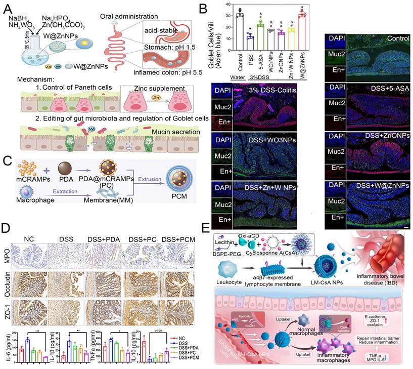
Beyond barrier repair, modulating the immune microenvironment via cell membrane coatings offers another indirect strategy. Through cell membrane coatings derived from extracted, non-viable cells and endowed with homing capabilities to inflammatory sites, the relevant nanosystems can achieve selective localised enrichment via inflammatory receptors on immune cell surfaces within gut-associated lymphoid tissue, thereby regulating immune homeostasis. Treg cells within the gut-associated lymphoid tissue are pivotal for maintaining intestinal immune homeostasis. Following intervention, they significantly upregulate the expression of the anti-inflammatory cytokine IL-10, thereby activating Treg cell function and enhancing their capacity to suppress abnormal inflammation [147]. The functional activation of Treg cells within the gut-associated lymphoid tissue is central to reversing immune dysregulation. Relevant systems can achieve local immunomodulation by targeting this microenvironment.
Representative studies demonstrate the efficacy of this membrane-coating approach. Bao et al. [147] developed macrophage membrane-coated antibacterial peptide-modified polydopamine nanoparticles (PCM NPs). PCM nanoparticles can target gut-associated lymphoid tissue via the inflammatory chemotactic properties of macrophage membranes (Figure 4C). PCM NPs inhibit the differentiation of naive T cells into Th17 cells via polydopamine, reduce expression of the Th17 cell marker IL-17A, and simultaneously upregulate the anti-inflammatory factor IL-10 to activate Treg cell function. This modulates the Treg/Th17 cell balance within the gut-associated lymphoid tissue, thereby improving local immune dysregulation in the intestine (Figure 4D). Li et al. [148] developed macrophage membrane/leukocyte membrane-coated cyclosporine A nanoparticles (MM-CsA NPs/LM-CsA NPs) for targeted therapy of active IBD. The particle achieves targeted delivery through specific binding to vascular cell adhesion molecule-1 (VCAM-1) and mucosal address cell adhesion molecule-1 (MadCAM-1) at inflammatory sites via integrin α4β1/α4β7. It exhibits ROS responsiveness to release cyclosporine A (CsA), which downregulates pro-inflammatory factors and reduces inflammatory infiltration. The cell membrane coating layer optimises particle uptake and retention while simultaneously upregulating barrier proteins to repair the mucosa (Figure 4E). In association with shifts in microbial balance and prolonged intestinal residence time, they indirectly modulate host intestinal barrier function, immune status, and metabolic levels [149].
Nanomaterial-microbe hybrids offer another route to indirectly modulate the microenvironment and host. Jiang et al. [150] constructed a multilayer-encapsulated Bifidobacterium longum biohybrid (Bif@FCSM(A)), whose metal-polyphenol/silica shell armed with MIL-101(Fe) disassembles in inflamed colonic tissue through transferrin-mediated Fe3+ chelation, co-releasing the probiotic and 5-aminosalicylic acid (5-ASA).
Importantly, nanomaterial effects on the microbiome are context dependent. Hyaluronic acid-bilirubin nanomedicine restored barrier integrity and enriched the microbiota in colitis, with its efficacy attenuated by antibiotic-mediated microbiota depletion [151], whereas orally administered TiO2 nanoparticles exacerbated DSS-induced colitis through NLRP3 inflammasome activation [152]. These contrasting outcomes indicate that nanomaterial effects depend on particle fate, barrier integrity, disease state, and ecological selectivity.
3.3. Reciprocal interactions
Traditional research in nanomedicine has largely focused on the unidirectional regulation of the gut microbiota by nanomaterials [153]. The gut microbiota, and the intestinal microenvironment it shapes, can in turn remodel the physicochemical properties, stability and drug-release behaviour of nanomaterials [154,155].
One important manifestation of this reverse influence is the formation of a “biocorona”. Once nanomaterials enter the body, biomolecules rapidly adsorb onto their surfaces to form a ‘biocorona’ [156]. The intrinsic surface properties of nanoparticles (size, surface charge and chemical functionality) determine the composition of their surface biocorona [157]. Critically, in the gastrointestinal tract the biomolecular pool available for corona formation is supplied jointly by the host (mucins, bile salts, phospholipids, digestive enzymes and plasma-derived proteins) and by the microbiota (bacterial surface proteins, extracellular polymeric substances, outer-membrane vesicles and metabolites) [158,159].
In inflamed intestinal tissue, the corona can be dominated by host-derived proteins. Zhang et al. [160] demonstrated that rigid, moderately hydrophobic PLGA nanoparticles selectively adsorb the inflammation-enriched S100A8 protein, forming a biocorona that targets macrophages via TLR4/RAGE and achieves superior targeting efficiency through disease microenvironment adaptation. The gut microbiota can also regulate corona formation systemically, even when the corona itself is composed of host proteins. Khetan et al. [161] modulated the gut microbiota of rats with antibiotics or prebiotics and found that the resulting changes in the plasma proteome produced markedly different protein coronas on intravenously administered lipid nanoparticles, which in turn altered their biodistribution and cellular uptake.
Microbiota-derived components also actively participate in corona formation. The R2ab autolysin from Staphylococcus epidermidis forms a corona on polystyrene nanoparticles, revealing specific protein-surface binding interactions at the molecular level [162]. Extracellular polymeric substances (EPS) represent another microbiota-derived corona component: studies on TiO2 nanoparticles have demonstrated that EPS from microbial sources can adsorb onto nanoparticle surfaces to form an “EPS corona”, with the crystalline phase and exposed facets of the nanoparticle influencing the extent and composition of this corona [163].
Beyond corona formation, direct microbiota-mediated transformation of nanomaterials is nevertheless well documented. Gut bacteria can even ferment engineered inorganic carbon nanomaterials into endogenous organic metabolites, demonstrating that the microbiota can actively degrade and remodel synthetic nanomaterials in vivo [164]. This capacity has been exploited for colon-targeted delivery. Microbial enzyme-responsive inulin nanoparticles are degraded by colonic bacteria to release 5-ASA for IBD therapy, and polysaccharide- or azo-based coatings are likewise cleaved by bacterial glycosidases and azoreductases [165,166]. Microbial esterases and lipases hydrolyse the ester bonds of polyester-based carriers, and microbial metabolites can act as chemical triggers that induce conformational changes or bond cleavage in nanomaterials [97,122,167].
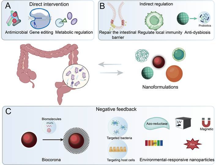
Nanomaterials can modulate the intestinal microbiota, and the intestinal microenvironment will, in turn, modify the properties of the nanomaterials (Figure 5). From a pharmacokinetic standpoint, these reciprocal interactions mean that the absorption, distribution, retention and release profile of a nanocarrier in the gut is co-determined by the microbiota itself. Nanomaterial-microbe hybrids add a further dimension, as their living component colonises, proliferates and metabolises in situ. The restoration of the gut microbiome is not the end goal. Dysbiosis of the gut microbiota is itself a driving factor behind various systemic diseases. Therefore, successful intervention in the gut microbiome will inevitably exert systemic effects on distant organs through “gut-X axis” signalling pathways. The next section will discuss these cross-organ therapeutic effects.
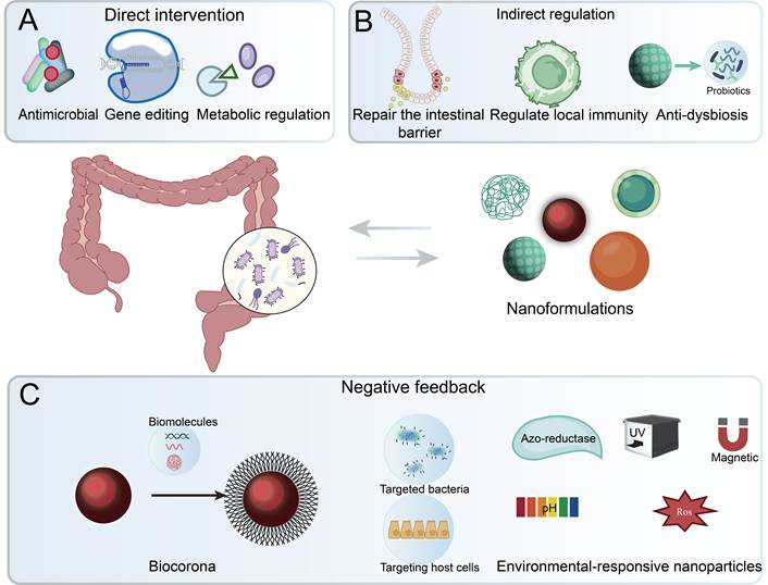
Nanomaterials-microbiota reciprocal interactions: a two-way street. (A) Direct intervention: nanomaterials act on the gut microbiota through antimicrobial activity, gene editing and metabolic regulation. (B) Indirect regulation: nanomaterials support microbiota homeostasis by repairing the intestinal barrier, regulating local immunity and protecting probiotics against dysbiosis. (C) Microbial feedback: in the reverse direction, biomolecule adsorption forms a biocorona that reshapes the recognition of targeted bacteria and host cells, while environment-responsive nanoparticles are modulated by microbiota-derived triggers together with external physical fields. Abbreviations: ROS, reactive oxygen species; UV, ultraviolet.
4. Beyond the intestine: regulating the gut-X axis for systemic disease therapy
Rationally designed delivery systems first overcome the gastrointestinal barriers to reach the intestine (section 2) and then remodel the local intestinal microecology (section 3). The intestine is not only a digestive organ but also the largest immune and a major endocrine organ in the human body: microecological imbalance can propagate to distant organs via microbiota-derived metabolites (e.g., SCFAs, LPS, TMAO), immune cell migration and neural signal transmission, so that a successful intestinal intervention can radiate along gut-X axis pathways to produce systemic effects. The two platform classes defined in Section 1 differ in how such effects are achieved, because their in vivo fates differ. Conventional engineered nanocarriers follow standard particle pharmacokinetics-gastrointestinal survival, mucosal distribution and retention, optional trans-epithelial absorption and eventual clearance, with dose-exposure relationships governed by size, charge, coating and environmental responsiveness-and their gut-X effects therefore arise either from gut-restricted action relayed by the above signals or from systemic biodistribution to the target organ. Nanomaterial-microbe hybrids follow a living fate instead: therapeutic exposure relies on gastric survival, intestinal colonisation and in situ metabolic output, and is thus self-sustaining and contingent on microbial viability rather than on the administered particle dose. A gut-X axis mechanism requires more than the co-occurrence of microbiota changes and distant-organ improvement-it requires a gut-origin signal that mechanistically links the two; the studies below are therefore grouped by whether this link is causally supported (e.g., by faecal microbiota transfer, microbiota depletion or metabolite manipulation) or remains associative, covering the brain, liver, skin, bone, lungs and tumours in a transition from local treatment to systemic therapy.
4.1. The gut-brain axis
Among the various gut-X axes, the gut-brain axis is the most extensively studied, with vagal neural signalling representing the fastest and most direct route. Vagal afferent terminals within the intestinal mucosa sense microbial metabolites and enteroendocrine signals and relay them via the nucleus tractus solitarius (NTS) to brain regions governing autonomic regulation, emotion, and cognition [168-171]. The principal microbe-derived neuroactive mediators include GABA, serotonin (5-HT), SCFAs, tryptophan metabolites, and BDNF [172]. These signals act either locally on vagal afferent terminals or systemically by suppressing intestinal inflammation and reinforcing gut barrier integrity, which limits the entry of LPS and pro-inflammatory cytokines into the circulation and protects blood-brain-barrier (BBB) integrity [173-179]. Conversely, neurotoxic proteins such as α-synuclein can propagate retrogradely along the vagus nerve from gut to brain, implicating this pathway in Parkinson’s disease (PD) and Alzheimer’s disease (AD) [180].
A critical yet often overlooked bottleneck is the BBB. Most gut-derived signals act on the brain indirectly via vagal afferents, enteroendocrine secretion, or immune modulation, so many oral gut-brain axis nanomedicines exert central effects without entering the brain [181,182]. When direct CNS delivery is required, BBB penetration can be enhanced by receptor- or adsorptive-mediated transcytosis, exploitation of endogenous nutrient transporters, transient tight-junction modulation, or biomimetic carriers such as exosomes and cell-membrane coatings, with particle size (<100-200 nm), surface charge, and ligand density as key design parameters [181,183,184]. Distinguishing indirect gut-to-brain signalling from direct BBB penetration is essential for the rational design of gut-brain axis nanomedicines.
Nanoparticle formulations can intervene in the gut-brain axis by reshaping microbial composition, boosting gut-derived neuroactive signals such as GABA and 5-HT, and containing intestinal inflammation. These effects are difficult to achieve with conventional oral probiotics or GABA owing to gastric degradation and poor mucosal adhesion [175,185-187]. Wang et al. [175] engineered magnetic field-responsive nano-probiotics (EcN-GadABC@Fe3O4-NE, a nanomaterial-microbe hybrid) that withstand gastric transit, colonise the intestine, and triple local GABA production upon magnetic triggering. This gut-derived GABA acts via vagal afferents, as shown by GABA-B antagonist blockade and vagal chemogenetic mimicry, thereby markedly alleviating anxiety-like behaviours in restraint-stressed mice (Figure 6A). Translating this magnetic-triggering design to the clinic, however, is constrained by the physics of field delivery. Field type (static, gradient, or alternating), frequency, source geometry, and source-to-target distance jointly determine the effective field strength and gradient at the target site. In the gastrointestinal tract, the field source must act across the abdominal wall and deep-lying loops of bowel, so generating sufficient and spatially confined fields remains a practical challenge [188,189]. In addition, sustained GABA output depends on stable maintenance of the GadABC expression cassette throughout intestinal colonisation [175]. In the absence of continuous antibiotic selection, which is impractical in the gut, engineered plasmids in probiotic chassis are subject to segregational instability and metabolic burden-driven loss during host colonisation [190]. Horizontal transfer of the gad gene to pathogens poses an ecological risk for which regulatory frameworks remain undeveloped [191-193].
(A) Schematic illustration of EcN-GadABC@Fe3O4-NE mechanism for regulating anxiety-like behaviours via gut-brain axis. Adapted with permission from Ref. [175]. Copyright 2025. BMC (B) Schematic illustration of the gut-brain axis. Gut dysbiosis increases intestinal permeability, allowing bacterial products (e.g., LPS) and pro-inflammatory cytokines (e.g., IL-6, TNF-α) to enter the circulation, promoting α-synuclein (α-syn) aggregation and neuroinflammation. Microbial metabolites also affect serotonin (5-HT) signalling along this axis. Adapted with permission from Ref. [194]. Copyright 2024. Elsevier. Abbreviations: 5-HT, 5-hydroxytryptamine; α-syn, α-synuclein; AMF, alternating magnetic field; BBB, blood-brain-barrier; GABA, γ-aminobutyric acid; IL-6, interleukin-6; LPS, lipopolysaccharide; TNF-α, tumour necrosis factor-α
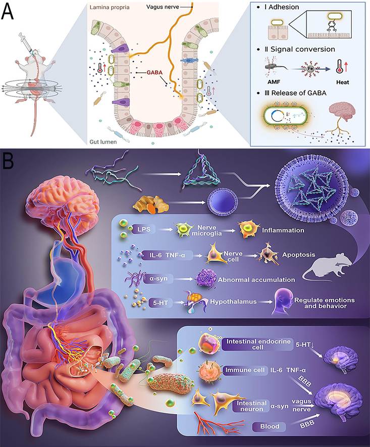
Beyond magnetic triggering, orally delivered biomimetic nanovesicles offer another intervention route. Cui et al. [194] developed ginger-derived exosome-like nanovesicles (Exo@tac) that selectively suppress PD-associated E. coli after oral delivery, with accompanying reductions in intestinal α-synuclein and LPS and enhanced enterochromaffin-cell 5-HT secretion. Motor dysfunction and neuropathology were ameliorated in two PD mouse models (Figure 6B). Whether the vesicles themselves reach the CNS was not directly examined, so the observed benefits are consistent with, but not proof of, gut-mediated action.
Such live biotherapeutic hybrids (EcN-GadABC@Fe3O4-NE) face constraints including plasmid instability, fitness costs, and horizontal gene transfer, which are absent from abiotic nanocarriers. By contrast, engineered nanocarrier systems such as Exo@tac face physicochemical rather than biological risks: carrier inactivation, premature payload release, burst kinetics, and non-specific microbiota disruption [30,195]. Chronically ingested food-grade nanomaterials can even reduce SCFAs and induce Alzheimer-like pathology [32,196-200].
4.2. The gut-liver axis
Microbiota-derived factors (LPS, TMAO, secondary bile acids, SCFAs) enter the portal circulation and mediate bidirectional gut-liver communication, while the liver counter-regulates the gut by secreting bile acids and antimicrobial molecules [201-206]. Disruption of this axis, for example, bacterial translocation following alcohol-induced barrier impairment, is a core pathological driver of ALD, NAFLD/NASH, cirrhosis, and HCC [202,207,208].
Nanoparticle formulations can intervene at both ends of this axis. Niu et al. [209] constructed vitamin A-modified cinnamaldehyde nanoemulsions (Va-Cin@NM) that remodelled the gut microbiota, increased SCFA production, and reinforced the intestinal barrier, while vitamin A-mediated hepatic targeting suppressed M1 macrophage polarisation and hepatic stellate cell activation, alleviating liver inflammation and fibrosis in bile duct ligation rats. This platform combines local gut remodeling with systemic hepatic accumulation, but the relative contribution of the two routes was not dissected, and no microbiota transfer or depletion experiments were performed.
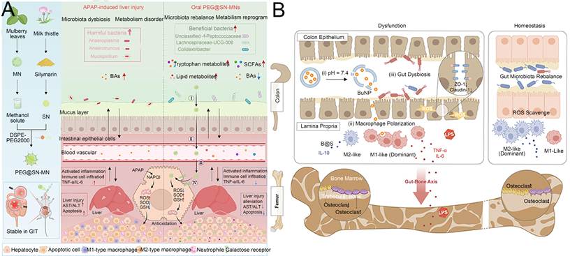
Applying the same dual-site logic to acute injury, Chen et al. [210] developed galactose-bearing, mulberry leaf-derived exosome-like nanoparticles loaded with silymarin nanocrystals (PEG@SN-MNs): by reshaping the microbiota and promoting butyrate production, they reinforce the intestinal barrier and cut off LPS influx, while galactose receptor-mediated uptake by hepatocytes and macrophages suppresses M1 polarisation, oxidative stress, and hepatocyte apoptosis, ultimately mitigating liver inflammation and necrosis in APAP-injured mice (Figure 7A). PEG@SN-MNs require trans-epithelial transport and systemic biodistribution; the absorption into the bloodstream and hepatic uptake were experimentally observed, consistent with the designed vitamin A-and galactose-mediated hepatic targeting. The hepatic benefits of these dual-site platforms therefore cannot be attributed to gut-liver axis signalling alone. Despite its dual-targeting design, the gut-liver contribution to its hepatic action remains associative.
(A) Schematic diagram of the protective effect and mechanism of oral PEG@SN-MNs on APAP-induced liver injury. PEG@SN-MNs are prepared by loading SNs into DSPE-PEG2000-modified MNs. After oral administration, PEG@SN-MNs maintain stability during the passage through the GIT. Adapted with permission from Ref. [210]. Copyright 2025. Wiley (B) Schematic of engineered postbiotics nanoparticles in the regulation of gut-bone axis. Adapted with permission from Ref. [229]. Copyright 2024. Springer. Abbreviations: ALT, alanine aminotransferase; APAP, acetaminophen; AST, aspartate aminotransferase; BA, bile acid; GSH, glutathione; GIT, gastrointestinal tract; IL, interleukin; LPS, lipopolysaccharide; NAPQI, N-acetyl-p-benzoquinone imine; PEG, polyethylene glycol; ROS, reactive oxygen species; SCFA, short-chain fatty acid; SOD, superoxide dismutase; TNF-α, tumour necrosis factor-α; ZO-1, zonula occludens-1.
In contrast to the associative evidence above, other platforms have established the gut microbiota as a causal mediator. Li et al. [211] developed honeysuckle-derived exosome-like nanovesicles (HNVs) that, upon oral administration, restored gut barrier integrity and remodelled the microbiota to suppress bile salt hydrolase, elevating taurochenodeoxycholic acid and inhibiting intestinal farnesoid X receptor (FXR)-fibroblast growth factor 15 (FGF15) thereby enhancing hepatic cholesterol catabolism in mice with high-fat-diet-induced MAFLD. Critically, the hepatoprotective effect was abolished by antibiotic-mediated microbiota depletion and was transferable to recipient mice by faecal microbiota transplantation from HNV-treated donors, establishing the gut microbiota as the causal mediator. HNVs act primarily through local gut action and gut-to-liver signalling, rather than through direct accumulation in the liver.
A distinct hybrid strategy assembles living probiotics into an engineered microsphere. Chen et al. [212] hierarchically assembled two probiotics into Ca2+-complexed polymer microspheres (LCA/LGG-LDB), in which an enteric outer coating releases L. rhamnosus GG in the intestine to activate intestinal FXR-FGF15 signalling and enrich bile-salt-hydrolase-active commensals, while entrapped L. delbrueckii subsp. bulgaricus together with the exposed chitosan shell adsorbs and fixes excess bile acids for faecal excretion, thereby dredging cholestasis in two drug-induced liver injury models. Component-wise dissection and pathway readouts resolved each partner’s contribution, although microbiota depletion or transplantation was not performed. This defined dual-strain formulation offers a standardized oral alternative to donor-dependent faecal microbiota transplantation, although preservation of strain viability and ratio during manufacturing and storage, and biocontainment of the living components, remain to be demonstrated.
After surviving gastrointestinal transit, absorbed nanoparticles face non-specific sequestration by Kupffer cells and the reticuloendothelial system, which can reduce the fraction available for hepatocyte targeting [144,213,214]. The examples in this axis thus span both pharmacokinetic patterns outlined above, ranging from gut-restricted action relayed by microbial signalling (HNVs) to systemic absorption with hepatic accumulation (Va-Cin@NM, PEG@SN-MNs), the latter subject to reticuloendothelial sequestration.
4.3. The gut-skin axis
The gut microbiota forms a bidirectional “gut-skin axis” with the skin through three major pathways: immune regulation, microbial metabolite signalling, and maintenance of intestinal barrier integrity [215,216]. Microbial metabolites such as SCFAs and neurotransmitters can modulate systemic immune responses, while outer membrane vesicles (OMVs) and other microbial-derived signals contribute to cross-organ communication and the transmission of inflammatory mediators [217,218]. When gut homeostasis is disrupted, toxins, inflammatory cytokines, and microbial metabolites may enter the circulation and reach the skin, thereby triggering or exacerbating inflammatory skin conditions such as psoriasis, atopic dermatitis, acne, rosacea, alopecia areata, and hidradenitis suppurativa [219,220]. Thus, the gut-skin axis provides an important mechanistic link between intestinal dysbiosis and cutaneous inflammation. Choi et al. [221] developed orally administered probiotic extracellular vesicles (LF216EV) that restored gut microbial homeostasis and enhanced serotonin-related signalling, reducing epidermal thickening and mast cell infiltration in DNCB-induced atopic dermatitis mice. Whether this cutaneous benefit is mediated by gut microbiota remodeling or by vesicles reaching the skin directly (the absorptive pattern) was not examined, leaving the platform unassigned between the two routes. Gut-skin axis studies remain largely correlative.
A recent study begins to close this gap. Wang et al. [222] showed that oral A. muciniphila-derived extracellular vesicles alleviate atopic dermatitis-like lesions by delivering microRNA (miR-21-5p), which directly targets TLR4 as validated by dual-luciferase assay and mimic/inhibitor perturbation, thereby suppressing NF-κB signalling in keratinocytes and promoting M2 macrophage polarisation. This work advances gut-skin communication research from correlative observations toward mechanistic understanding. However, it remains unclear whether these protective effects arise from gut-localised signalling or from direct vesicle delivery to the skin, so the pharmacokinetic route cannot be definitively assigned.
4.4. The gut-bone axis
The gut-bone axis refers to the regulatory connection between intestinal homeostasis and bone metabolism [223]. Gut microbiota and their metabolites can influence bone remodeling by modulating systemic inflammation, intestinal barrier integrity, and immune cell activity [224]. When the intestinal barrier is compromised, microbial products such as lipopolysaccharide (LPS) and lipoteichoic acid (LTA) can enter the circulation and promote inflammatory signalling, which may enhance osteoclastogenesis and disrupt the balance between osteoblast and osteoclast activity [225]. Conversely, restoration of microbial homeostasis and barrier function can reduce systemic inflammatory burden and support bone remodeling [226-228].
Colon-targeted oral nanomedicines offer a promising strategy to intervene in this axis by delivering bioactive agents to specific intestinal sites. These systems can regulate intestinal flora, repair the intestinal barrier, and limit the translocation of inflammatory factors and microbial toxins into the bloodstream, thereby restoring osteoblast-osteoclast balance through gut-bone signalling pathways [229].
Representative nanocarrier strategies illustrate this approach. Yu et al. [229] engineered colon-targeted postbiotic nanoparticles (B@S) with a shellac shell and butyrate core, enabling gastric tolerance and colonic release at pH 7.4. Oral B@S reshaped the microbiota, reinforced tight junctions, reduced ROS, and promoted M2 macrophage polarisation, alleviating osteoporosis in DSS- and OVX-induced mouse models (Figure 7B). Biodistribution confirmed colonic retention with negligible systemic absorption, but whether the skeletal benefits are causally mediated by microbiota remodeling was not tested by transfer or depletion experiments. This negligible systemic absorption exemplifies the gut-restricted pattern. The skeletal benefit is conferred entirely through gut-derived signals rather than through carrier distribution to bone.
Moreover, a distinct nanomaterial-microbe hybrid strategy leverages living probiotics. Li et al. [230] coated Lacticaseibacillus rhamnosus with a quercetin-Ca2+ metal-phenolic network and a polydopamine shell (L@Q-Ca/PDA); the nanoarmor protected the bacteria through gastric transit and was shed in the intestine, where quercetin and the colonising probiotic jointly suppressed inflammation, repaired the mucosal barrier and raised acetate, alleviating DSS-induced colitis and ovariectomy-induced osteoporosis. This exposure is gut-restricted by design: the skeletal benefit is relayed by gut-derived acetate rather than by distribution of the bacterium or coating material to bone, although translocation of the living component was not directly assessed. Component comparisons confirmed the coating’s contribution to survival and efficacy, but the gut-bone link remains associative, as no microbiota-depletion or transplantation experiments were performed. Such nanoarmor coatings directly target the central viability bottleneck of oral live biotherapeutics, although scalable and uniform coating of living cells, and preservation of viability during manufacturing and storage, remain to be demonstrated.
4.5. The gut-lung axis
The gut-lung axis is a bidirectional pathway. Gut microbiota and their metabolites influence immune function in the lungs, whilst lung inflammation, in turn, alters the gut microbiota [231]. Dysbiosis is associated with asthma, chronic obstructive pulmonary disease, pulmonary fibrosis and respiratory infections [232]. Microbial tryptophan metabolites are among the most extensively studied mediators: gut-derived indole derivatives signal through the aryl hydrocarbon receptor (AhR) to modulate pulmonary immunity and suppress airway inflammation [233,234].
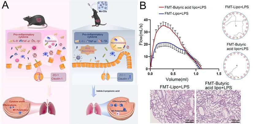
Peng et al. [235] designed oral manganese-doped carbon dots (Mn-CDs) with multi-enzyme activities. In mice with sepsis, manganese-carbon dots enriched Clostridium and Bacteroides in the gut, leading to increased production of indole-3-propionic acid (IPA). This gut-derived IPA activates the AhR in pulmonary macrophages, enhancing phagocytosis and ultimately alleviating lung injury. Faecal transplantation from donors treated with manganese-carbon dots replicated this protective effect, whereas gut decontamination abolished it, confirming a causal, microbiota-dependent mechanism (Figure 8A). The pulmonary benefit was attributed to gut-derived IPA entering the circulation rather than to direct pulmonary targeting by the carrier. Zhu et al. [236] encapsulated butyric acid in lipid nanoparticles for oral delivery; in an ARDS mouse model, these nanoparticles restored gut microbiota diversity, increased butyrate-producing Bacteroides, and reduced pulmonary PTPN1 expression, improving endothelial barrier function and pulmonary mechanics (Figure 8B). The pulmonary benefit was attributed to gut-derived butyrate rather than direct pulmonary targeting, although this link remains associative without transfer or depletion experiments. In both cases, efficacy followed the gut-restricted pattern. The therapeutically relevant exposure was the circulating microbial metabolite (IPA or butyrate), not pulmonary accumulation of the carrier itself. Gut-lung axis modulation should therefore be viewed as a promising but still early-stage complementary approach [237].
(A) Mechanism of Mn-CDs in alleviating sepsis-induced lung injury via the gut-lung axis. Orally administered Mn-CDs, endowed with multienzyme-mimetic activities and ROS-scavenging capacity, restored sepsis-induced gut dysbiosis in mice. This microbiota remodeling enhanced systemic delivery of IPA synthesized by g-Clostridium and g-Bacteroides, which subsequently alleviated pulmonary injury by enhancing macrophage efferocytosis-associated polarization in lung tissues. Adapted with permission from Ref. [235]. Copyright 2025. ACS (B) Liposomes loaded with butyric acid alleviate ARDS symptoms by modulating the gut microbiota. Adapted with permission from Ref. [236]. Copyright 2025. Wiley. Abbreviations: ARDS, acute respiratory distress syndrome; FMT, faecal microbiota transplantation; H&E, haematoxylin and eosin; IL, interleukin; LPS, lipopolysaccharide; Mn-CDs, manganese-doped carbon dots; ROS, reactive oxygen species; TNF-α, tumour necrosis factor-α; ZO-1, zonula occludens-1; g-Clostridium, genus-level Clostridium; g-Bacteroides, genus-level Bacteroides.
4.6. The gut-cancer axis
Interdisciplinary research on drug delivery systems and the gut-cancer axis is emerging as one of the most promising areas in precision oncology [238]. Its central premise is that gut microbiota-derived metabolites enter the circulation to regulate anti-tumor immunity and chemotherapeutic drug metabolism [239,240]. This provides the route by which an oral, gut-restricted intervention can be converted into a systemic anti-tumor effect, provided the carrier survives gastric transit and releases its payload in the colon [238,241].
For engineered nanocarriers, pH-responsive designs are commonly employed to achieve this. Liu et al. [242] illustrated this chain with chitosan/pectin-based Astragalus polysaccharide nanoparticles (APs-CS/PT-NPs): released in the colon at pH 6.5-7.5, the polysaccharides remodel the gut microbiota and boost endogenous SCFA production, particularly acetate, which activates G-protein-coupled receptor 43 (GPR43) and suppresses NF-κB signalling, thereby markedly inhibiting the development of NAFLD-driven liver cancer. This anti-tumor effect is achieved without the carrier itself leaving the intestine; it is attributed to gut-derived metabolites. Furthering this nanocarrier approach, locally acting nanomedicines can synergize with immunotherapy. Ma et al. [243] used an oral dextran-aspirin nanomedicine (P3C-Asp) that expanded Lactobacillus and Akkermansia and synergised with αPD-L1 blockade, extending median survival by 29 days (Figure 9A). Because the tumours reside in the colon, P3C-Asp acts at the same site where it modulates the microbiota, with no transport to distant organs involved. Both examples demonstrate that conventional nanocarriers can achieve anti-tumor efficacy by acting locally on the gut microbiota without systemic biodistribution.
(A) Schematic illustration of oral P3C-Asp in shrinking primary CRC. The P3C-Asp was fabricated by conjugating PBAsp (aspirin prodrug) with DEX-N3 (dextran-graft-3-azido-1-propylamine) via “click reaction”, the obtained P3C-Asp could easily self-assemble into stable nanoparticles. After oral administration, P3CAsp could accumulate into primary CRC tissues and realize the assembly dissociation with SA and the prebiotics (dextran) release while scavenging tumoral ROS, resulting in immunosuppressive TME relief, characterized by reduced M2 macrophages, MDSCs and COX-2, and increased CD8+ T cells. Due to the synergistic effect of inflammation relief and the homeostasis recovery, P3C-Asp efficiently inhibited CRC. Adapted with permission from Ref. [243]. Copyright 2024. Elsevier (B) Preparation and effective synergistic effect of chemo-immunotherapy of supraLBT. Adapted with permission from Ref. [244]. Copyright 2025. ACS. Abbreviations: COX-2, cyclooxygenase-2; CRC, colorectal cancer; EcN, Escherichia coli Nissle 1917; EGCG, epigallocatechin gallate; MDSC, myeloid-derived suppressor cell; ROS, reactive oxygen species; SA, salicylic acid; TME, tumour microenvironment; Treg, regulatory T cell.
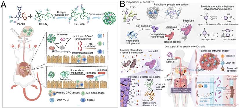
Nanomaterial-microbe hybrids incorporate living microorganisms as an integral therapeutic component, which determines their in vivo fate and mechanisms. Liu et al. [244] encapsulated the probiotic E. coli Nissle 1917 in green tea polyphenol/milk protein supraparticles (supraLBT), shielding it from chemotherapy-induced killing and restoring chemosensitivity and anti-tumour immunity, with a 2.35-fold increase in tumour suppression when combined with doxorubicin (Figure 9B). The benefit appears to come mainly from preserving the gut flora, since unarmoured EcN killed by chemotherapy provided no benefit. This dependence on bacterial viability illustrates the distinct in vivo fate of nanomaterial-microbe hybrids in this therapeutic setting. Therapeutic exposure is governed by the survival and intestinal persistence of a living organism rather than by particle distribution and clearance, and is therefore lost once the microbial component is inactivated. Moving beyond passive protection, a more sophisticated hybrid strategy achieves active tumour targeting and spatiotemporal control. Fan et al. [245] anchored Staphylococcus aureus membrane-coated piezoelectric BaTiO3 nanocubes onto Veillonella atypica (VA-SAM@BTO). After oral administration, the hybrid homed to orthotopic colorectal tumours via bacterial membrane inflammatory tropism and bacterial hypoxia tropism, and under ultrasound the nanocubes generated ROS and CO while the bacteria consumed tumour-derived lactic acid. Component ablation confirmed causality, as removing the bacteria, nanocubes or ultrasound each abolished most of the benefit, and only the complete, ultrasound-activated hybrid achieved ~90% tumour inhibition and reshaped the immunosuppressive microenvironment. The key barriers to the clinical translation of these platforms are analysed in the next section.
Table 3 summarises the key metabolites/mediators of the gut-X axis (gut-brain, gut-liver, gut-skin, gut-lung, gut-cancer) system and their cross-organ action mechanisms and regulatory strategies. Table 4 summarises key examples targeting the gut-brain, gut-liver, gut-skin, gut-bone, gut-lung and gut-cancer axes. Figure 10 classifies the two platform classes by their design logic: engineered nanocarriers by therapeutic target (intratumoural bacteria and tumour cells, host immune cells, and bacterial signalling or microbe-host crosstalk), and nanomaterial-microbe hybrids by microbial component (engineered or protected strains, and community- or metabolism-level platforms). Both classes converge on a shared interface of biological mediators (metabolite, neuroendocrine, immune and vesicle routes) and engineering entry points (barrier modulation and spatiotemporal control), through which they engage the six gut-X axes. Table 5 further summarises their typical targeting sites, response mechanisms, strength of mechanistic evidence (causal vs. associative), therapeutic benefits, and safety profiles.
Metabolites/mediators of key bacterial communities in the gut-X axis system and their cross-organ action mechanisms
| Gut-X axis | Key microbial metabolites/substrates | Strategies to overcome the barrier | Refs |
|---|---|---|---|
| Gut-brain | SCFAs, GABA, 5-HT, BDNF, tryptophan metabolites | Vagus nerve stimulation, modulation of the blood-brain barrier, regulation of neuroinflammation | [172,246] |
| Gut-Liver | LPS, TMAO, secondary BAs, ethanol metabolites | Portal vein circulation, activation of Kupffer cells, activation of HSCs | [203-205] |
| Gut-skin | Microbial metabolites, OMVs, SCFAs | Systemic immune regulation and cross-organ transmission of inflammatory cytokines | [217,218] |
| Gut-Bone | SCFAs, Vitamin D, estrogen | Maintain the integrity of the intestinal barrier, regulate the systemic immune and endocrine signalling axes | [247-249] |
| Gut-Lung | SCFAs, Tryptophan metabolites, Bile acids | The systemic circulation of intestinal microbial metabolites, Immune cell homing triggered by the intestine | [234,250-252] |
| Gut-Cancer | Secondary BAs, butyrates, TMAO | Gut microbiota metabolites regulate anti-tumor immunity and influence the metabolism of chemotherapeutic drugs | [239,240] |
Representative examples of engineering strategies targeting the gut-X axis via oral administration
| Gut-X Axis | Nanoengineering strategy | Disease Model | Refs |
|---|---|---|---|
| Gut-Brain | Oxyberberine-hydroxypropyl-β-cyclodextrin nanoparticles remodel the gut microbiota and suppress the CXCL10/CXCR3 axis | 3×Tg AD mouse model | [253] |
| Chiral Au nanoparticles - L-handed (L-) and D-handed (D-) chiral gold nanoparticles | AD mouse model | [254] | |
| A. muciniphila OMVs | Old smoking mice (with cognitive decline) | [255] | |
| Gut-Liver | Honeysuckle-derived exosome-like nanovesicles (HNVs) (~158.5 nm, -15.97 mV) | Acute liver failure (ALF) mouse (LPS/D-galactosamine, LPS/GalN, hepatocyte necrosis) | [256] |
| Broccoli-derived exosome-like nanoparticles (BDENs, ~100-200 nm) | Metabolic dysfunction-related fatty liver disease (MASLD) mice | [257] | |
| Bovine milk-derived extracellular vesicles | Chronic liver injury mice | [258] | |
| Gut-Bone | CBD/LNPs (≤170 nm, -16.57 mV, 98.78% EE) | Chronic restraint stress rat (trabecular bone loss, osteocalcin reduction) | [259] |
| Gut-Lung | Platinum-doped emodin carbon dots (Pt-ECDs, ~2.4 nm) | Sepsis-induced acute lung injury | [260] |
| Polylysine-bilirubin conjugate nanoparticles (BRNPs, ~100-300 nm) | Lung adenocarcinoma | [261] | |
| Gut-Cancer | Oral dual-targeted (CRC cell-specific+gut microbiota-targeted) GSH-responsive nanocarrier for GSDMD-N mRNA delivery | Obese mouse model with colorectal cancer (CRC) | [262] |
| Neutrophil membrane-coated PLGA nanoparticles with dual-drug encapsulation | Colorectal cancer liver metastasis | [263] | |
| Microbiota-bile acid-Kupffer cell axis modulation | Colon/breast/melanoma/pancreatic tumor models | [264] |
Comparison of representative strategies for targeted gut microbiome modulation
| Strategy category | Typical targeting site | Microbial/host target, specificity & response mechanism | Mechanistic evidence | Therapeutic benefits | Safety & limitations | |
|---|---|---|---|---|---|---|
| Nanocarriers | Photothermal/photodynamic nanosystems (e.g., IR/CP@BNP@HA) | Lesion/tumour site | Intratumoral bacteria + tumour cells (species-selective); light-triggered ROS/heat | Causal (killing assays) | Synergistic chemo-immunotherapy | Requires external irradiation [140] |
| Enzyme/pH-responsive carriers (e.g., W@ZnNPs, B@S) | Colon / inflamed sites | Host barrier + microbiota; pH-, enzyme-, or inflammation-responsive release | Mechanistic to associative | Site-specific release; barrier repair | Response varies with host microbiota [128,229] | |
| Inorganic multifunctional NPs (e.g., Mn-CDs) | Gut + distant organ | Metabolite-producing commensals (community-level); multi-enzyme ROS scavenging | Causal (FMT + decontamination) in select systems | Cross-organ protection via metabolites | Metal exposure; early-stage evidence [235] | |
| Microbial/plant-derived vesicles (e.g., Exo@tac, LF216EV) | Intestine | Microbiota signalling; EV-mediated cargo delivery | Associative | Cross-axis benefits (brain, skin) | Biodistribution beyond gut often unexamined [194,221] | |
| Dual-site gut-organ platforms (e.g., PEG@SN-MNs) | Gut + distant organ | Microbiota + organ-resident cells (low specificity); receptor-mediated organ targeting | Associative + biodistribution | Simultaneous gut and organ action | RES sequestration; absorption variability [210] | |
| Biomimetic membrane-coated NPs (e.g., PCM NPs, MM-CsA NPs) | Inflamed colon / GALT | Host immune cells (lesion-directed); chemotactic targeting and triggered release | Mechanistic | Immune rebalance; barrier repair | Manufacturing complexity [147,148] | |
| Nanomaterial-microbe hybrids | Engineered probiotics (e.g., EcN-GadABC@Fe3O4-NE) | Intestine | Engineered strain metabolite output (strain-level); magnetic trigger | Causal (vagal blockade) in select systems | Programmable metabolite production | Gene transfer risk; genetic stability [175] |
| Microbiota-protective armours (e.g., supraLBT) | Intestine | Shielding probiotics from chemotherapy (strain-protective) | Associative | Preserved chemosensitivity and anti-tumour immunity | Viability-dependent; live-bacterium risks (translocation, containment) not assessed [244] | |
| Probiotic-nanozyme hybrids (e.g., MnCe@LR/AMs) | Inflamed colon | Probiotic metabolism (community-level) + ROS scavenging | Associative | Combined antioxidant and microbiota restoration | Off-target metal ion release; batch consistency [145] | |
Integrated framework linking therapeutic platform design to gut-X axis engagement. (A) Engineered nanocarriers, organised by therapeutic target: intratumoural bacteria and tumour cells, host immune cells, or bacterial signalling and microbe-host crosstalk. (B) Nanomaterial-microbe hybrids, organised instead by microbial component: engineered or protected strains versus community- and metabolism-level platforms. (C) Although designed differently, both classes act through a shared interface that converts local microbiome modulation into systemic effects, comprising biological mediators that carry the signal and engineering entry points that determine where and when it is delivered. (D) Through this interface, they engage six gut-X axes. Abbreviations: 5-HT, 5-hydroxytryptamine; AhR, aryl hydrocarbon receptor; GABA, γ-aminobutyric acid; GLP-1, glucagon-like peptide-1; GVB, gut vascular barrier; LPS, lipopolysaccharide; NP, nanoparticle; SCFA, short-chain fatty acid; Th17, T helper 17 cell; TLR4, Toll-like receptor 4; TMAO, trimethylamine N-oxide; Treg, regulatory T cell; Trp, tryptophan.
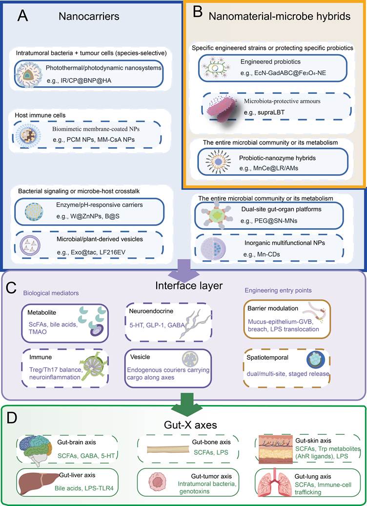
5. Current developments and future prospects: Challenges and opportunities from concept to clinical practice
The previously mentioned gut-X axis regulatory strategy has demonstrated the broad potential of nanotechnology in the treatment of systemic diseases. However, translating these strategies from animal experiments to clinical applications still requires overcoming a series of fundamental obstacles. These obstacles not only involve technical aspects (such as the ecological validity of animal models, batch consistency in large-scale production), but also safety assessment, individual differences (individualised characteristics of the microbiome), and regulatory systems. This section will analyze these key challenges.
5.1. Key challenges
Currently, strategies for regulating the gut-X axis are shifting from traditional probiotics, FMT and dietary interventions towards targeted intervention using engineered nanocarriers [50]. Clinically, purely microbiological interventions have yielded encouraging but inconsistent progress: in the phase 2 FMT-LUMINate trial, oral FMT capsules combined with first-line immunotherapy achieved objective response rates of 80% in advanced non-small cell lung cancer and 75% in melanoma, yet clinical benefit correlated with the loss of unfavorable baseline taxa rather than simple donor-strain engraftment [265]. Chen et al. [266] further demonstrated that phylogenetically distinct strains within the same species can exert opposite effects on anti-PD-1 efficacy, providing a mechanistic explanation for such variability. Meanwhile, a defined 15-strain live biotherapeutic product achieved engraftment and recurrence-free rates comparable to same-donor FMT in recurrent Clostridioides difficile infection, highlighting the feasibility of standardized formulations [267]. Nevertheless, common bottlenecks remain: poor survival against gastric acid and bile, limited and strain-specific colonisation, donor dependency and lack of standardisation (for FMT), and unpredictable responses arising from inter-individual and strain-level variability, which together preclude targeted regulation of the gut-X axis [12,268]. Nanotechnology offers an integrated solution to these bottlenecks, yet its clinical translation still faces challenges in animal model selection, safety assessment, large-scale production and regulatory pathways. Table 6 systematically compares the distinct translational challenges faced by engineering nanocarriers and nanomaterial-microbe hybrids at each development stage.
Distinct translational challenges of engineering nanocarriers vs. nanomaterial-microbe hybrids across development stages
| Challenge dimension | Engineering nanocarriers | Nanomaterial-microbe hybrids | Refs |
|---|---|---|---|
| In vivo fate / PK | Size, charge, mucus interaction, RES clearance; AUC/Cmax | Colonisation, metabolic fitness, plasmid stability; CFU kinetics and metabolite output | [213,214,304] |
| Preclinical models | Germ-free mice isolate material-intrinsic toxicity | Germ-free mice lack a resident community; SPF or humanised models required | [269-272,305] |
| Safety | Material persistence, degradation products, dysbiosis (e.g., TiO2, Ag) | Adds HGT, plasmid loss and ecological risks; needs biocontainment (auxotrophy, kill switches) | [191,193,195, 283,285] |
| Scale-up & CMC | Size, coating, loading, sterility | Adds strain identity, CFU potency, viability, plasmid retention, cold chain | [286,288, 289] |
| Individual variability | Microbiota-dependent carrier metabolism and release | Colonisation resistance makes engraftment itself individually variable | [99,292, 304] |
| Clinical endpoints | PK/PD plus clinical outcomes | Strain-level tracking and engraftment readouts linked to outcomes | [288,297] |
| Regulatory | Drug/biological/combination product under nanomedicine guidance | LBP × nanomedicine combination: no approved precedent | [286,288, 299] |
5.1.1 The dilemma of selecting preclinical animal models
In preclinical studies of nanoengineering strategies targeting microbiota-host interactions, the selection of animal models requires a balance between precision and ecological validity. Although germ-free mice eliminate microbial interference and allow independent assessment of nanomaterial host toxicity, their skin barrier and immune response are underdeveloped due to the absence of regulation by the skin’s resident microbiota in local immunity [269-272]. Moreover, the inherent differences between mice and humans in terms of microbiome composition and physiological characteristics limit the model’s ability to accurately predict the combined toxicity of nanomaterials in real-world conditions [272,273].
Mice with humanised microbiota, created through the transplantation of human gut microbiota, are more closely aligned with clinical translation and are suitable for evaluating microbiota-dependent therapeutic effects. However, they suffer from issues such as microbiota convergence, poor colonisation stability and a high degree of donor dependence, which limit their reproducibility [274,275]. Colonisation of germ-free mice with specific bacterial strains forms the basis for gradient models ranging from germ-free states to monomicrobial and complex microbial communities, allowing precise analysis of individual strains within the neuro-immune axis under strictly controlled conditions. However, attention must be paid to the potential impact of congenital developmental defects in germ-free mice on the extrapolation of results to normal physiological conditions [276-278]. The humanised germ-free mouse model combines microbial genomic homology with controllable conditions, enabling precise mechanistic studies of microorganism-host interactions. However, these models are complex to establish and expensive to maintain, making them unsuitable for high-throughput screening applications [279,280].
Therefore, model selection should be purpose-driven. HMA mice are suited for translational studies of microbiota-dependent efficacy, despite their inherent limitations, whereas humanised gnotobiotic platforms are reserved for dissecting complex multi-system mechanisms. Complementary validation across these models mitigates the limitations of any single system, consistent with the approach of progressing from minimal models to complex systems [274,281]. Figure 11 illustrates the selection of animal models for different research objectives - such as assessing the toxicity of nanomaterials, evaluating the efficacy of microbiome-dependent therapies, investigating the functions of specific bacterial strains, and modelling humanised microbiomes - along with their respective advantages and limitations.
Selection of animal models: an analysis of suitability and limitations based on different research objectives.
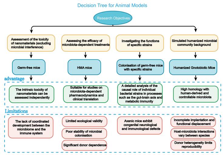
5.1.2 Safety
Following oral or transdermal administration, nanomaterials may persist locally in the gut or skin and degrade slowly; the long-term effects of both the materials themselves and their degradation products on the microbiota-host interaction are a key concern in preclinical safety assessments [282]. Long-term exposure to titanium dioxide nanoparticles (TiO2 NPs) significantly reduces the number of beneficial gut bacteria (such as bifidobacteria and lactobacilli), disrupts the microbial balance, and exacerbates DSS-induced chronic colitis and immune responses [195]. Exposure to silver nanoparticles during development may lead to persistent intestinal flora imbalance and neurobehavioural changes, suggesting that early exposure may have long-term effects through the “gut-brain-microbiota axis” [283]. The biological transformation of nanomaterials is bidirectional: the intestinal microbiota can alter the surface chemical properties, solubility, and reactivity of nanomaterials through metabolic pathways such as redox reactions, while the metal ions or active metabolites produced by nanomaterial degradation also reshape the composition of the microbiota, promote the colonisation of opportunistic pathogens, and inhibit the growth of beneficial bacteria [284]. Therefore, the safety assessment of nanomaterials must go beyond acute toxicity endpoints to include long-term monitoring of the microbial community, identification of degradation products, and assessment of cross-generational effects.
For nanomaterial-microbe hybrids incorporating live engineered bacteria, safety assessment must extend beyond these physicochemical endpoints to encompass biological containment criteria: plasmid segregational stability over the colonisation period, horizontal gene transfer frequency to resident faecal bacteria, in vivo metabolic fitness relative to commensal competitors, and efficacy of any biocontainment safeguard [190-193,285]. These biological risk endpoints are categorically distinct from the biodistribution and degradation profiles required for abiotic nanocarriers, including biomimetic membrane-coated nanoparticles, yet they are equally critical for clinical translation [286-288].
5.1.3 Scale-up and standardisation
The complex physical and chemical properties of nanomaterials, including particle size distribution, surface modification and drug-loading efficiency pose significant challenges for their industrial-scale production. When processes optimized in the laboratory, such as self-assembly, nano-precipitation or single/double emulsion methods, are scaled up to cGMP standards, the nonlinear changes in quality and momentum transfer rates often lead to increased particle size heterogeneity, deviations in drug loading, and uneven surface coating, resulting in significant batch-to-batch variability [289]. Biologically derived nano-carriers such as exosomes are even more challenging: their diverse cell sources preclude a homogeneous product, cultivation conditions introduce batch-to-batch differences, and their inherent instability makes them prone to rapid degradation, all of which has hindered the establishment of a consistent regulatory assessment framework [290].
The GMP compliance requirements mandate end-to-end monitoring of key quality attributes (CQAs) and key process parameters (CPPs), including integrated purification processes involving tangential flow filtration (TFF) and size exclusion chromatography (SEC), as well as the validation of aseptic filling and freeze-drying processes. However, there is still a severe shortage of GMP facilities globally capable of synthesizing complex nanomedicines in the late stages of clinical development, and the production costs remain prohibitively high [291].
In addition, regulatory authorities have not yet established uniform classification standards for nanomedicines. The FDA assesses such products on a “case-by-case” basis, whilst the European Medicines Agency (EMA) requires early scientific consultation; the lack of internationally harmonised guidelines means that developers must repeat safety and quality control validation across multiple jurisdictions, further increasing both the costs and time required for translation [289].
Live biotherapeutic hybrids introduce additional manufacturing complexities, including fermentation-scale-up under plasmid-selective pressure, CFU-based dose quantification, viability maintenance during nanomaterial co-assembly, and cold-chain logistics, all of which lack standardised GMP guidance [267,288].
5.1.4 Individual variability
The individual characteristics of the gut microbiota constitute a natural barrier to the targeted delivery of nanomedicines, significant differences exist between individuals in terms of microbial composition, metabolic activity and the state of the intestinal mucosal barrier, leading to the phenomenon of “the same drug, different effects” [98,99].
McCoubrey et al. [292] found that low-molecular-weight PLGA can be metabolised by the colonic microbiota into lactic acid, thereby promoting the synthesis of SCFAs, and that the rate of this metabolism varies significantly among different host microbiota. Furthermore, this polymer can also influence the expression of intestinal mucosal inflammatory markers (such as IL-8 and IL-10), indicating that microbiota composition directly determines the degradation fate of carriers. Renukadevi et al. [293] note that gut microbiota can degrade carrier matrices by secreting enzymes such as azoreductase and β-glucuronidase, thereby enabling colon-specific drug release. However, there are significant variations in microbial composition and enzyme activity between individuals, making the drug release behaviour of a given formulation difficult to predict. For example, only individuals carrying the cardiac glycoside reductase operon of Eggerthella lenta can inactivate digoxin, confirming that functional microbial genes determine drug fate [294]. Accordingly, a practical strategy is to first profile the patient’s microbiota enzyme spectrum via metagenomic sequencing and then select the carrier trigger based on this profile. Enzyme-sensitive polymeric shells (such as carriers linked via azo or glycosidic bonds) are preferable for individuals with a high abundance of enzyme-producing bacteria, whereas pH-or redox-responsive systems suit those with lower bacterial metabolic activity [293,295]. Building on this, the future integration of microbiota functional typing with machine learning algorithms holds promise for achieving closed-loop optimisation, from “microbiota diagnosis” to “formulation adaptation” [296].
5.1.5 Specific aspects of clinical endpoint design
The clinical evaluation of microbiome-targeted delivery systems faces a fundamental dilemma: whether changes in the microbiome structure can in themselves serve as a clinical endpoint. Although many clinical trials of microbiome interventions report statistically significant changes in the microbiota, these have not consistently translated into clinically meaningful outcomes; the root cause lies in an over-reliance on descriptive microbiota markers as surrogate endpoints [297].
Patients with cognitive impairment following ischaemic stroke exhibit characteristic alterations in gut microbiota composition; a diagnostic model based on these characteristic microbiota features achieved an AUC of 0.84, suggesting that gut microbiota characteristics may serve as biomarkers of disease status [298]. For nanomedicines, endpoint design should adopt a tiered approach: using changes in the microbiome as a mechanistic basis, whilst prioritising host-related biomarkers and clinical outcomes as primary endpoints [297].
5.1.6 Regulatory considerations
Nanomedicines do not fit neatly into a single regulatory category: depending on composition and primary mode of action, a nanoformulation may be regulated as a drug, a biological product, or a combination product, and this classification ambiguity directly increases regulatory uncertainty [299]. Both the FDA and the EMA have issued nanotechnology-specific guidance, which consistently emphasises comprehensive physicochemical characterisation-particle size and distribution, morphology, surface charge, coating integrity, and drug-loading and release behaviour-as the basis of quality control [286]. For orally administered, gut-targeted nanomedicines, regulators additionally require data on gastrointestinal stability, mucus interaction, local versus systemic exposure, and the long-term fate of non-degradable inorganic carriers [286]. From a chemistry, manufacturing and controls (CMC) perspective, batch-to-batch reproducibility, scalable manufacturing, sterility and endotoxin control, and validated analytical methods must be established early, and early engagement with regulatory authorities is advised [286].
For nanomaterial-microbe hybrids incorporating living engineered bacteria, an additional regulatory layer applies under the live biotherapeutic product (LBP)/advanced therapy medicinal product (ATMP) frameworks in the EU and the investigational new drug (IND)/biologics license application (BLA) pathways in the US, requiring evidence on genetic stability, horizontal gene transfer risk, colonisation kinetics, and strain-level characterisation [287,288]. These biological endpoints complement, but do not replace, the physicochemical CQAs (size, charge, drug release) and systemic exposure metrics (AUC, Cmax) required for abiotic nanocarriers [286]. By contrast, biomimetic membrane-coated nanoparticles are regulated as conventional nanomedicines, since their membrane components are non-viable and do not raise the biological containment issues associated with live hybrids. Internationally harmonised standards for these emerging combination products remain to be established.
5.2 Future directions
AI-driven rational design is accelerating the prediction of interactions between nanomaterials, microbiota and hosts. Deep learning models such as LOCATE have been able to predict host phenotypes with greater accuracy than traditional methods by establishing latent variable relationships between the microbiota and metabolites [300]. In the screening of nanomedicines, algorithms such as Random Forest and XGBoost can predict carrier degradation rates based on microbial community characteristics, thereby optimising material selection [301]. This nanoscale platform is not merely a passive delivery vehicle; rather, it is an intelligent hub that integrates biological sensing, micro-environment reprogramming, and signal pathway regulation [17]. However, when applied in the body, such hubs do not operate independently but rather function within a complex network of organs. Therefore, the axis-based framework can clearly monitor the status of the intestinal microbiota along each intestinal axis, the integrity of the intestinal barrier, and the functions of distant organs [302]. After undergoing engineering modifications, the probiotics (possibly with the aid of functional nanomaterials) can be designed to be capable of sensing the metabolic signals of specific systems and responding by releasing customised therapeutic molecules, thereby achieving programmable and targeted regulation for specific systems [175,303].
In the future, the integration of real-time biological sensors and feedback controllers is expected to enable these systems to achieve true closed-loop operation. The Phase I clinical trial should assess the tolerable dose range of LBP, the colonisation kinetics, and the changes in the microbial ecosystem, and monitor the colonisation and clearance cycles through faecal sampling. The Phase II clinical trial will test two to three doses to determine the dose-response relationship, while monitoring the changes in microbial composition as an indicator of pharmacodynamics [192]. In addition, a comprehensive risk management plan must be established, which includes setting up an independent data monitoring committee, formulating dose reduction rules, and pre-developing a bacterial susceptibility test protocol, in order to address potential adverse events [285].
6. Conclusion
Nanotechnology is driving the shift in microbiome interventions from “broad-spectrum modulation” to “targeted modulation”. Nanocarriers and nanomaterial-microbe hybrids can protect therapeutic drugs as they pass through the gastrointestinal tract, releasing the drugs on demand in response to local signals such as pH levels, reactive oxygen species or microbial enzymes, and extending their range of action from the gut to distant organs such as the brain, liver, skin, bones, lungs and tumours. This implies that future treatment strategies will no longer be confined to a single organ or target, but will instead focus on restoring the dynamic balance between the host and the microbiome. However, the transition from laboratory concepts to clinical applications still faces significant challenges, including the limitations of animal models in terms of ecological validity, the uncertain long-term safety of nanomaterials and their degradation products, and the additional biosafety concerns raised by the living components of hybrids (horizontal gene transfer, loss of engineered function and ecological disruption of the resident microbiota), the bottlenecks in achieving batch consistency during large-scale production, the unpredictability of treatment effects due to individual differences in the microbiome, and the lagging regulatory framework. To overcome these transformational barriers, it is necessary to enhance interdisciplinary cooperation among materials science, microbiology, immunology and clinical medicine.
Acknowledgements
This study was supported by the Liaoning Provincial Science and Technology Programme General Project (2025-MSLH-310), the Science and Technology Project of Wenzhou (YC20250096), the Basic Scientific Research Project of the Liaoning Provincial Department of Education (LJ212610140012), the NSFC Excellent Young Scientists Fund (Overseas), the Open Access made possible with partial support from the Open Access Publishing Fund of the City University of Hong Kong, and the RGC Junior Research Fellow Scheme (JRFS2526-4S03).
Competing Interests
The authors have declared that no competing interest exists.
References
1. Zielińska K, Udekwu KI, Rudnicki W, Frolova A, Łabaj PP. Healthy microbiome-moving towards functional interpretation. GigaScience. 2025;14:giaf015
2. Joos R, Boucher K, Lavelle A, Arumugam M, Blaser MJ, Claesson MJ. et al. Examining the healthy human microbiome concept. Nat Rev Microbiol. 2024;23:192-205
3. Santana PT, Rosas SLB, Ribeiro BE, Marinho Y, de Souza HSP. Dysbiosis in inflammatory bowel disease: pathogenic role and potential therapeutic targets. Int J Mol Sci. 2022;23:3464
4. Guo S, Al-Sadi R, Said HM, Ma TY. Lipopolysaccharide causes an increase in intestinal tight junction permeability in vitro and in vivo by inducing enterocyte membrane expression and localization of tlr-4 and cd14. Am J Pathol. 2013;182:375-387
5. Scarlata GGM, Abenavoli L. Gut microbiota: the pathogenetic bridge between inflammatory bowel disease and metabolic-associated steatotic liver disease. Expert Rev Gastroenterol Hepatol. 2025;19:85-88
6. Ahmad S, Assaf M, Thomas B, Ibrahim H, Muppuri G, Chand P. et al. Fecal short-chain fatty acids (SCFAs) and their role in metabolic disorders: a systematic review. Cureus. 2025;17:e91579
7. Boicean A, Ichim C, Sasu SM, Todor SB. Key insights into gut alterations in metabolic syndrome. J Clin Med. 2025;14:2678
8. Weiner HL. Immune mechanisms and shared immune targets in neurodegenerative diseases. Nat Rev Neurol. 2024;21:67-85
9. Keane L, Cryan JF, Gleeson JP. Exploiting the gut microbiome for brain tumour treatment. Trends Mol Med. 2025;31:213-223
10. Mukherjee S, Joardar N, Sengupta S, Sinha Babu SP. Gut microbes as future therapeutics in treating inflammatory and infectious diseases: lessons from recent findings. J Nutr Biochem. 2018;61:111-128
11. Najim RAJ. Probiotics and the microbiome: therapeutic potential and mechanisms of action. Int J Med Sci Dent Health. 2025;11:115-125
12. Carolak E, Czajkowska J, Stypułkowska A, Waszczuk W, Dutkiewicz A, Grzymajlo K. Being a better version of yourself: genetically engineered probiotic bacteria as host defense enhancers in the control of intestinal pathogens. Gut Microbes. 2025;17:2519696
13. Ramirez J, Guarner F, Bustos Fernandez L, Maruy A, Sdepanian VL, Cohen H. Antibiotics as major disruptors of gut microbiota. Front Cell Infect Microbiol. 2020;10:572912
14. D. Goldenberg S, Merrick B. The role of faecal microbiota transplantation: looking beyond Clostridioides difficile infection. Ther Adv Infect Dis. 2021;8:2049936120981526
15. Xu B, Shaoyong W, Wang L, Yang C, Chen T, Jiang X. et al. Gut-targeted nanoparticles deliver specifically targeted antimicrobial peptides against Clostridium perfringens infections. Sci Adv. 2023;9:eadf8782
16. Zhang J, Zhang Y, Lu X, Han J, Wang L, He Y. et al. Colon-targeted microbiota-modulating nanoparticles amplify azathioprine efficacy via gut microbiota axis remodeling in inflammatory bowel disease. J Nanobiotechnology. 2026;24:256
17. Yang Y, Feng S, Cheng Y, Zhang M. Nanomaterial-mediated modulation of gut microbiota for precision diagnosis and treatment of colorectal cancer: a comprehensive review. Front Pharmacol. 2026;17:1758617
18. Chen A, Gong Y, Wu S, Du Y, Liu Z, Jiang Y. et al. Navigating a challenging path: precision disease treatment with tailored oral nano-armor-probiotics. J Nanobiotechnology. 2025;23:72
19. Qu N, Guo J, Zhang M, Liu M, Yu C, Li Y. et al. STING-targeted nanomedicine in combination cancer immunotherapy: from rational design to formulation engineering to clinical translation. J Control Release. 2026;398:115245
20. Stevens CA, Sevarika B, Wilson BK, Wang CM, Cárcamo-Oyarce G, Romeo J. et al. Polymer blend controls nanoparticles' surface charge for improved mucus penetration and epithelial cell adhesion. Nano Lett. 2025;25:16963-16969
21. Zhydzetski A, Głowacka-Grzyb Z, Chlebicka K, Władyka B. Detection and identification of pathogens using agents targeting the bacterial cell wall. Folia Microbiol (Praha). 2025;71:267-280
22. Geng Z, Cao Z, Liu J. Recent advances in targeted antibacterial therapy basing on nanomaterials. Exploration. 2023;3:20210117
23. Khalifa HO, Alkhoori H. Beyond the glitter: gold nanoparticles as powerful weapons against multi-drug resistant pathogens. Front Mol Biosci. 2025;12:1612526
24. Mayorga-Ramos A, Carrera-Pacheco SE, Barba-Ostria C, Guamán LP. Bacteriophage-mediated approaches for biofilm control. Front Cell Infect Microbiol. 2024;14:1428637
25. Lam KN, Spanogiannopoulos P, Soto-Perez P, Alexander M, Nalley MJ, Bisanz JE. et al. Phage-delivered CRISPR-cas9 for strain-specific depletion and genomic deletions in the gut microbiome. Cell Rep. 2021;37:109930
26. Wu L, Li Z, Zhu J, Sun Z, Yan L, Luo M. et al. Engineering the gut microbiome: emerging genome-editing strategies and therapeutic applications. Microorganisms. 2026;14:1174
27. Brödel AK, Charpenay LH, Galtier M, Fuche FJ, Terrasse R, Poquet C. et al. In situ targeted base editing of bacteria in the mouse gut. Nature. 2024;632:877-884
28. Song K, Ren L, Guo X, Ren G, Sun Y, Tian X. et al. Microbiota-responsive oral nano-amifostine enables colorectal-specific radioprotection and tumor immunity via gut microenvironment reprogramming. Adv Compos Hybrid Mater. 2025;8:432
29. Cao L, Duan D, Peng J, Li R, Cao Q, Li X. et al. Oral enzyme-responsive nanoprobes for targeted theranostics of inflammatory bowel disease. J Nanobiotechnology. 2024;22:484
30. Zhang Y, Wang Y, Lu Y, Quan H, Wang Y, Song S. et al. Advanced oral drug delivery systems for gastrointestinal targeted delivery: the design principles and foundations. J Nanobiotechnology. 2025;23:400
31. Wang ZH, Huang W, Zhang S, Chu M, Yin N, Zhu C. et al. Self-thermophoretic nanoparticles enhance intestinal mucus penetration and reduce pathogenic bacteria interception in colorectal cancer. Adv Funct Mater. 2023;33:2212013
32. Zheng G, Zhang B, Yu H, Song Z, Xu X, Zheng Z. et al. Therapeutic applications and potential biological barriers of nano-delivery systems in common gastrointestinal disorders: a comprehensive review. Adv Compos Hybrid Mater. 2025;8:227
33. Li X, Ouyang Z, Hetjens L, Ni M, Lin K, Hu Y. et al. Functional dendrimer nanogels for DNA delivery and gene therapy of tumours. Angew Chem Int Ed Engl. 2025;64:e202505669
34. Li X, Zhang W, Li H, Shuai Q, Zhang X, Pich A. Sprayed aqueous microdroplets for spontaneous synthesis of functional microgels. Angew Chem Int Ed Engl. 2025;64:e202420926
35. Li X, Hu Y, Zhang X, Shi X, Parak WJ, Pich A. Transvascular transport of nanocarriers for tumor delivery. Nat Commun. 2024;15:8172
36. Ensign LM, Cone R, Hanes J. Oral drug delivery with polymeric nanoparticles: the gastrointestinal mucus barriers. Adv Drug Deliv Rev. 2012;64:557-570
37. Hua S. Advances in oral drug delivery for regional targeting in the gastrointestinal tract - influence of physiological, pathophysiological and pharmaceutical factors. Front Pharmacol. 2020;11:524
38. Wathoni N, Nguyen AN, Rusdin A, Umar AK, Mohammed AFA, Motoyama K. et al. Enteric-coated strategies in colorectal cancer nanoparticle drug delivery system. Drug Des Devel Ther. 2020;14:4387-4405
39. Gustafsson JK, Hansson GC. Immune regulation of goblet cell and mucus functions in health and disease. Annu Rev Immunol. 2025;43:169-189
40. Fan F, Guo R, Pan K, Xu H, Chu X. Mucus and mucin: changes in the mucus barrier in disease states. Tissue Barriers. 2025;13:2499752
41. Ribeiro A, Nunes R. Selective nanoparticulate systems for drug delivery in inflammatory bowel disease. Pharmaceutics. 2025;18:55
42. Tan X, Zhang Y, Wang Q, Ren T, Gou J, Guo W. et al. Cell-penetrating peptide together with peg-modified mesostructured silica nanoparticles promotes mucous permeation and oral delivery of therapeutic proteins and peptides. Biomater Sci. 2019;7:2934-2950
43. Zhu Y, Fang Z, Bai J, Wang L, Chen J, Zhang Z. et al. Orally administered functional polyphenol-nanozyme-armored probiotics for enhanced amelioration of intestinal inflammation and microbiota dysbiosis. Adv Sci. 2025;12:e2411939
44. Mahadevegowda SH, Hou S, Ma J, Keogh D, Zhang J, Mallick A. et al. Raman-encoded, multivalent glycan-nanoconjugates for traceable specific binding and killing of bacteria. Biomater Sci. 2018;6:1339-1346
45. Lee JH, Park S, Hyun H, Bordo MW, Oketokoun R, Nasr KA. et al. High-throughput screening of small molecule ligands targeted to live bacteria surface. Anal Chem. 2013;85:3508-3514
46. Choudhury S, Mishra RR, Maji S, Arora K, Singh M, Mishra J. et al. LPS-focused nanomedicine for potent antibacterial therapy. Nanomedicine. 2025;20:2971-2992
47. Ma N, Cai K, Zhao J, Liu C, Li H, Tan P. et al. Mannosylated mof encapsulated in Lactobacillus biofilm for dual-targeting intervention against mammalian Escherichia coli infections. Adv Mater. 2025;37:e2503056
48. Zhu J, Liu Y, Chen T, Feng F, Fang T, Zhang W. et al. "Precision-guided killer" engineered phage for combating carbapenem-resistant Klebsiella pneumoniae induced inflammatory bowel disease. Adv Funct Mater. 2025;35:e00754
49. Raval YS, Stone R, Fellows B, Qi B, Huang G, Mefford OT. et al. Synthesis and application of glycoconjugate-functionalized magnetic nanoparticles as potent anti-adhesion agents for reducing enterotoxigenic Escherichia coli infections. Nanoscale. 2015;7:8326-8331
50. Ray S, Moonshi SS, Ta HT. "Therapies through gut:" targeted drug delivery for non-gastrointestinal diseases by oral administration. Adv Healthc Mater. 2025;14:2570105
51. Abed OA, Attlassy Y, Xu J, Han K, Moon JJ. Emerging nanotechnologies and microbiome engineering for the treatment of inflammatory bowel disease. Mol Pharm. 2022;19:4393-4410
52. Tang S, Wang W, Wang Y, Gao Y, Dai K, Zhang W. et al. Sustained release of 5-aminosalicylic acid from azoreductase-responsive polymeric prodrugs for prolonged colon-targeted colitis therapy. J Nanobiotechnology. 2024;22:468
53. Li H, Cheng Y, Cui L, Yang Z, Wang J, Zhang Z. et al. Combining gut microbiota modulation and enzymatic-triggered colonic delivery by prebiotic nanoparticles improves mouse colitis therapy. Biomater Res. 2024;28:0062
54. Forier K, Raemdonck K, De Smedt SC, Demeester J, Coenye T, Braeckmans K. Lipid and polymer nanoparticles for drug delivery to bacterial biofilms. J Control Release. 2014;190:607-623
55. Izquierdo N, Gamez E, Alejo T, Mendoza G, Arruebo M. Antimicrobial photodynamic therapy using encapsulated protoporphyrin IX for the treatment of bacterial pathogens. Materials. 2024;17:1717
56. Kumar Singh A, Cabral C, Kumar R, Ganguly R, Kumar Rana H, Gupta A. et al. Beneficial effects of dietary polyphenols on gut microbiota and strategies to improve delivery efficiency. Nutrients. 2019;11:2216
57. Bouckaert J, Berglund J, Schembri M, De Genst E, Cools L, Wuhrer M. et al. Receptor binding studies disclose a novel class of high-affinity inhibitors of the Escherichia coli FimH adhesin. Mol Microbiol. 2004;55:441-455
58. Hatton NE, Nabarro J, Yates NDJ, Parkin A, Wilson LG, Baumann CG. et al. Mannose-presenting "glyco-colicins" convert the bacterial cell surface into a multivalent adsorption site for adherent bacteria. JACS Au. 2024;4:2122-2129
59. Chou S, Zhang S, Guo H, Chang YF, Zhao W, Mou X. Targeted antimicrobial agents as potential tools for modulating the gut microbiome. Front Microbiol. 2022;13:879207
60. Easwaran M, Abdelrahman F, El-Shibiny A, Venkidasamy B, Thiruvengadam M, Sivalingam P. et al. Exploring bacteriophages to combat gut dysbiosis: a promising new frontier in microbiome therapy. Microb Pathog. 2025;208:108008
61. Baker ZR, Zhang Y, Zhang H, Franklin HC, Serpa PBS, Southard T. et al. Sustained in situ protein production and release in the mammalian gut by an engineered bacteriophage. Nat Biotechnol. 2026;44:90-99
62. Ye Z, Chen H, Weinans H, van der Wal B, Rios JL. Novel aptamer strategies in combating bacterial infections: from diagnostics to therapeutics. Pharmaceutics. 2024;16:1140
63. Zhang Y, Xing H, Bolotnikov G, Krämer M, Gotzmann N, Knippschild U. et al. Enriched aptamer libraries in fluorescence-based assays for Rikenella microfusus-specific gut microbiome analyses. Microorganisms. 2023;11:2266
64. Jing M, Wang X, Li Y, He L, Tan W, Zhang Y. et al. The utilization of a magneto-fluorescence sequential labeling strategy for the visualization and tracking of intestinal translocation of oral microbiota. Mater Today Bio. 2026;38:103062
65. Liu H, Wu T, Yu Y, Xin Y, Lu H, Zhao S. et al. A peptide-derived aggregation-induced emission nanobioprobe: unlocking selective detection of Fusobacterium nucleatum and noninvasive screening of colorectal cancer. Aggregate. 2025;6:e740
66. Lei P, Yu H, Ma J, Du J, Fang Y, Yang Q. et al. Cell membrane nanomaterials composed of phospholipids and glycoproteins for drug delivery in inflammatory bowel disease: a review. Int J Biol Macromol. 2023;249:126000
67. Shrestha N, Xu Y, Prévost JRC, McCartney F, Brayden D, Frédérick R. et al. Impact of pegylation on an antibody-loaded nanoparticle-based drug delivery system for the treatment of inflammatory bowel disease. Acta Biomater. 2022;140:561-572
68. Du W, Fan Y, Zheng N, He B, Yuan L, Zhang H. et al. Transferrin receptor specific nanocarriers conjugated with functional 7peptide for oral drug delivery. Biomaterials. 2013;34:794-806
69. Li T, Li Q, Liu S, Cao J, Mei J, Gong J. et al. Targeted v-type peptide-decorated nanoparticles prevent colitis by inhibiting endosomal tlr signalling and modulating intestinal macrophage polarization. Biomaterials. 2025;314:122843
70. Shi J, Zhou J, Liu B, Lin K, Xie X, Han X. et al. Enzyme/ROS dual-sensitive nanoplatform with on-demand celastrol release capacity for enhanced ulcerative colitis therapy by ros scavenging, microbiota rebalancing, inflammation alleviating. J Nanobiotechnology. 2024;22:437
71. Yang J, des Rieux A, Malfanti A. Stimuli-responsive nanomedicines for the treatment of non-cancer related inflammatory diseases. ACS Nano. 2025;19:15189-15219
72. Zhang Y, Wang T, Sun M, Song Y, Huang X, Zhang S. et al. Advanced nanomedicine: redefining therapeutic paradigms for inflammatory bowel disease. Adv Healthc Mater. 2023;12:e2300069
73. Cho H, Huh KM, Cho HJ, Kim B, Shim MS, Cho YY. et al. Beyond nanoparticle-based oral drug delivery: transporter-mediated absorption and disease targeting. Biomater Sci. 2024;12:3045-3067
74. Hu S, Zhao R, Xu Y, Gu Z, Zhu B, Hu J. Orally-administered nanomedicine systems targeting colon inflammation for the treatment of inflammatory bowel disease: latest advances. J Mater Chem B. 2024;12:13-38
75. Kotla NG, Singh R, Baby BV, Rasala S, Rasool J, Hynes SO. et al. Inflammation-specific targeted carriers for local drug delivery to inflammatory bowel disease. Biomaterials. 2022;281:121364
76. Hlaing SP, Cao J, Lee J, Kim J, Saparbayeva A, Kwak D. et al. Hyaluronic acid-conjugated plga nanoparticles alleviate ulcerative colitis via cd44-mediated dual targeting to inflamed colitis tissue and macrophages. Pharmaceutics. 2022;14:2118
77. Xiao Q, Li X, Li Y, Wu Z, Xu C, Chen Z. et al. Biological drug and drug delivery-mediated immunotherapy. Acta Pharm Sin B. 2021;11:941-960
78. Le T, Aguilar B, Mangal JL, Acharya AP. Oral drug delivery for immunoengineering. Bioeng Transl Med. 2021;7:e10243
79. Marotti V, Xu Y, Bohns Michalowski C, Zhang W, Domingues I, Ameraoui H. et al. A nanoparticle platform for combined mucosal healing and immunomodulation in inflammatory bowel disease treatment. Bioact Mater. 2024;32:206-221
80. Zhou HC, Yu WW, Yan XY, Liang XQ, Ma XF, Long JP. et al. Lactate-driven macrophage polarization in the inflammatory microenvironment alleviates intestinal inflammation. Front Immunol. 2022;13:1013686
81. Shen J, Du S, Zhang Y, Li H, Liu X, Jing J. Bidirectional crosstalk between intestinal epithelium and immune microenvironment in inflammatory bowel disease: mechanisms and therapeutic implications. J Inflamm Res. 2026;19:1-20
82. Wu L, Liu M, Shan W, Zhu X, Li L, Zhang Z. et al. Bioinspired butyrate-functionalized nanovehicles for targeted oral delivery of biomacromolecular drugs. J Control Release. 2017;262:273-283
83. Wu L, Bai Y, Liu M, Li L, Shan W, Zhang Z. et al. Transport mechanisms of butyrate modified nanoparticles: insight into “easy entry, hard transcytosis” of active targeting system in oral administration. Mol Pharm. 2018;15:4273-4283
84. Chen R, Lin X, Wang Q, An X, Zhao X, Lin Y. et al. Dual-targeting celecoxib nanoparticles protect intestinal epithelium and regulate macrophage polarization for ulcerative colitis treatment. Chem Eng J. 2023;452:139445
85. Yang R, Xu K, Li H, Feng Y, Xiang G, Zhou X. et al. Laminarin-mediated oral delivery of miRNA-223 for targeted macrophage polarization in inflammatory bowel disease. Int J Biol Macromol. 2025;311:143052
86. Quan L, Ouyang Y, Liang W, Chen Z, Miao D, Zheng B. et al. Probiotic-enhanced porous bio-hybrids with inflammatory targeting, ROS scavenging, and long-term drug release for ulcerative colitis treatment. Adv Sci. 2025;12:e04802
87. Xu Y, Xu J, Shan W, Liu M, Cui Y, Li L. et al. The transport mechanism of integrin α v β 3 receptor targeting nanoparticles in caco-2 cells. Int J Pharm. 2016;500:42-53
88. Azevedo C, Nilsen J, Grevys A, Nunes R, Andersen JT, Sarmento B. Engineered albumin-functionalized nanoparticles for improved fcrn binding enhance oral delivery of insulin. J Control Release. 2020;327:161-173
89. Li J, Zhang K, Yan F, Wang Y, Wu J, Xu L. et al. Aptamer-chitosan modified chlorogenic acid derived nanoparticles for enhancing target treatment in diarrhea-predominant irritable bowel syndrome. Int J Biol Macromol. 2025;320:145729
90. Cheng F, Soleimani Samarkhazan H, Khazaei Y. CRISPR-engineered microbiome: living therapeutics revolutionize blood cancer immunotherapy. NPJ Biofilms Microbiomes. 2025;12:17
91. Lin Z, Zhao Z, Lin X, Yang Z, Wang L, Xi R. et al. Advances in oral treatment of inflammatory bowel disease using protein-based nanoparticle drug delivery systems. Drug Deliv. 2025;32:2544689
92. Shinn J, Lee J, Lee SA, Lee SJ, Choi AH, Kim JS. et al. Oral nanomedicines for siRNA delivery to treat inflammatory bowel disease. Pharmaceutics. 2022;14:1969
93. Lamson NG, Berger A, Fein KC, Whitehead KA. Anionic nanoparticles enable the oral delivery of proteins by enhancing intestinal permeability. Nat Biomed Eng. 2019;4:84-96
94. Li S, Jin M, Wu Y, Jung S, Li D, He N. et al. An efficient enzyme-triggered controlled release system for colon-targeted oral delivery to combat dextran sodium sulfate (DSS)-induced colitis in mice. Drug Deliv. 2021;28:1120-1131
95. Fang Y, Zhao J, Chen Y, Luo L, Qu J, Yi Z. et al. Engineered nanoplatform with dual anti-inflammatory and microbiota-modulating actions for targeted therapy in chronic inflammatory bowel disease. Small. 2026;22:e10165
96. Tai Y, Zhang M, Han Y, Hu H, Lin S, Zhai F. et al. Synthetic niclosamide-loaded controlled-release nanospheres with high solubility and stability exerting multiple effects against clostridioides difficile. Front Microbiol. 2025;16:1617631
97. Ma ZG, Ma R, Xiao XL, Zhang YH, Zhang XZ, Hu N. et al. Azo polymeric micelles designed for colon-targeted dimethyl fumarate delivery for colon cancer therapy. Acta Biomater. 2016;44:323-331
98. Wang X, Zhong Y, Zhu Z, Zhang N, Chen X, Wang F. et al. Gut microbiota: a new perspective for bioavailability of selenium and human health. NPJ Sci Food. 2025;9:228
99. Jain MS, Prasanthi S, Bommala ND, Goudanavar P, Naveen NR. Harnessing the human microbiome for innovative drug delivery systems: exploring pharmacomicrobiomics and targeted therapies. Curr Pharm Des. 2025;31:2959-2971
100. Cai WQ, Liang W, Li D, Dai W, Li Z, Wei X. et al. Reactive oxygen species-responsive polymer drug delivery system targeted oxidative stressed colon cells to ameliorate colitis. ACS Nano. 2025;19:17287-17308
101. Wang D, Jiang Q, Shen R, Peng L, Zhou W, Meng T. et al. ROS-responsive nanoparticles targeting inflamed colon for synergistic therapy of inflammatory bowel disease via barrier repair and anti-inflammation. Nano Res. 2024;17:5409-5423
102. Zhao Y, Yao Y, Fan S, Shen X, Chai X, Li Z. et al. Oral delivery of pH-sensitive nanoparticles loaded celastrol targeting the inflammatory colons to treat ulcerative colitis. J Tissue Eng. 2024;15:20417314241265892
103. Zhang Y, Xing J, Jiang J, Liao M, Pan G, Wang Y. Hypoxia-responsive nanoparticles for fluorescence diagnosis and therapy of cancer. Theranostics. 2025;15:1353-1375
104. Gvozdeva Y, Staynova R. pH-dependent drug delivery systems for ulcerative colitis treatment. Pharmaceutics. 2025;17:226
105. Zeeshan M, Ali H, Khan S, Khan SA, Weigmann B. Advances in orally-delivered pH-sensitive nanocarrier systems; an optimistic approach for the treatment of inflammatory bowel disease. Int J Pharm. 2019;558:201-214
106. Sun W, Zhang X, Zhao Y, Ye B, Gao W, Jiang G. et al. pH/NIR dual-responsive implantable patch for combined photothermal and chemotherapy of tumours. Int J Pharm X. 2026;11:100582
107. Zhang X, Sun W, Li X, Li Y, Xiang S, Zhang L. et al. Cyano-modulated zn(II)-schiff base complexes for lysosome-targeted fluorescence imaging and ph-responsive camptothecin delivery. Int J Pharm X. 2026;12:100594
108. Silva PBVd, Rosales TKO, Fabi JP. Nanoencapsulation of biotics: feasibility to enhance stability and delivery for improved gut health. Pharmaceutics. 2025;17:1180
109. Shi J, Zhang Y, Wei X, Liu X, Li Z, Huang H. et al. Sequential responsive single-cell encapsulated probiotics for targeted therapy of inflammatory bowel diseases. Nano Res. 2025;18:94908003
110. Gan R, Ni E, Li G, Chen W. A new paradigm for precision drug delivery in inflammatory bowel disease: effective transfer, enhanced retention, and pathology-targeting treatment via biomaterials and engineered platforms. MedComm Biomater Appl. 2025;4:e70022
111. Wang ZH, Zeng XJ, Zhang SH, Li HL, Zhou JC, Yang YF. et al. Acoustic-magnetic responsive nanomotor augments oral drug delivery by gastrointestinal site navigation and mucus layer penetration. ACS Nano. 2025;19:17228-17246
112. Li H, Song F, Hu X, Jing L, Shuai Q, Su W. et al. Tailored nanogel network topology enables clinical ultrasound-induced mechanochemical activation for in vivo therapy. Angew Chem Int Ed Engl. 2026;65:e4591365
113. Liang X, Yu X, Liu Y, He X, Ding K, Tan X. et al. NIR-controlled photothermal intelligent nanozyme synergizes catalysis and gas therapy for multidimensional treatment of inflammatory bowel disease. J Nanobiotechnology. 2025;23:800
114. Wang ZH, Zeng X, Huang W, Yang Y, Zhang S, Yang M. et al. Bioactive nanomotor enabling efficient intestinal barrier penetration for colorectal cancer therapy. Nat Commun. 2025;16:1678
115. Lu H, Zhang Y, Huang W, Jiang Y, Chen L, Zhu Z. et al. Red-light-activated living bacterial electron generator for on-demand drug release in colonic inflammation. J Control Release. 2026;393:114836
116. Mehmood A, Javaid S, Rehman SU, Ahmed N, Kanwal S, Baig MM. Exploring drug administration routes using chitosan-based polymeric nanoparticles: a comprehensive review. J Drug Deliv Sci Technol. 2025;113:107347
117. Nguyen Trinh QN, Trinh KXT, Trinh NT, Vo VT, Li N, Nagasaki Y. et al. A silica-based antioxidant nanoparticle for oral delivery of camptothecin which reduces intestinal side effects while improving drug efficacy for colon cancer treatment. Acta Biomater. 2022;143:459-470
118. Rodrigues JA, Correia JH. Photodynamic therapy for colorectal cancer: an update and a look to the future. Int J Mol Sci. 2023;24:12204
119. Soares GA, Faria JVC, Pinto LA, Prospero AG, Pereira GM, Stoppa EG. et al. Long-term clearance and biodistribution of magnetic nanoparticles assessed by AC biosusceptometry. Materials. 2022;15:2121
120. Spleis H, Sandmeier M, Claus V, Bernkop-Schnürch A. Surface design of nanocarriers: key to more efficient oral drug delivery systems. Adv Colloid Interface Sci. 2023;313:102848
121. Guo S, Liang Y, Liu L, Yin M, Wang A, Sun K. et al. Research on the fate of polymeric nanoparticles in the process of the intestinal absorption based on model nanoparticles with various characteristics: size, surface charge and pro-hydrophobics. J Nanobiotechnology. 2021;19:32
122. Li S, Lu S, Xiong J, Chen L, Lin Y, Chen X. et al. Hydroxyethyl starch-curcumin nanoparticles ameliorate dss-induced ulcerative colitis in mice via synergistic tlr4/nf-κB suppression, nrf2 activation, intestinal barrier restoration, and gut microbiota modulation. Front Pharmacol. 2025;16:1610711
123. Feng Y, Liu Y, Liu L, Yang Q, An M, Yang H. Magnetite micro/nanorobots for efficient targeted alleviation of inflammatory bowel disease. Adv Sci. 2025;12:e2503307
124. Salem W, Leitner DR, Zingl FG, Schratter G, Prassl R, Goessler W. et al. Antibacterial activity of silver and zinc nanoparticles against Vibrio cholerae and enterotoxic Escherichia coli. Int J Med Microbiol. 2015;305:85-95
125. Hao W, Cha R, Wang M, Zhang P, Jiang X. Impact of nanomaterials on the intestinal mucosal barrier and its application in treating intestinal diseases. Nanoscale Horiz. 2022;7:6-30
126. Liu T, Chai S, Li M, Chen X, Xie Y, Zhao Z. et al. A nanoparticle-based sonodynamic therapy reduces helicobacter pylori infection in mouse without disrupting gut microbiota. Nat Commun. 2024;15:844
127. Wang ZH, Zhu C, Zhao F, Shi X, Liu J, Shi J. Selective adsorption to pathogenic bacteria augments antibacterial activity via adjusting the physicochemical property of nanoparticles. Adv Funct Mater. 2025;35:2416594
128. Qin Y, Wang Z, Chen H, Nie G, Zhao R. Oral nanoparticle therapy for inflammatory bowel disease by Paneth cell regulation and mucus layer remodeling. Matter. 2025;8:102084
129. Xu H, Wang Y, Liu G, Zhu Z, Shahbazi MA, Reis RL. et al. Nano-armed limosilactobacillus reuteri for enhanced photo-immunotherapy and microbiota tryptophan metabolism against colorectal cancer. Adv Sci. 2024;12:e2410011
130. Wang Y, Fu X, Zhu Y, Lin M, Cai R, Zhu Y. et al. An intratumor bacteria-targeted DNA nanocarrier for multifaceted tumor microenvironment intervention. Mater Today Bio. 2024;27:101144
131. Sabeel Z, Yang Z. Microbiome-targeted nanoplatforms and engineering approaches in breast cancer therapy. Mol Cancer. 2025;24:276
132. Yuksekdag Z, Kilickaya R, Kara F, Acar BC. Biogenic-synthesized silver nanoparticles using the Ligilactobacillus salivarius KC27L postbiotic: antimicrobial, anti-biofilm, and antioxidant activity and cytotoxic effects. Probiotics Antimicrob Proteins. 2025;17:3685-3699
133. Yoo A, Lin M, Mustapha A. Zinc oxide and silver nanoparticle effects on intestinal bacteria. Materials. 2021;14:2489
134. Wang M, Li Y, Yang J, Shi R, Xiong L, Sun Q. Effects of food-grade inorganic nanoparticles on the probiotic properties of Lactobacillus plantarum and Lactobacillus fermentum. Lwt. 2021;139:110540
135. Skalny AV, Aschner M, Lei XG, Gritsenko VA, Santamaria A, Alekseenko SI. et al. Gut microbiota as a mediator of essential and toxic effects of zinc in the intestines and other tissues. Int J Mol Sci. 2021;22:13074
136. Zorraquín-Peña I, Cueva C, Bartolomé B, Moreno-Arribas MV. Silver nanoparticles against foodborne bacteria. Effects at intestinal level and health limitations. Microorganisms. 2020;8:132
137. Ma Y, Zhang J, Yu N, Shi J, Zhang Y, Chen Z. et al. Effect of nanomaterials on gut microbiota. Toxics. 2023;11:384
138. Mercier-Bonin M, Despax B, Raynaud P, Houdeau E, Thomas M. Mucus and microbiota as emerging players in gut nanotoxicology: the example of dietary silver and titanium dioxide nanoparticles. Crit Rev Food Sci Nutr. 2018;58:1023-1032
139. Elbehiry A, Abalkhail A. Antimicrobial nanoparticles against superbugs: mechanistic insights, biomedical applications, and translational frontiers. Pharmaceuticals. 2025;18:1195
140. Li M, Liu Y, Han D, Wu S, Gong J. Multifunctional nanoplatform combining photothermal therapy and chemoimmunotherapy for the treatment of Escherichia coli-colonized colorectal cancer. Biomacromolecules. 2025;26:6203-6218
141. Chen YH, Li TJ, Tsai BY, Chen LK, Lai YH, Li MJ. et al. Vancomycin-loaded nanoparticles enhance sporicidal and antibacterial efficacy for clostridium difficile infection. Front Microbiol. 2019;10:1141
142. Chen W, Guo Q, Li H, Chi X, Ma X, Tang Y. et al. Engineered probiotics mitigate gut barrier dysfunction induced by nanoplastics. Adv Sci. 2025;12:e2417283
143. Jiang Z, Mei L, Li Y, Guo Y, Yang B, Huang Z. et al. Enzymatic regulation of the gut microbiota: mechanisms and implications for host health. Biomolecules. 2024;14:1638
144. Khurana A, Hartmann P. Gut microbiome-specific nanoparticle-based therapeutics for liver diseases. World J Gastroenterol. 2025;31:109105
145. Zhou P, Sun Q, Huang L, Xia Y, Wang J, Mo D. et al. Dual-targeting Mn@CeO2 nanozyme-modified probiotic hydrogel microspheres reshape gut homeostasis in inflammatory bowel disease. ACS Nano. 2025;19:31619-31642
146. Wang X, Yu M, Ye J, Liu T, Jian L, Zheng X. et al. Research advances on encapsulation of probiotics with nanomaterials and their repair mechanisms on intestinal barriers. Food Sci Hum Wellness. 2024;13:3095-3109
147. Bao M, Wang K, Li J, Li Y, Zhu H, Lu M. et al. ROS scavenging and inflammation-directed polydopamine nanoparticles regulate gut immunity and flora therapy in inflammatory bowel disease. Acta Biomater. 2023;161:250-264
148. Li S, Chen L, Wu T, Wu J, Yang H, Ju Q. et al. Cell membrane-coated nanotherapeutics for the targeted treatment of acute and chronic colitis. Biomater Res. 2024;28:0102
149. Hu B, Wang Y, Yu L, Cao L, Liu S, Zhong L. et al. Biomimetic wrinkled prebiotic microspheres with enhanced intestinal retention for hyperphosphatemia and vascular calcification. Sci Adv. 2025;11:eads5286
150. Jiang J, Dong J, Tian Z, Yang J, Li H, Zhang X. et al. Programmable biohybrid probiotics with long-term storage stability for enhanced intestinal microbiota regulation and ulcerative colitis treatment. Small. 2026;22:e12736
151. Lee Y, Sugihara K, Gillilland MG, Jon S, Kamada N, Moon JJ. Hyaluronic acid-bilirubin nanomedicine for targeted modulation of dysregulated intestinal barrier, microbiome and immune responses in colitis. Nature Materials. 2019;19:118-126
152. Ruiz PA, Morón B, Becker HM, Lang S, Atrott K, Spalinger MR. et al. Titanium dioxide nanoparticles exacerbate DSS-induced colitis: role of the NLRP3 inflammasome. Gut. 2017;66:1216-1224
153. Chowdhury A, Ghosh M. Engineered nanomaterials and the microbiome: assessing disruptions in environmental and human microbial communities. Front Nanotechnol. 2025;7:1666431
154. Soliman MG, Martinez-Serra A, Antonello G, Dobricic M, Wilkins T, Serchi T. et al. Understanding the role of biomolecular coronas in human exposure to nanomaterials. Environ Sci Nano. 2024;11:4421-4448
155. Wojciechowska O, Costabile A, Kujawska M. The gut microbiome meets nanomaterials: exposure and interplay with graphene nanoparticles. Nanoscale Adv. 2023;5:6349-6364
156. Nierenberg D, Khaled AR, Flores O. Formation of a protein corona influences the biological identity of nanomaterials. Rep Pract Oncol Radiother. 2018;23:300-308
157. Zhao T, Ren M, Shi J, Wang H, Bai J, Du W. et al. Engineering the protein corona: strategies, effects, and future directions in nanoparticle therapeutics. Biomed Pharmacother. 2024;175:116627
158. Aljabbari A, Kihara S, Rades T, Boyd BJ. The biomolecular gastrointestinal corona in oral drug delivery. J Control Release. 2023;363:536-549
159. Kihara S, Aljabbari A, Bērziņš K, Krog LS, Mota-Santiago P, Terry A. et al. The “gut” corona at the surface of nanoparticles is dependent on exposure to bile salts and phospholipids. J Colloid Interface Sci. 2025;680:797-807
160. Wu J, Ni M, Xing L, Wang Y, Liu X, Zheng Y. et al. Surface hydrophobicity and rigidity determines protein corona on orally delivered nanoparticles treating colitis. Nat Commun. 2026;17:2497
161. Khetan R, Donnellan L, Collins K, Kamath S, Ariaee A, Rajapaksha W. et al. The gut microbiota regulates the protein corona formation, biodistribution, and cellular uptake of lipid nanoparticles. J Control Release. 2026;395:114995
162. Somarathne RP, Misra SK, Kariyawasam CS, Kessl JJ, Sharp JS, Fitzkee NC. Exploring residue-level interactions between the biofilm-driving r2ab protein and polystyrene nanoparticles. Langmuir. 2024;40:1213-1222
163. Du T, Meng R, Qian L, Wang Z, Li T, Wu L. Formation of extracellular polymeric substances corona on TiO2 nanoparticles: roles of crystalline phase and exposed facets. Water Res. 2024;249:120990
164. Cui X, Wang X, Chang X, Bao L, Wu J, Tan Z. et al. A new capacity of gut microbiota: fermentation of engineered inorganic carbon nanomaterials into endogenous organic metabolites. Proc Natl Acad Sci U S A. 2023;120:e2218739120
165. Park H, Son P, Oh C, Oh H, Hwang JE, Kim SY. et al. Microbial enzyme-responsive inulin nanoparticles for colon-targeted oral delivery of 5-asa toward improved therapy of inflammatory bowel disease. Theranostics. 2026;16:8892-8913
166. Xie Z, Wu J, Xie J, Chen H, Wang X, Huang J. et al. Mesoporous nanomaterial-based smart nanoplatforms for precise therapies of ulcerative colitis: strategies and challenges. Theranostics. 2025;15:7872-7901
167. Kemme M, Prokesch I, Heinzel-Wieland R. Comparative study on the enzymatic degradation of poly(lactic-co-glycolic acid) by hydrolytic enzymes based on the colorimetric quantification of glycolic acid. Polym Test. 2011;30:743-748
168. Yan M, Man S, Sun B, Ma L, Guo L, Huang L. et al. Gut liver brain axis in diseases: the implications for therapeutic interventions. Signal Transduct Target Ther. 2023;8:443
169. Góralczyk-Bińkowska A, Szmajda-Krygier D, Kozłowska E. The microbiota-gut-brain axis in psychiatric disorders. Int J Mol Sci. 2022;23:11245
170. Bonaz B, Bazin T, Pellissier S. The vagus nerve at the interface of the microbiota-gut-brain axis. Front Neurosci. 2018;12:49
171. Breit S, Kupferberg A, Rogler G, Hasler G. Vagus nerve as modulator of the brain-gut axis in psychiatric and inflammatory disorders. Front Psychiatry. 2018;9:44
172. Kurhaluk N, Kołodziejska R, Kamiński P, Tkaczenko H. Integrative neuroimmune role of the parasympathetic nervous system, vagus nerve and gut microbiota in stress modulation: a narrative review. Int J Mol Sci. 2025;26:11706
173. López-Ojeda W, Hurley RA. The vagus nerve and the brain-gut axis: implications for neuropsychiatric disorders. J Neuropsychiatry Clin Neurosci. 2024;36:278-282
174. Hwang YK, Oh JS. Interaction of the vagus nerve and serotonin in the gut-brain axis. Int J Mol Sci. 2025;26:1160
175. Wang X, Zhang Y, Chang Y, Zhu Y, Xiang G, Chen K. et al. Alternating magnetic field-responsive engineered probiotics for anxiety therapy via gut-brain axis modulation. J Nanobiotechnology. 2025;23:463
176. Liu N, He J, Yang Y, Wang Y, Zhang L, Xiao Z. et al. Enteric gabaergic neuron-derived γ-aminobutyric acid initiates expression of Igfbp7 to sustain ilc3 homeostasis. Nat Immunol. 2025;26:404-415
177. Matsuda S, Fujita T, Kajiya M, Kashiwai K, Takeda K, Shiba H. et al. Brain-derived neurotrophic factor prevents the endothelial barrier dysfunction induced by interleukin-1β and tumor necrosis factor-α. J Periodontal Res. 2014;50:444-451
178. Jin H, Li M, Jeong E, Castro-Martinez F, Zuker CS. A body-brain circuit that regulates body inflammatory responses. Nature. 2024;630:695-703
179. Ge X, Zheng M, Hu M, Fang X, Geng D, Liu S. et al. Butyrate ameliorates quinolinic acid-induced cognitive decline in obesity models. J Clin Invest. 2023;133:e154612
180. Xiang J, Tang J, Kang F, Ye J, Cui Y, Zhang Z. et al. Gut-induced alpha-synuclein and tau propagation initiate parkinson's and alzheimer's disease co-pathology and behaviour impairments. Neuron. 2024;112:3585-3601.e5
181. Liu HJ, Xu P. Strategies to overcome/penetrate the BBB for systemic nanoparticle delivery to the brain/brain tumor. Adv Drug Deliv Rev. 2022;191:114619
182. Kasarello K, Cudnoch-Jedrzejewska A, Czarzasta K. Communication of gut microbiota and brain via immune and neuroendocrine signalling. Front Microbiol. 2023;14:1118529
183. Qu Z, Luo J, Li Z, Yang R, Zhao J, Chen X. et al. Advancements in strategies for overcoming the blood-brain barrier to deliver brain-targeted drugs. Front Aging Neurosci. 2024;16:1353003
184. Yuan S, Hu D, Gao D, Butch CJ, Wang Y, Zheng H. et al. Recent advances of engineering cell membranes for nanomedicine delivery across the blood-brain barrier. J Nanobiotechnology. 2025;23:493
185. Braga JD, Yang Y, Nagao T, Kato N, Yanaka N, Nishio K. et al. Fructooligosaccharides and Aspergillus enzymes increase brain GABA and homocarnosine by modulating microbiota in adolescent mice. NPJ Sci Food. 2025;9:48
186. Li S, Li Y, Xue C, Zhang Y, Tong T, Ouyang Z. et al. Progress in research on the mechanism of GABA in improving sleep. Foods. 2025;14:3856
187. Ma T, Gan G, Cheng J, Shen Z, Zhang G, Liu S. et al. Engineered probiotics enable targeted gut delivery of dual gasotransmitters for inflammatory bowel disease therapy. Angew Chem Int Ed Engl. 2025;64:e202502588
188. Price PM, Mahmoud WE, Al-Ghamdi AA, Bronstein LM. Magnetic drug delivery: where the field is going. Front Nanotechnol. 2018;6:619
189. Yang Z, Yang H, Cao Y, Cui Y, Zhang L. Magnetically actuated continuum medical robots: a review. Adv Intell Syst. 2023;5:2200416
190. Hung JJC, Tsai TPJ, Lan EI. Escherichia coli nissle 1917 as a probiotic microbial cell factory: from genetic engineering to fermentation. Fermentation. 2026;12:265
191. Plavec TV, Berlec A. Safety aspects of genetically modified lactic acid bacteria. Microorganisms. 2020;8:297
192. Tseng CH, Wong S, Yu J, Lee YY, Terauchi J, Lai HC. et al. Development of live biotherapeutic products: a position statement of Asia-Pacific Microbiota Consortium. Gut. 2025;74:706-713
193. Tóth AG, Csabai I, Judge MF, Maróti G, Becsei Á, Spisák S. et al. Mobile antimicrobial resistance genes in probiotics. Antibiotics. 2021;10:1287
194. Cui W, Guo Z, Chen X, Yan R, Ma W, Yang X. et al. Targeting modulation of intestinal flora through oral route by an antimicrobial nucleic acid-loaded exosome-like nanovesicles to improve Parkinson’s disease. Sci Bull. 2024;69:3925-3935
195. Mu W, Wang Y, Huang C, Fu Y, Li J, Wang H. et al. Effect of long-term intake of dietary titanium dioxide nanoparticles on intestine inflammation in mice. J Agric Food Chem. 2019;67:9382-9389
196. Hassan AAA, Ramadan E, Kristó K, Regdon G, Sovány T. Lipid-polymer hybrid nanoparticles as a smart drug delivery system for peptide/protein delivery. Pharmaceutics. 2025;17:797
197. Sutton SC, Hills RD. Role of nanoplastics in decreasing the intestinal microbiome ratio: a review of the scope of polystyrene. Toxics. 2025;13:1036
198. Farooq U, Muhammad Z, Yang Q, Usman M, Qu Z, Zou B. et al. Microplastics and the gut-brain axis: unraveling neurotoxic mechanisms and health implications. Innov Med. 2026;4:100190
199. Du L, Wang B, Wang X, Wang L, Wang R, Zhang Y. et al. Gastrointestinal exposure to silica nanoparticles induced alzheimer’s disease-like neurotoxicity in mice relying on gut microbiota and modulation through tlr4/nf-κB and hdac. J Nanobiotechnology. 2025;23:406
200. Shou R, Wang Z, Han Z, Li A, Shang J, Lou H. et al. Dietary silver nanoparticle supplementation induces alzheimer-like lesions through bifidobacterium deficiency-dominated gut microbiota dysbiosis and neuroinflammation. NPJ Sci Food. 2026;10:174
201. Pabst O, Hornef MW, Schaap FG, Cerovic V, Clavel T, Bruns T. Gut-liver axis: barriers and functional circuits. Nat Rev Gastroenterol Hepatol. 2023;20:447-461
202. Lau HCH, Zhang X, Yu J. Gut microbiome in metabolic dysfunction-associated steatotic liver disease and associated hepatocellular carcinoma. Nat Rev Gastroenterol Hepatol. 2025;22:619-638
203. Zhou J, Zhu B, Bing Z, Wang T, Zhao Y. The gut-liver axis in masld: from host-microbiome crosstalk to precision therapeutics. Microorganisms. 2026;14:471
204. Zhang D, Liu Z, Bai F. Roles of gut microbiota in alcoholic liver disease. Int J Gen Med. 2023;16:3735-3746
205. Li X, Hong J, Wang Y, Pei M, Wang L, Gong Z. Trimethylamine-n-oxide pathway: a potential target for the treatment of mafld. Front Mol Biosci. 2021;8:733507
206. Zhang S, Wang Q, Tan DEL, Sikka V, Ng CH, Xian Y. et al. Gut-liver axis: potential mechanisms of action of food-derived extracellular vesicles. J Extracell Vesicles. 2024;13:e12466
207. Zhang X, Lau HCH, Ha S, Liu C, Liang C, Lee HW. et al. Intestinal TM6SF2 protects against metabolic dysfunction-associated steatohepatitis through the gut-liver axis. Nat Metab. 2025;7:102-119
208. Llorente C, Raya Tonetti F, Bruellman R, Brea R, Pell N, Hartmann P. et al. mAChR4 suppresses liver disease via GAP-induced antimicrobial immunity. Nature. 2025;646:180-189
209. Niu X, Chang G, Xu N, Li R, Niu B, Mao R. et al. Vitamin a-integrated cinnamaldehyde nanoemulsion: a nanotherapeutic approach to counteract liver fibrosis via gut-liver axis modulation. ACS Nano. 2025;19:10433-10451
210. Chen L, Zu M, Cao Y, Wang Y, Jiang A, Bao S. et al. Oral plant-derived nanomedicines mitigate acetaminophen-induced liver injury by modulating the gut-liver axis and intestinal microbiota metabolism. Small. 2025;21:e2502001
211. Li P, Wu Q, Zhou Y, Hu X, Tang Y, Wang Y. et al. Honeysuckle-derived exosome-like nanovesicles ameliorate metabolic-associated fatty liver disease by modulating gut microbiota and its metabolites. Nano Res. 2025;18:94907986
212. Chen QW, Li QR, Cao MW, Yan JH, Zhang XZ. Hierarchy-assembled dual probiotics system ameliorates cholestatic drug-induced liver injury via gut-liver axis modulation. Adv Sci. 2022;9:e2200986
213. Han X, Du L, Dou Y, Wang H, Lv M, Wang L. et al. Orally ingested nanosilica causes liver-specific accumulation and induces liver senescence and fibrosis via the microbiota-gut-liver axis. J Nanobiotechnology. 2025;23:645
214. Chen Z, Zhou D, Han S, Zhou S, Jia G. Hepatotoxicity and the role of the gut-liver axis in rats after oral administration of titanium dioxide nanoparticles. Part Fibre Toxicol. 2019;16:48
215. Andrzejczak K, Kucharczyk E, Sternak A, Busłowicz T, Ponikowska M. Gut dysbiosis and the molecular landscape of the gut-skin axis: comparative insights and therapeutic implications for atopic dermatitis and psoriasis. Cells. 2026;15:594
216. Bhutta NK, Xu X, Jian C, Wang Y, Liu Y, Sun J. et al. Gut microbiota mediated T cells regulation and autoimmune diseases. Front Microbiol. 2024;15:1477187
217. Su D, Li M, Xie Y, Xu Z, Lv G, Jiu Y. et al. Gut commensal bacteria parabacteroides goldsteinii-derived outer membrane vesicles suppress skin inflammation in psoriasis. J Control Release. 2025;377:127-145
218. Munteanu C, Turti S, Marza SM. Unraveling the gut-skin axis: the role of microbiota in skin health and disease. Cosmetics. 2025;12:167
219. Mahmud MR, Akter S, Tamanna SK, Mazumder L, Esti IZ, Banerjee S. et al. Impact of gut microbiome on skin health: gut-skin axis observed through the lenses of therapeutics and skin diseases. Gut Microbes. 2022;14:2096995
220. De Pessemier B, Grine L, Debaere M, Maes A, Paetzold B, Callewaert C. Gut-skin axis: current knowledge of the interrelationship between microbial dysbiosis and skin conditions. Microorganisms. 2021;9:353
221. Choi H, Kwak MJ, Choi Y, Kang AN, Mun D, Eor JY. et al. Extracellular vesicles of Limosilactobacillus fermentum slam216 ameliorate skin symptoms of atopic dermatitis by regulating gut microbiome on serotonin metabolism. Gut Microbes. 2025;17:2474256
222. Wang J, Zhang Y, Yuan S, Zhang M, Zhang X, Chen J. et al. Gut probiotic Akkermansia muciniphila-derived extracellular vesicles deliver miR-21-5p to alleviate atopic dermatitis by targeting tlr4 signalling. Nano Res. 2026;19:94908421
223. Qi P, Chen X, Liu H, Ma J, Qi Z, Xie X. Regulatory mechanisms of gut homeostasis and bone metabolism interplay in osteoporosis. Phenomics. 2025;5:435-445
224. Basak S, Hridayanka KSN, Duttaroy AK. Roles of gut metabolites on bone mineralization and remodeling: impact of metabolic dysregulation on the gut-immune-bone axis. Front Endocrinol. 2026;17:1774824
225. Xie X, Chen X, Wang Z, Chen Y, Li J. The role of gut microbiota-immune-endocrine crosstalk in the pathogenesis of osteoporosis. Front Immunol. 2026;17:1813653
226. Chelakkot C, Ghim J, Ryu SH. Mechanisms regulating intestinal barrier integrity and its pathological implications. Exp Mol Med. 2018;50:1-9
227. Schepper JD, Collins FL, Rios-Arce ND, Raehtz S, Schaefer L, Gardinier JD. et al. Probiotic Lactobacillus reuteri prevents postantibiotic bone loss by reducing intestinal dysbiosis and preventing barrier disruption. J Bone Miner Res. 2019;34:681-698
228. Wei J, Liu Q, Yuen HY, Lam ACH, Jiang Y, Yang Y. et al. Gut-bone axis perturbation: mechanisms and interventions via gut microbiota as a primary driver of osteoporosis. J Orthop Translat. 2025;50:373-387
229. Yu T, Bai R, Wang Z, Qin Y, Wang J, Wei Y. et al. Colon-targeted engineered postbiotics nanoparticles alleviate osteoporosis through the gut-bone axis. Nat Commun. 2024;15:10893
230. Li Z, Deng X, Qiu C, Wang Z, Tang F, Pi Y. et al. Orally administered degradable nanoarmor-assisted probiotics for remodeling the gut microenvironment and treating osteoporosis. J Nanobiotechnology. 2025;23:660
231. Özçam M, Lynch SV. The gut-airway microbiome axis in health and respiratory diseases. Nat Rev Microbiol. 2024;22:492-506
232. Budden KF, Gellatly SL, Wood DLA, Cooper MA, Morrison M, Hugenholtz P. et al. Emerging pathogenic links between microbiota and the gut-lung axis. Nat Rev Microbiol. 2016;15:55-63
233. Kullberg RFJ, van Linge CCA, Haak BW, Paul PS, Butler JM, Wolff N. et al. Effect of the gut microbiota-derived tryptophan metabolite indole-3-acetic acid in pneumonia. Nat Commun. 2025;16:8565
234. Liu J, Hong W, Sun Z, Zhang S, Xue C, Dong N. The gut-lung axis: effects and mechanisms of gut microbiota on pulmonary diseases. Front Immunol. 2026;16:1693964
235. Peng L, Song H, Shi H, Wu L, Ma Y, Fan X. et al. Oral multi-enzymatic manganese-carbon dots alleviate sepsis-associated lung injury via the gut-lung axis. ACS Nano. 2025;19:37758-37782
236. Zhu W, Wu Y, Huang X, Qiu S, Shao H. Innovative butyric acid nanoparticle therapy restores gut-lung axis and suppresses PTPN1-mediated inflammation in acute respiratory distress syndrome. FASEB J. 2025;39:e71203
237. Qiu FS, Wang JF, Guo MY, Li XJ, Shi CY, Wu F. et al. Rgl-exomiR-7972, a novel plant exosomal microRNA derived from fresh rehmanniae radix, ameliorated lipopolysaccharide-induced acute lung injury and gut dysbiosis. Biomed Pharmacother. 2023;165:115007
238. Ying K, Bai B, Gao X, Xu Y, Wang H, Xie B. Orally administrable therapeutic nanoparticles for the treatment of colorectal cancer. Front Bioeng Biotechnol. 2021;9:670124
239. Sadeghloo Z, Sadeghi A. Gut microbiota as a hidden modulator of chemotherapy: implications for colorectal cancer treatment. Discov Oncol. 2025;16:1717
240. Toner-Bartelds C, Mimpen IL, Parra-Martinez M, Burgering BMT, Voest EE. Microbiota-derived metabolites as modulators of cancer immunotherapy response. Nat Commun. 2026;16:1569152
241. Al-Zihaymee RMM, Mohammadnejad J, Narmani A, Jafar H, Özbolat G, Daemi A. et al. Sodium butyrate entrapped chitosan-PAMAM for suppression of colorectal cancer cells. BMC Gastroenterol. 2025;25:875
242. Liu D, Li R, Li M, Liang Y, Wang Z, Sun Y. et al. Colon-targeted astragalus polysaccharide nanoparticles prevent NAFLD-driven hepatocarcinogenesis via microbiota remodeling and NF-κB inhibition. J Exp Clin Cancer Res. 2025;44:330
243. Ma S, Yao H, Si X, Huang Z, Wang R, Wan R. et al. Orally available dextran-aspirin nanomedicine modulates gut inflammation and microbiota homeostasis for primary colorectal cancer therapy. J Control Release. 2024;370:528-542
244. Liu Q, Wu Y, Fan Q, Liu J, Chen Y, He Y. et al. Oral nanoarmored live bacterial biotherapeutics bearing polyphenol-based supraparticles enhance chemotherapy via reestablishing immuno-oncology-microbiome axis. ACS Nano. 2025;19:23575-23591
245. Fan Y, Ye J, Kang Y, Niu G, Shi J, Yuan X. et al. Biomimetic piezoelectric nanomaterial-modified oral microrobots for targeted catalytic and immunotherapy of colorectal cancer. Sci Adv. 2024;10:eadm9561
246. Park KJ, Gao Y. Gut-brain axis and neurodegeneration: mechanisms and therapeutic potentials. Front Neurosci. 2024;18:1481390
247. Liang X, Wang L. The mediating role of bioactive molecules in gut microbiota-bone metabolism crosstalk. Nutrients. 2025;17:3421
248. Gu C, Du H, Li N, Zhou Y, Li S, Sun Y. et al. The gut-bone axis in osteoporosis: a multifaceted interaction with implications for bone health. Front Endocrinol. 2025;16:1569152
249. Gong Y, Ma X, Wang L, Zhang P, Liu T, Li Y. et al. Gut microbiota orchestrates bone homeostasis: a multi-pathway network from intestine to skeleton. Front Endocrinol. 2026;17:1767726
250. Zhang J, Zheng X, Luo W, Sun B. Cross-domain microbiomes: the interaction of gut, lung and environmental microbiota in asthma pathogenesis. Front Nutr. 2024;11:1346923
251. Lv J, Zhang Y, Liu S, Wang R, Zhao J. Gut-lung axis in allergic asthma: microbiota-driven immune dysregulation and therapeutic strategies. Front Pharmacol. 2025;16:1617546
252. Liu X, Yang S, Yan Y, Zhang L, Yang X, Liu L. The gut-lung axis: pathological crosstalk and inter-organ communication in chronic obstructive pulmonary disease and inflammatory bowel disease. Front Immunol. 2026;17:1796944
253. Wu YL, Xu QQ, Qin ZH, Wu X, Hu Z, Yang W. et al. Oxyberberine-nanoparticle attenuates the cognitive deficits in a transgenic mouse model of Alzheimer’s disease via modulating gut microbiota through suppressing CXCL10/CXCR3 pathway. Acta Pharmacol Sin. 2026 https://doi.org/10.1038/s41401-026-01896-5
254. Guo X, Li C, Zhang J, Sun M, Xu J, Xu C. et al. Chiral nanoparticle-remodeled gut microbiota alleviates neurodegeneration via the gut-brain axis. Nat Aging. 2023;3:1415-1429
255. Zhu B, Huang J, Zhang H, Lin H, Chen T, Min L. et al. Akkermansia muciniphila vesicles attenuate smoking-induced cognitive decline via ILA-mediated AhR-dependent microglial reprogramming. J Nanobiotechnology. 2026;24:170
256. Li P, Tang Y, Chen Y, Fan W, Yao J, Yu K. et al. Honeysuckle-derived exosome-like nanovesicles protect against acute liver failure by modulating gut microbiota. Int J Nanomedicine. 2025;20:12975-12992
257. Zhang F, Liu R, Xu T, Xu W, Huang K, He X. Broccoli-derived exosome-like nanoparticles alleviates metabolic dysfunction-associated steatotic liver disease through modulating the gut-liver axis. Nutrients. 2026;18:953
258. Wang D, Dai S, Li D, Du P, Zhao Y, Chen Y. et al. Bovine milk-derived extracellular vesicles attenuate liver injury by modulating the gut-liver axis via Faecalibaculum-mediated SCFA production. J Agric Food Chem. 2026;74:2157-2171
259. Lapmanee S, Lumsutti J, Charoenphon N, Inchan A, Boonmuen N, Wongchitrat P. et al. Cannabidiol lipid nanoparticles stabilize gut-brain-bone axis integrity and enhance neuroplasticity in stressed rats: a comparison with atomoxetine and escitalopram. Int J Mol Sci. 2025;26:9318
260. Song H, Ma Y, Peng L, Gao F, Fan X, Yang M. et al. Platinum-doped emodin carbon dots mitigate sepsis-induced lung injury by targeting the gut-lung axis. J Nanobiotechnology. 2025;24:84
261. Zhu X, Tang W, Fan Z, Sun S, Tan X. Bilirubin nanoparticles modulate Treg/Th17 cells and functional metabolism of gut microbiota to inhibit lung adenocarcinoma. Biochim Biophys Acta Mol Basis Dis. 2025;1871:167641
262. Song M, Wang L, Lin X, Shi Y, Ou Y, Cai Q. et al. Oral GSDMD-N mRNA nanoparticle-induced pyroptosis for the treatment of colorectal cancer. Small. 2026;22:e74889
263. Niu Y, Zhao X, Li Y, Ma X, Yang W, Ma J. et al. Neutrophil-mimicking nanomedicine eliminates tumor intracellular bacteria and enhances chemotherapy on liver metastasis of colorectal cancer. Adv Sci. 2025;12:e04188
264. Chang M, Wang Y, Ha J, Neale ZR, Ajami NJ, Diggs LP. et al. A transferable gut microbiota-bile acid pathway programs nanomedicine pharmacokinetics and therapeutic response. Nat Mater. 2026;25:1634-1645
265. Duttagupta S, Messaoudene M, Hunter S, Desilets A, Jamal R, Mihalcioiu C. et al. Fecal microbiota transplantation plus immunotherapy in non-small cell lung cancer and melanoma: the phase 2 FMT-LUMINate trial. Nat Med. 2026;32:1337-1350
266. Chen K, Liu Y, Rong J, Dai N, Xu C, Li H. et al. Strain-level genetic heterogeneity and colonization dynamics drive microbiome therapeutic efficacy. Cell Host Microbe. 2026;34:393-405.e5
267. Bethlehem L, Bartu L, Marke G, Mar P, Feldman S, Eggers J. et al. 15-strain live biotherapeutic product or same donor fecal microbiota transplant for recurrent Clostridioides difficile infection: a randomized phase 1b trial. Nat Med. 2026;32:2546-2555
268. Liu Y, Li X, Chen Y, Yao Q, Zhou J, Wang X. et al. Fecal microbiota transplantation: application scenarios, efficacy prediction, and factors impacting donor-recipient interplay. Front Microbiol. 2025;16:1556827
269. Uberoi A, Bartow-McKenney C, Zheng Q, Flowers L, Campbell A, Knight SAB. et al. Commensal microbiota regulates skin barrier function and repair via signalling through the aryl hydrocarbon receptor. Cell Host Microbe. 2021;29:1235-1248.e8
270. Coates M, Lee MJ, Norton D, MacLeod AS. The skin and intestinal microbiota and their specific innate immune systems. Front Immunol. 2019;10:2950
271. Xia J, Hochgerner M, Wei Q, Zhong XG, Wang J. Redefining the skin barrier: a microbiome-integrated multilayered defense model. Barrier Immun. 2025;1:27-44
272. Lunjani N, Ahearn-Ford S, Dube FS, Hlela C, O’Mahony L. Mechanisms of microbe-immune system dialogue within the skin. Genes Immun. 2021;22:276-288
273. Chung H, Pamp SJ, Hill JA, Surana NK, Edelman SM, Troy EB. et al. Gut immune maturation depends on colonization with a host-specific microbiota. Cell. 2012;149:1578-1593
274. Aguanno D, Metwaly A, Coleman OI, Haller D. Modeling microbiota-associated human diseases: from minimal models to complex systems. Microbiome Res Rep. 2022;1:17
275. Gray SM, Moss AD, Herzog JW, Kashiwagi S, Liu B, Young JB. et al. Mouse adaptation of human inflammatory bowel diseases microbiota enhances colonization efficiency and alters microbiome aggressiveness depending on the recipient colonic inflammatory environment. Microbiome. 2024;12:147
276. Cheng AG, Ho PY, Aranda-Díaz A, Jain S, Yu FB, Meng X. et al. Design, construction, and in vivo augmentation of a complex gut microbiome. Cell. 2022;185:3617-3636.e19
277. Bai X, De Palma G, Boschetti E, Nishiharo Y, Lu J, Shimbori C. et al. Vasoactive intestinal polypeptide plays a key role in the microbial-neuroimmune control of intestinal motility. Cell Mol Gastroenterol Hepatol. 2024;17:383-398
278. Sturgeon H, Castillo-Ruiz A, Ukpong E, Praveen L, Leyva G, Chassaing B. et al. Effects of a highly simplified microbiota on postnatal intestinal, immune, and brain development. Brain Behav Immun. 2025;130:106085
279. Park JC, Im SH. Of men in mice: the development and application of a humanised gnotobiotic mouse model for microbiome therapeutics. Exp Mol Med. 2020;52:1383-1396
280. Bose D, Saha P, Roy S, Trivedi A, More M, Klimas N. et al. A double-humanised mouse model for studying host gut microbiome-immune interactions in Gulf War Illness. Int J Mol Sci. 2024;25:6093
281. Arrieta MC, Walter J, Finlay BB. Human microbiota-associated mice: a model with challenges. Cell Host Microbe. 2016;19:575-578
282. Sanati S, Bakhti A, Mohammadipanah F. Long-term toxic effects of nanoparticles on human microbiota. J Trace Elem Med Biol. 2025;91:127723
283. Lyu Z, Ghoshdastidar S, Rekha KR, Suresh D, Mao J, Bivens N. et al. Developmental exposure to silver nanoparticles leads to long term gut dysbiosis and neurobehavioural alterations. Sci Rep. 2021;11:6558
284. Lamas B, Martins Breyner N, Houdeau E. Impacts of foodborne inorganic nanoparticles on the gut microbiota-immune axis: potential consequences for host health. Part Fibre Toxicol. 2020;17:19
285. Rouanet A, Bolca S, Bru A, Claes I, Cvejic H, Girgis H. et al. Live biotherapeutic products, a road map for safety assessment. Frontiers in Medicine. 2020;7:237
286. [Internet] U.S. Food and Drug Administration. Drug Products, Including Biological Products, that Contain Nanomaterials: Guidance for Industry. 2022. Docket No. FDA-2017-D-0759. Accessed October 15, 2026.
287. [Internet] European Medicines Agency. Concept paper on the development of a reflection paper on the non-clinical development and evaluation of microbiome-based medicinal products. 2026. EMA/CHMP/30023/2026. Accessed October 15, 2026.
288. [Internet] U.S. Food and Drug Administration. Early Clinical Trials with Live Biotherapeutic Products: Chemistry, Manufacturing, and Control Information. 2016. Docket No. FDA-2010-D-0500. Accessed October 15, 2026.
289. Stucchi F, Li M, Castellano G, Cellesi F. Regulatory framework for polymer-based nanotherapeutics in clinical translation. Front Bioeng Biotechnol. 2026;14:1735885
290. Chigozie VU. Exosomal bioactive molecules: molecular regulation, functional heterogeneity, and translational implications in disease diagnostics and therapeutics. Clinical and Experimental Medicine. 2026;26:179
291. Huang Q, Wang S, Liu Z, Rao L, Cheng K, Mao X. Engineering exosomes for targeted neurodegenerative therapy: innovations in biogenesis, drug loading, and clinical translation. Theranostics. 2026;16:545-579
292. McCoubrey LE, Ferraro F, Seegobin N, Verin J, Alfassam HA, Awad A. et al. Poly(D,l-lactide-co-glycolide) particles are metabolised by the gut microbiome and elevate short chain fatty acids. J Control Release. 2024;369:163-178
293. Renukadevi J, Ranjani M, Sneha P, Hussain JS, Shakthi H. Gut-targeted nutraceutical delivery: engineering microbiome-responsive nutraceutical interfaces. J Ferment Bioeng. 2025;4:248-261
294. Haiser HJ, Gootenberg DB, Chatman K, Sirasani G, Balskus EP, Turnbaugh PJ. Predicting and manipulating cardiac drug inactivation by the human gut bacterium Eggerthella lenta. Science. 2013;341:295-298
295. Zimmermann M, Zimmermann-Kogadeeva M, Wegmann R, Goodman AL. Mapping human microbiome drug metabolism by gut bacteria and their genes. Nature. 2019;570:462-467
296. Qi Y, Wang Y, Wang X, Zheng H, Lu Y. Tuning gut microbiota by advanced nanotechnology. Mater Futur. 2025;4:012302
297. Cano RdJ, García Menéndez G. Why clinical trials of microbiome-targeted interventions often fail to support health claims: a commentary on probiotics and translational design. Microorganisms. 2026;14:470
298. Ling Y, Gong T, Zhang J, Gu Q, Gao X, Weng X. et al. Gut microbiome signatures are biomarkers for cognitive impairment in patients with ischemic stroke. Front Aging Neurosci. 2020;12:511562
299. Desai N, Rana D, Patel M, Bajwa N, Prasad R, Vora LK. Nanoparticle therapeutics in clinical perspective: classification, marketed products, and regulatory landscape. Small. 2025;21:e2502315
300. Shtossel O, Koren O, Shai I, Rinott E, Louzoun Y. Gut microbiome-metabolome interactions predict host condition. Microbiome. 2024;12:24
301. Ayivi-Tosuh SM, Dofuor AK, Yamoah JAA, Gayi BK, Aiduenu AF, Akomea A. et al. Gut-microbiome interactions: characterization, therapeutic implications and machine learning. Sage Open Pathol. 2026;19:30502098251415109
302. Zhang Y, Sun C, Wang Y, Zhang H, Fan Y, Zhao H. et al. Targeting gut-liver-kidney axis: microbiota-derived metabolites and therapeutic implications. Cell Commun Signal. 2026;24:107
303. Aggarwal N, Shen H, Lee LT, Zhou L, Zhu MT, Koh XQ. et al. Engineered commensals for metabolic modulation of the gut-liver-brain axis. Cell. 2026;189:3950-3967.e12
304. Dsouza M, Menon R, Crossette E, Bhattarai SK, Schneider J, Kim Y-G. et al. Colonization of the live biotherapeutic product VE303 and modulation of the microbiota and metabolites in healthy volunteers. Cell Host Microbe. 2022;30:583-598.e8
305. Walter J, Armet AM, Finlay BB, Shanahan F. Establishing or exaggerating causality for the gut microbiome: lessons from human microbiota-associated rodents. Cell. 2020;180:221-232
Author contact
Corresponding authors: xinli435edu.hk (X. Li), zhengchenguo80com (C. Zheng), liqiaocn (Q. Li).