Impact Factor
 Global reach, higher impact
Theranostics 2018; 8(15):4152-4154. doi:10.7150/thno.28368 This issue Cite
Erratum
A smart bilayered scaffold supporting keratinocytes and muscle cells in micro/nano-scale for urethral reconstruction
XiangGuo Lv1*, Chao Feng2*, YiDong Liu1*, XuFeng Peng2, ShiYan Chen3, DongDong Xiao1, HuaPing Wang3, Zhe Li4 ![]() , YueMin Xu2
, YueMin Xu2 ![]() , MuJun Lu1
, MuJun Lu1 ![]()
1. Department of Urology and Andrology, Shanghai Renji Hospital, Shanghai Jiao Tong University, School of Medicine, Shanghai, China;
2. Department of Urology, Shanghai Jiao Tong University Affiliated Sixth People's Hospital, Shanghai, China;
3. State Key Laboratory for Modification of Chemical Fibers and Polymer Materials, College of Materials Science and Engineering, Donghua University, Shanghai, China;
4. College of Materials and Textile Engineering, JiaXing University, Zhejiang, China.
*Co-first author: These authors contributed equally to the work
Published 2018-7-27
Corrected-article in Theranostics, Volume 8, 3153
Erratum 1
In our paper [1], Figure 5 and Figure 6 should be corrected as follows.
Erratum 2
Sentence of page 3156:
“After the urethral caliber was assessed with retrograde urethrograms, five animals from each group were killed at one and three months post-implantation.”.
should be modified to be:
“"After the urethral caliber was assessed with retrograde urethrograms, five animals from each group were killed at three and six months post-implantation".
Erratum 3
Sentence of page 3159:
“At 1 month, retrograde urethrograms showed a wide urethral caliber without a fistula or stricture in group A (Fig. 5 G). However, group B revealed mild strictures in all dogs, and bladder distension occurred after catheter removal in 3 dogs at 1 month (Fig. 5 I).
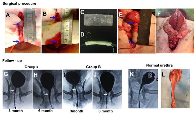
(A-F) During the surgical procedure in a dog model, the urethra between the bladder and the pubic symphysis was exposed, and a 5 cm long urethra section was transected and removed. Then, the scaffold was sutured onto the urethral defect. (G-L) Comparison of urethrography images in each group at 3 and 6 months after operation. The arrow indicates the urethroplasty site of the urethra.
At 3 months, retrograde urethrography revealed the maintenance of a wide urethral caliber without any sign of strictures in group A (Fig. 5 H).
Histological assays were performed on all groups at 1 and 3 months post-operation. At 1 month, all canines in group A had intact epithelial cellular layers: 2-3 layers of well-developed, stratified epithelium (Fig. 6 A, C) and increased numbers of organized muscle bundles were observed (Fig. 6 B, D). At 1 and 3 months, a large amount of new growth vascular formation was observed in groups A and B (Fig. 6 E, J, O, T).”
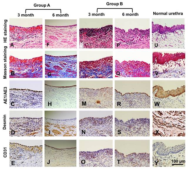
Histologic analysis of reconstructed urethras at 3 and 6 months post-implantation. Evaluation of epithelium, smooth muscle and vessels with AE1/AE3, desmin and CD31 immunohistochemical (IHC) staining in the retrieved urethra; H&E: hematoxylin and eosin.
should be modified to be:
" At 3 month, retrograde urethrograms showed a wide urethral caliber without a fistula or stricture in group A (Fig. 5 G). However, group B revealed mild strictures in all dogs, and bladder distension occurred after catheter removal in 3 dogs at 3 month (Fig. 5 I).
At 6 months, retrograde urethrography revealed the maintenance of a wide urethral caliber without any sign of strictures in group A (Fig. 5 H).
Histological assays were performed on all groups at 3 and 6 months post-operation. At 3 month, all canines in group A had intact epithelial cellular layers: 2-3 layers of well-developed, stratified epithelium (Fig. 6 A, C) and increased numbers of organized muscle bundles were observed (Fig. 6 B, D). At 3 and 6 months, a large amount of new growth vascular formation was observed in groups A and B (Fig. 6 E, J, O, T).".
References
1. Lv X, Feng C, Liu Y. et al. A smart bilayered scaffold supporting keratinocytes and muscle cells in micro/nano-scale for urethral reconstruction. Theranostics. 2018;8(11):3153-63 doi: 10.7150/thno.22080
Author contact
![]() Corresponding author: MuJun Lu MD. PhD; E-mail: lumujuncom; Zhe Li PhD; E-mail: lizhe830817com; YueMin Xu MD. PhD; E-mail: xuyueminnet
Corresponding author: MuJun Lu MD. PhD; E-mail: lumujuncom; Zhe Li PhD; E-mail: lizhe830817com; YueMin Xu MD. PhD; E-mail: xuyueminnet